Review
. 2017 Feb 17:10:38.
doi: 10.3389/fnmol.2017.00038. eCollection 2017.

Molecular Mechanisms Involved in Schwann Cell Plasticity

Affiliations
Review

Molecular Mechanisms Involved in Schwann Cell Plasticity

Angélique Boerboom et al. Front Mol Neurosci. .

Abstract

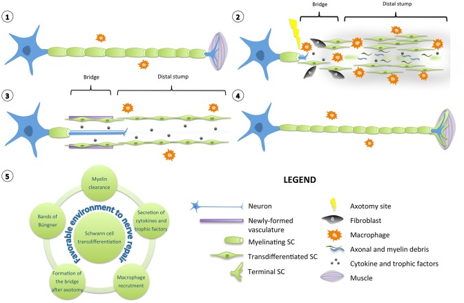
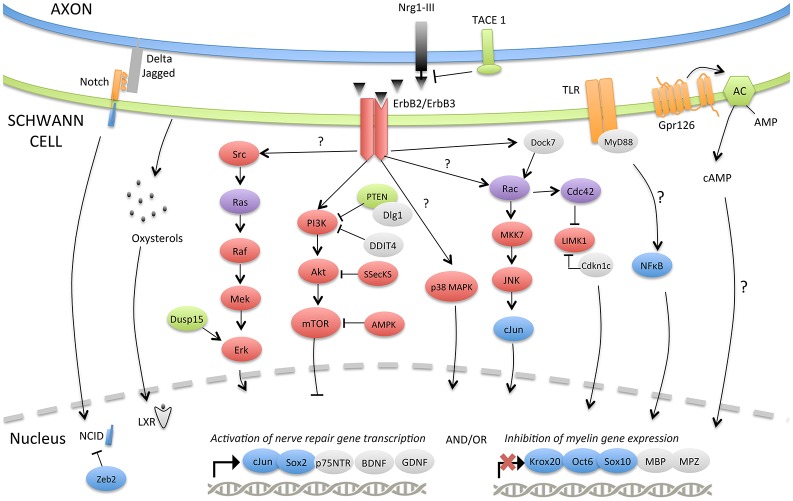
Schwann cell incredible plasticity is a hallmark of the utmost importance following nerve damage or in demyelinating neuropathies. After injury, Schwann cells undergo dedifferentiation before redifferentiating to promote nerve regeneration and complete functional recovery. This review updates and discusses the molecular mechanisms involved in the negative regulation of myelination as well as in the reprogramming of Schwann cells taking place early following nerve lesion to support repair. Significant advance has been made on signaling pathways and molecular components that regulate SC regenerative properties. These include for instance transcriptional regulators such as c-Jun or Notch, the MAPK and the Nrg1/ErbB2/3 pathways. This comprehensive overview ends with some therapeutical applications targeting factors that control Schwann cell plasticity and highlights the need to carefully modulate and balance this capacity to drive nerve repair.

Keywords: Schwann cell; molecular mechanisms; nerve injury; peripheral neuropathy; plasticity.


Figures

Figure 1
Figure 1
Diagram of Schwann cell response to nerve injury. (1) Schematic representation of a single neuron with myelinating SCs and resident macrophages. For simplification, the basal lamina around SCs is not shown. (2) After injury, the nerve distal to the injury site degenerates and undergoes a series of complex multicellular and molecular events in which SCs play a role of orchestrator. SCs transdifferentiate into repair-promoting cells, creating a permissive and favorable environment for nerve regeneration. SCs downregulate pro-myelinating genes and clear their myelin sheaths. They proliferate, secrete several pro-inflammatory cytokines and trophic factors that support glial and neuronal survival and regrowth. Axonal and myelin debris are also phagocyted by resident and blood-derived macrophages recruited by SCs. SCs interact with fibroblasts to build a bridge between the two stumps of the nerve over the lesion site. (3) Newly formed vasculature guides the SCs and the growing axons through the lesion site. In the distal stump, SCs align in tracts named bands of Büngner to provide a trophic and physical support for axons to regrow. (4) After axonal regeneration, transdifferentiated SCs readily exit the cell cycle, differentiate again and remyelinate the axons to support the complete functional recovery. However, the newly-formed myelin sheaths are most of the time shorter and thinner than expected based on axonal diameter. Specialized terminal SCs direct reinnervation by helping the axons to find their paths toward their initial targets. (5) Diagram displaying the various roles played by a transdifferentiated SC to create a favorable environment for nerve repair.
Figure 2
Figure 2
Molecular signaling pathways involved in SC plasticity, negative regulation of myelination and nerve repair. The injury-induced reprogramming of SCs in regeneration-promoting cells involves a down-regulation of pro-myelinating genes including Krox-20, Oct-6, Sox10, MPZ, or MBP as well as an up-regulation of markers characteristic of immature, de-differentiated SCs such as c-Jun and p75NTR but also specific repair-supportive proteins like BDNF or GDNF. The signaling pathways or molecular components activating (c-Jun, Notch, MAPKs, TLRs, Gpr126, NF-κB) or inhibiting (PI3K/Akt/mTOR) this reprogramming are represented. Some negative regulators of myelination during development (TACE1, LXRs, Dusp15, PTEN, Dlg1, DDIT4, Dock7, Zeb2) and possibly playing a role after injury are also shown.

Similar articles

Cited by

References

    1. Agthong S., Kaewsema A., Tanomsridejchai N., Chentanez V. (2006). Activation of MAPK ERK in peripheral nerve after injury. BMC Neurosci. 7:45. 10.1186/1471-2202-7-45 - DOI - PMC - PubMed
    1. Ammoun S., Flaiz C., Ristic N., Schuldt J., Hanemann C. O. (2008). Dissecting and targeting the growth factor-dependent and growth factor-independent extracellular signal-regulated kinase pathway in human schwannoma. Cancer Res. 68, 5236–5245. 10.1158/0008-5472.CAN-07-5849 - DOI - PubMed
    1. Ammoun S., Provenzano L., Zhou L., Barczyk M., Evans K., Hilton D. A., et al. . (2014). Axl/Gas6/NFκB signalling in schwannoma pathological proliferation, adhesion and survival. Oncogene 33, 336–346. 10.1038/onc.2012.587 - DOI - PubMed
    1. Andorfer B., Kieseier B. C., Mathey E., Armati P., Pollard J., Oka N., et al. . (2001). Expression and distribution of transcription factor NF-κB and inhibitor IκB in the inflamed peripheral nervous system. J. Neuroimmunol. 116, 226–232. 10.1016/S0165-5728(01)00306-X - DOI - PubMed
    1. Anliker B., Choi J. W., Lin M.-E., Gardell S. E., Rivera R. R., Kennedy G., et al. . (2013). Lysophosphatidic acid (LPA) and its receptor, LPA1, influence embryonic schwann cell migration, myelination, and cell-to-axon segregation. Glia 61, 2009–2022. 10.1002/glia.22572 - DOI - PMC - PubMed