. 2017 Feb 14:8:43.
doi: 10.3389/fphar.2017.00043. eCollection 2017.

Ginsenoside Rg5 Inhibits Succinate-Associated Lipolysis in Adipose Tissue and Prevents Muscle Insulin Resistance

Affiliations

Ginsenoside Rg5 Inhibits Succinate-Associated Lipolysis in Adipose Tissue and Prevents Muscle Insulin Resistance

Na Xiao et al. Front Pharmacol. .

Abstract

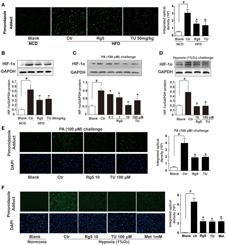
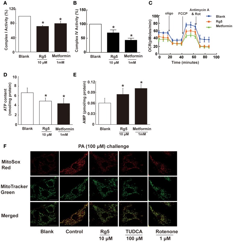
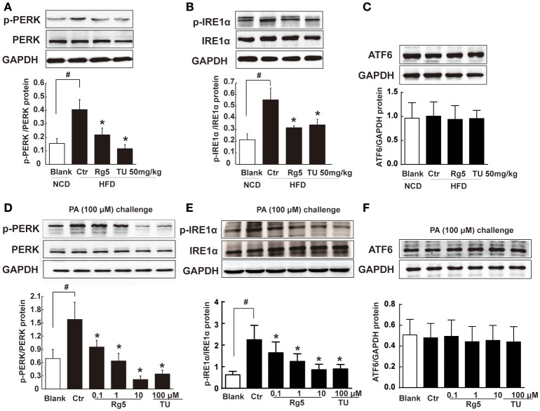
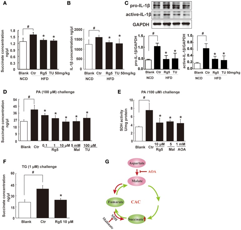
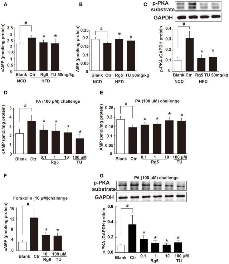
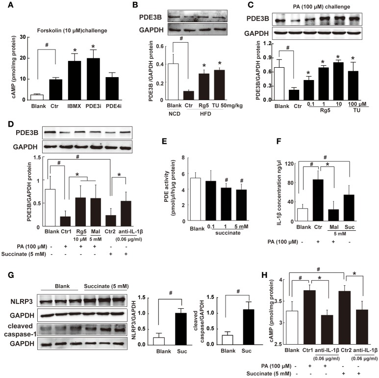
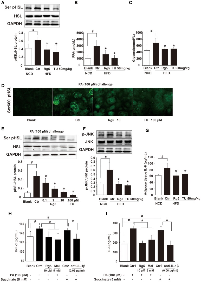
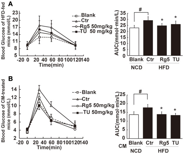
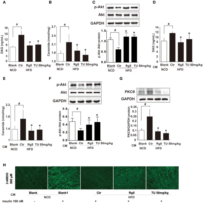
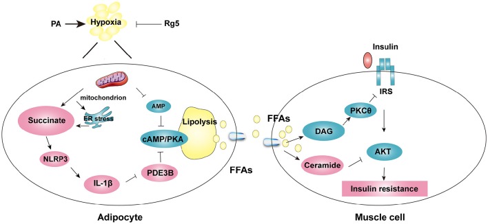
Endoplasmic reticulum (ER) stress, inflammation, and lipolysis occur simultaneously in adipose dysfunction and contribute to insulin resistance. This study was designed to investigate whether ginsenoside Rg5 could ameliorate adipose dysfunction and prevent muscle insulin resistance. Short-term high-fat diet (HFD) feeding induced hypoxia with ER stress in adipose tissue, leading to succinate accumulation due to the reversal of succinate dehydrogenase (SDH) activity. Rg5 treatment reduced cellular energy charge, suppressed ER stress and then prevented succinate accumulation in adipose tissue. Succinate promoted IL-1β production through NLRP3 inflammasome activation and then increased cAMP accumulation by impairing PDE3B expression, leading to increased lipolysis. Ginsenoside Rg5 treatment suppressed NLRP3 inflammasome activation, preserved PDE3B expression and then reduced cAMP accumulation, contributing to inhibition of lipolysis. Adipose lipolysis increased FFAs trafficking from adipose tissue to muscle. Rg5 reduced diacylglycerol (DAG) and ceramides accumulation, inhibited protein kinase Cθ translocation, and prevented insulin resistance in muscle. In conclusion, succinate accumulation in hypoxic adipose tissue acts as a metabolic signaling to link ER stress, inflammation and cAMP/PKA activation, contributing to lipolysis and insulin resistance. These findings establish a previously unrecognized role of ginsenosides in the regulation of lipid and glucose homeostasis and suggest that adipose succinate-associated NLRP3 inflammasome activation might be targeted therapeutically to prevent lipolysis and insulin resistance.

Keywords: ER stress; Ginsenoside Rg5; Lipolysis; hypoxia; insulin resistance; succinate.


Figures

Figure 1
Figure 1
Effects of Rg5 on adipose hypoxia. (A) Adipose hypoxia examination with pimonidazole staining in HFD mice. (B) HIF-1α protein expression in adipose tissue of HFD-fed mice. (C,D) HIF-1α protein expression in differentiated adipocytes treated with palmitate (PA), 1% O2 for 8 h. (E,F) Pimonidazole staining in differentiated adipocytes treated with PA for 8 h or 1% O2 for 4 h. The results were expressed as the mean ± SD of three independent experiments. *p < 0.05 vs. Control (Ctr), #p < 0.05 vs. Blank. TU, Tauroursodeoxycholic Acid; Met, Metformin.
Figure 2
Figure 2
Rg5 reduced cellular energy charge in differentiated adipocytes. (A,B) Mitochondrial respiratory complex I and IV activity in differentiated adipocytes (n = 3). (C) Oxygen consumption ratio (OCR) in differentiated adipocytes (n = 4). (D,E) ATP and AMP contents in differentiated adipocytes (n = 6). (F) Mitochondrial ROS levels were measured by MitoSox Red (red) using confocal microscopy (n = 3). MitoSox Red was colocalized with MitoTracker Green. Scale bar, 20 μM. Data above were expressed as the mean ± SD. *p < 0.05 vs. Blank. TUDCA, Tauroursodeoxycholic Acid.
Figure 3
Figure 3
Rg5 suppressed ER stress in adipose tissue. (A–C) PERK, IRE1α phosphorylations (n = 3) and ATF6 protein expression (n = 4) in adipose tissue of HFD-fed mice. (D–F) PERK (n = 3), IRE1α (n = 4) phosphorylations and ATF6 (n = 4) protein expression in differentiated adipocytes incubated with palmitate (PA) for 8 h. The results were expressed as the mean ± SD. *p < 0.05 vs. Control (Ctr), #p < 0.05 vs. Blank. TU, Tauroursodeoxycholic Acid; oligo, oligomycin; rot, rotenone.
Figure 4
Figure 4
Rg5 reduced succinate accumulation in adipose tissue. (A,B) Succinate and IL-1β contents in adipose tissue of HFD mice (n = 6–8). (C) Protein expression of pro-IL-1β and active IL-1β in adipose tissue was determined by immunoblot analysis (n = 3). (D,F) Succinate accumulation in differentiated adipocytes treated with palmitate (PA) or thapsigargin (TG) for 8 h (n = 6). (E) SDH activity in differentiated adipocytes treated with PA for 8 h. (G) Summary of MAS pathway as potential drivers for the reversal of succinate accumulation. The results were expressed as the mean ± SD (n = 4). *p < 0.05 vs. Control (Ctr), #p < 0.05 vs. Blank. TU, Tauroursodeoxycholic Acid; AOA, aminooxyacetate; Mal, dimethyl malonate.
Figure 5
Figure 5
Rg5 blocked cAMP/PKA signaling. (A,B) cAMP and AMP contents in adipose tissue of HFD-fed mice (n = 8). (C) PKA phosphorylation in adipose tissue of HFD-fed mice (n = 3). (D,E) cAMP and AMP contents in differentiated adipocytes incubated with palmitate (PA) for 8 h (n = 6). (F) cAMP contents in differentiated adipocytes pretreated with agents for 4 h, and stimulated with forskolin for 0.5 h (n = 8). (G) PKA phosphorylation in differentiated adipocytes treated with PA for 8 h (n = 3). The results were expressed as the mean ± SD. *p < 0.05 vs. Control (Ctr), #p < 0.05 vs. Blank. TU, Tauroursodeoxycholic Acid.
Figure 6
Figure 6
Succinate increased cAMP accumulation. (A) Differentiated adipocytes were incubated with 20 μM IBMX, 20 μM Cilostamide (PDE3Bi), or 50 μM Ro-20-1724 (PDE4i) for 0.5 h. Cells were treated with 10 μM forskolin for 0.5 h, lysed, and total cellular cAMP assayed. (B,C) PDE3B expression in adipose tissue of HFD-fed mice (n = 3) or in palmitate (PA)-treated adipocytes (n = 4) were determined with western blot. (D) PDE3B expression in PA- or succinate-treated differentiated adipocytes (n = 3). (E) PDE activity in differentiated adipocytes incubated with succinate for 8 h (n = 4). (F) IL-1β contents in differentiated adipocytes were determined with ELISA (n = 6). (G) NLRP3 and cleaved caspase-1 expression in differentiated adipocytes were determined with western blot (n = 3). (H) cAMP contents in adipocytes were determined with ELISA (n = 6). The results were expressed as the mean ± SD. *p < 0.05 vs. Control (Ctr), #p < 0.05 vs. Blank. TU, Tauroursodeoxycholic Acid; Mal, dimethyl malonate; Suc, dimethyl succinate.
Figure 7
Figure 7
Rg5 inhibited FFAs release and inflammation in adipose tissue. (A) HSL phosphorylation in adipose tissue of HFD-fed mice (n = 3). (B,C) FFAs, Glycerol release from adipose tissue of HFD-fed mice (n = 6–8). (D,E) HSL recruitment and phosphorylation in adipocytes treated with palmitate (PA) (n = 3) for 8 h. (F,G) JNK expression (n = 3) and IL-6 (n = 6) contents in adipose tissue of HFD mice. (H,I) TNF-α and IL-6 contents in adipocytes (n = 6). GAPDH band was duplicated for (A,F) as a result of the experiments having been performed at the same time. Data were expressed as the mean ± SD. *p < 0.05 vs. Control (Ctr), #p < 0.05 vs. Blank. TU, Tauroursodeoxycholic Acid; Mal, dimethyl malonate.
Figure 8
Figure 8
Rg5 improved glucose tolerance in HFD-fed mice. (A) Oral glucose tolerance in HFD mice. (B) Oral glucose tolerance in normal mice treated with CM. Data were expressed as the mean ± SD (n = 10). *p < 0.05 vs. Control (Ctr), #p < 0.05 vs. Blank. TU, Tauroursodeoxycholic Acid.
Figure 9
Figure 9
Rg5 reduced DAG and ceramide contents and improved insulin signaling in the muscle. (A,B) DAG and ceramide contents in the muscle of HFD mice (n = 6–8). (C) Akt phosphorylation in response to glucose load in the muscle of HFD mice (western blot, n = 4). (D–F) Normal mice were injected with CM, DAG, ceramide contents (n = 6–8) and glucose load-induced Akt phosphorylation (n = 5) in the muscle were determined with ELISA or western blot. (G,H) Differentiated C2C12 myotubes were incubated with CM, and membrane PKCθ and glucose uptake were examined. Data were expressed as the mean ± SD (n = 3). *p < 0.05 vs. Control (Ctr), #p < 0.05 vs. Blank. TU, Tauroursodeoxycholic Acid.
Figure 10
Figure 10
The proposed working pathway for ginsenoside Rg5 action in hypoxic adipose tissue. Saturated fatty acid PA induces hypoxia with ER stress in adipose tissue, and then increases succinate formation. Succinate promotes IL-1β production through NLRP3 inflammasome activation, and then induces cAMP/PKA signaling by inactivation of PDE3B, leading to lipolysis. PA also decreases AMP concentration, and then increases cAMP accumulation. The release of FFAs increases cytosolic DAG, PKC translocation, ceramide accumulation and inhibits insulin-mediated Akt phosphorylation, thus leading to insulin resistance in muscle. Rg5 inhibits lipolysis by suppression of succinate accumulation in hypoxic adipocytes and thus prevents insulin resistance in muscle.

Similar articles

Cited by

References

    1. Bogdanovic E., Kraus N., Patsouris D. (2015). Endoplasmic reticulum stress in adipose tissue augments lipolysis. J. Cell. Mol. Med. 19, 82–91. 10.1111/jcmm.12384 - DOI - PMC - PubMed
    1. Chaurasia B., Summers S. A. (2015). Ceramides - lipotoxic inducers of metabolic disorders. Trends Endocrinol. Metab. 26, 538–550. 10.1016/j.tem.2015.07.006 - DOI - PubMed
    1. Chen W., Wang J., Luo Y., Wang T., Li X., Li A., et al. . (2016). Ginsenoside Rb1 and compound K improve insulin signaling and inhibit ER stress-associated NLRP3 inflammasome activation in adipose tissue. J. Ginseng Res. 40, 351–358. 10.1016/j.jgr.2015.11.002 - DOI - PMC - PubMed
    1. Chouchani E. T., Pell V. R., Gaude E., Aksentijević D., Sundier S. Y., Robb E. L., et al. . (2014). Ischaemic accumulation of succinate controls reperfusion injury through mitochondrial ROS. Nature 515, 431–435. 10.1038/nature13909 - DOI - PMC - PubMed
    1. Coppack S. W., Evans R. D., Fisher R. M., Frayn K. N., Gibbons G. F., Humphreys S. M., et al. . (1992). Adipose tissue metabolism in obesity: lipase action in vivo before and after a mixed meal. Metab. Clin. Exp. 41, 264–272. 10.1016/0026-0495(92)90269-G - DOI - PubMed