

Sixth Nerve Palsy from Cholesterol Granuloma of the Petrous Apex
- PMID: 28261154
- PMCID: PMC5309254
- DOI: 10.3389/fneur.2017.00048
Sixth Nerve Palsy from Cholesterol Granuloma of the Petrous Apex
Abstract
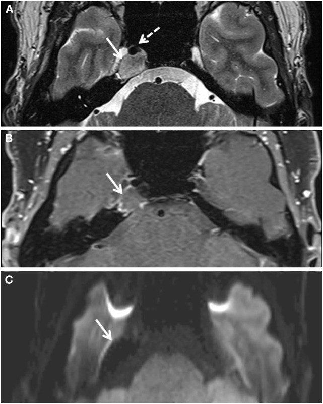
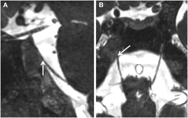
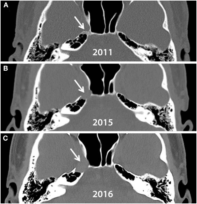
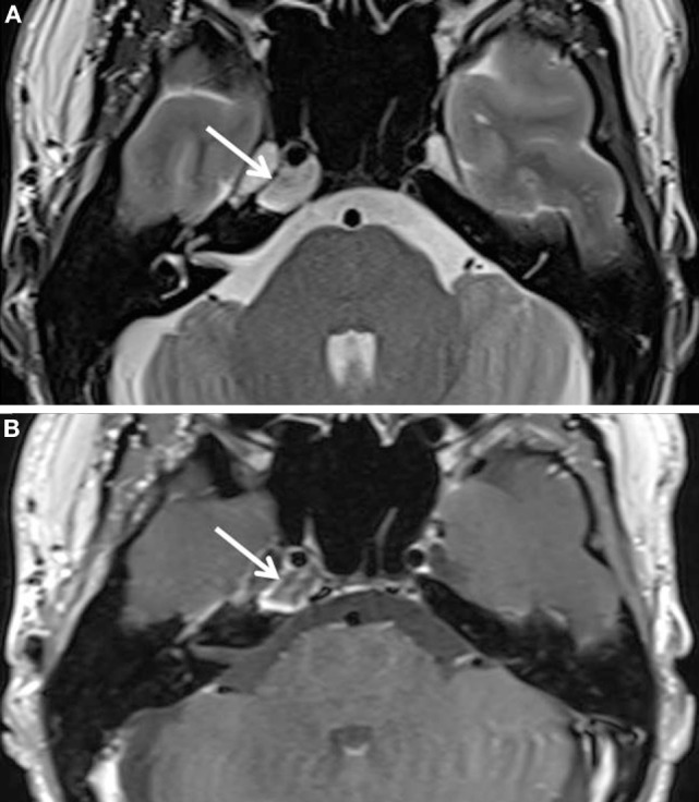
Herein, we report a patient who had an isolated sixth nerve palsy due to a petrous apex cholesterol granuloma. The sixth nerve palsy appeared acutely and then spontaneously resolved over several months, initially suggesting a microvascular origin of the palsy. Subsequent recurrences of the palsy indicated a different pathophysiologic etiology and MRI revealed the lesion at the petrous apex. Surgical resection improved the compressive effect of the lesion at Dorello's canal and clinical improvement was observed. A relapsing-remitting sixth nerve palsy is an unusual presentation of this rare lesion.
Keywords: abducens palsy; cholesterol granuloma; diplopia; esotropia; petrous apex tumor; sixth nerve palsy; skull-base tumor.
Figures
Similar articles
-
Gruber, Gradenigo, Dorello, and Vail: key personalities in the historical evolution and modern-day understanding of Dorello's canal.J Neurosurg. 2016 Jan;124(1):224-33. doi: 10.3171/2014.12.JNS14835. Epub 2015 Jun 26. J Neurosurg. 2016. PMID: 26115474
-
Abducens nerve palsy due to inferior petrosal sinus thrombosis.J Clin Neurosci. 2017 Jun;40:69-71. doi: 10.1016/j.jocn.2017.02.018. Epub 2017 Feb 24. J Clin Neurosci. 2017. PMID: 28242132
-
Isolated Sixth Nerve Palsy as a First Presentation of Nasopharyngeal Carcinoma: A Case Series.Int Med Case Rep J. 2021 Nov 23;14:801-808. doi: 10.2147/IMCRJ.S334476. eCollection 2021. Int Med Case Rep J. 2021. PMID: 34849037 Free PMC article.
-
Cholesterol granuloma and other petrous apex lesions.Otolaryngol Clin North Am. 2015 Apr;48(2):361-73. doi: 10.1016/j.otc.2014.12.009. Epub 2015 Jan 31. Otolaryngol Clin North Am. 2015. PMID: 25650229 Review.
-
Unique Diagnostic Features and Surgical Strategy for Intracranial Carotid Sympathetic Plexus Schwannoma: Case Report and Literature Review.World Neurosurg. 2017 Feb;98:876.e1-876.e8. doi: 10.1016/j.wneu.2016.11.105. Epub 2016 Dec 1. World Neurosurg. 2017. PMID: 27916722 Review.
Cited by
-
Endoscopic transsphenoidal resection of a recurrent petrous apex cholesterol granuloma: Operative video.Surg Neurol Int. 2020 Apr 25;11:83. doi: 10.25259/SNI_40_2020. eCollection 2020. Surg Neurol Int. 2020. PMID: 32844048 Free PMC article.
References
-
- Graham MD, Kemink JL, Latack JT, Kartush JM. The giant cholesterol cyst of the petrous apex: a distinct clinical entity. Laryngoscope (1985) 95:1401–6. - PubMed
-
- Ojala L. Pneumatization of the bone and environmental factors: experimental studies on chick humerus. Acta Otolaryngol Suppl (1957) 133:1–28. - PubMed
-
- Beaumont GD. The effects of exclusion of air from pneumatized bones. J Laryngol Otol (1966) 80:236–49. - PubMed
-
- Jackler RK, Cho M. A new theory to explain the genesis of petrous apex cholesterol granuloma. Otol Neurotol (2003) 24:96–106. - PubMed
-
- Mafee MF, Kumar A, Heffner DK. Epidermoid cyst (cholesteatoma) and cholesterol granuloma of the temporal bone and epidermoid cysts affecting the brain. Neuroimaging Clin N Am (1994) 4:561–78. - PubMed
Publication types
LinkOut - more resources
Full Text Sources
Other Literature Sources
Miscellaneous