

Current Stance of Magnetic Resonance Imaging in the Diagnosis and Monitoring of Hepatic Encephalopathy
- PMID: 28261361
- PMCID: PMC5327831
Current Stance of Magnetic Resonance Imaging in the Diagnosis and Monitoring of Hepatic Encephalopathy
Abstract
Objectives: Hepatic encephalopathy is a complex of neuropsychiatric manifestations in patients with acute or chronic liver insufficiency and/or porto-systemic shunts.
Material and methods: The diagnostic can be sustained by various elements, clinical and paraclinical. Selected patients with hepatic encephalopathy have been investigated by Magnetic Resonance, in parallel with specific biochemical analysis.
Outcomes: This paper emphasizes the importance of Magnetic Resonance Imaging in an accurate diagnosis and patient monitoring after treatment.
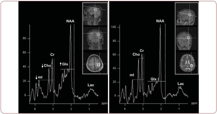
Conclusions: Magnetic Resonance Spectroscopy has a substantial role, showing even minute metabolite ratio changes, with a potential in investigating minimal hepatic encephalopathy.
Figures
Similar articles
-
Usefulness of magnetic resonance spectroscopy for diagnosis of hepatic encephalopathy in a patient with relapsing confusional syndrome.Dig Dis Sci. 2001 Nov;46(11):2451-5. doi: 10.1023/a:1012323819757. Dig Dis Sci. 2001. PMID: 11713951
-
Does magnetic resonance spectroscopy identify patients with minimal hepatic encephalopathy?Neurol Neurochir Pol. 2012 Sep-Oct;46(5):436-42. doi: 10.5114/ninp.2012.31353. Neurol Neurochir Pol. 2012. PMID: 23161187 Clinical Trial.
-
[Cerebral magnetic resonance spectroscopy in patients with hepatic encephalopathy: analysis before and after liver transplantation].Rev Assoc Med Bras (1992). 2009 Jan-Feb;55(1):35-9. doi: 10.1590/s0104-42302009000100012. Rev Assoc Med Bras (1992). 2009. PMID: 19360275 Portuguese.
-
Pathogenesis, diagnosis, and treatment of hepatic encephalopathy.J Clin Exp Hepatol. 2011 Sep;1(2):77-86. doi: 10.1016/S0973-6883(11)60126-6. Epub 2011 Nov 9. J Clin Exp Hepatol. 2011. PMID: 25755319 Free PMC article. Review.
-
Brain magnetic resonance in hepatic encephalopathy.Semin Ultrasound CT MR. 2014 Apr;35(2):136-52. doi: 10.1053/j.sult.2013.09.008. Semin Ultrasound CT MR. 2014. PMID: 24745889 Review.
Cited by
-
Recent advances in hepatic encephalopathy.F1000Res. 2017 Sep 4;6:1637. doi: 10.12688/f1000research.11938.1. eCollection 2017. F1000Res. 2017. PMID: 29026534 Free PMC article. Review.
References
LinkOut - more resources
Full Text Sources