

Improved accuracy in periodontal pocket depth measurement using optical coherence tomography
- PMID: 28261520
- PMCID: PMC5332330
- DOI: 10.5051/jpis.2017.47.1.13
Improved accuracy in periodontal pocket depth measurement using optical coherence tomography
Abstract
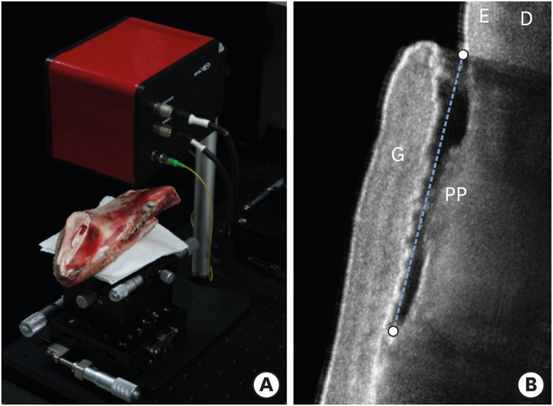
Purpose: The purpose of this study was to examine whether periodontal pocket could be satisfactorily visualized by optical coherence tomography (OCT) and to suggest quantitative methods for measuring periodontal pocket depth.
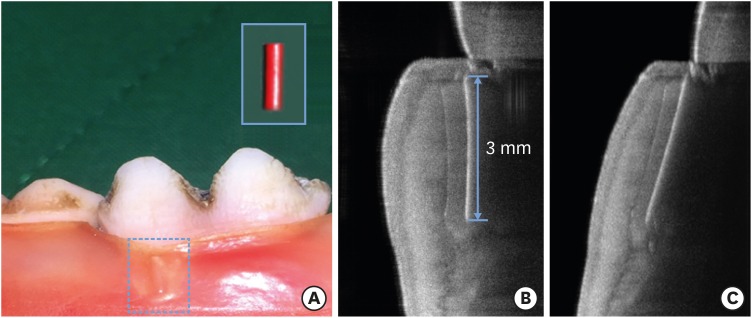
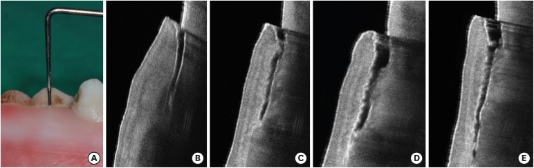
Methods: We acquired OCT images of periodontal pockets in a porcine model and determined the actual axial resolution for measuring the exact periodontal pocket depth using a calibration method. Quantitative measurements of periodontal pockets were performed by real axial resolution and compared with the results from manual periodontal probing.
Results: The average periodontal pocket depth measured by OCT was 3.10±0.15 mm, 4.11±0.17 mm, 5.09±0.17 mm, and 6.05±0.21 mm for each periodontal pocket model, respectively. These values were similar to those obtained by manual periodontal probing.
Conclusions: OCT was able to visualize periodontal pockets and show attachment loss. By calculating the calibration factor to determine the accurate axial resolution, quantitative standards for measuring periodontal pocket depth can be established regardless of the position of periodontal pocket in the OCT image.
Keywords: Computer-assisted image interpretation; Gingiva; Optical coherence tomography; Periodontal pocket.
Conflict of interest statement
Conflict of Interest: No potential conflict of interest relevant to this article was reported.
Figures
Similar articles
-
Observation and determination of periodontal tissue profile using optical coherence tomography.J Periodontal Res. 2018 Apr;53(2):188-199. doi: 10.1111/jre.12506. Epub 2017 Oct 24. J Periodontal Res. 2018. PMID: 29063599
-
Non-Invasive Periodontal Probing Through Fourier-Domain Optical Coherence Tomography.J Periodontol. 2015 Sep;86(9):1087-94. doi: 10.1902/jop.2015.150047. Epub 2015 Apr 16. J Periodontol. 2015. PMID: 25879790
-
In vivo assessment of periodontal structures and measurement of gingival sulcus with Optical Coherence Tomography: a pilot study.J Biophotonics. 2017 Jun;10(6-7):862-869. doi: 10.1002/jbio.201600082. Epub 2016 Aug 9. J Biophotonics. 2017. PMID: 27503608
-
Periodontal probing: what does it mean?J Clin Periodontol. 1980 Jun;7(3):165-76. doi: 10.1111/j.1600-051x.1980.tb01960.x. J Clin Periodontol. 1980. PMID: 7000852 Review.
-
Optical Coherence Tomography.Dent Clin North Am. 2018 Jul;62(3):421-434. doi: 10.1016/j.cden.2018.03.004. Dent Clin North Am. 2018. PMID: 29903559 Review.
Cited by
-
Quantitative measurement of peri-implant bone defects using optical coherence tomography.J Periodontal Implant Sci. 2018 Apr 30;48(2):84-91. doi: 10.5051/jpis.2018.48.2.84. eCollection 2018 Apr. J Periodontal Implant Sci. 2018. PMID: 29770237 Free PMC article.
-
Impact of Optical Coherence Tomography (OCT) for Periodontitis Diagnostics: Current Overview and Advances.Dent J (Basel). 2025 Jul 4;13(7):305. doi: 10.3390/dj13070305. Dent J (Basel). 2025. PMID: 40710150 Free PMC article. Review.
-
Evaluation Through the Optical Coherence Tomography Analysis of the Influence of Non-Alcoholic Fatty Liver Disease on the Gingival Inflammation in Periodontal Patients.Diabetes Metab Syndr Obes. 2021 Jun 29;14:2935-2942. doi: 10.2147/DMSO.S310314. eCollection 2021. Diabetes Metab Syndr Obes. 2021. PMID: 34234491 Free PMC article.
-
Chlamydia trachomatis in the gingival sulcus and pharynx in patients of Northeast Mexico.Clin Exp Dent Res. 2020 Aug;6(4):415-419. doi: 10.1002/cre2.290. Epub 2020 Mar 27. Clin Exp Dent Res. 2020. PMID: 32220009 Free PMC article.
-
Microbiota and Metatranscriptome Changes Accompanying the Onset of Gingivitis.mBio. 2018 Apr 17;9(2):e00575-18. doi: 10.1128/mBio.00575-18. mBio. 2018. PMID: 29666288 Free PMC article.
References
LinkOut - more resources
Full Text Sources
Other Literature Sources
