

Automatic detection of tooth cracks in optical coherence tomography images
- PMID: 28261523
- PMCID: PMC5332334
- DOI: 10.5051/jpis.2017.47.1.41
Automatic detection of tooth cracks in optical coherence tomography images
Abstract
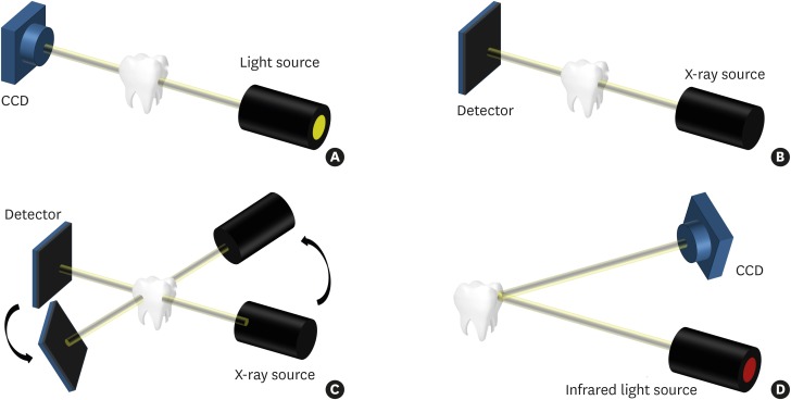
Purpose: The aims of the present study were to compare the image quality and visibility of tooth cracks between conventional methods and swept-source optical coherence tomography (SS-OCT) and to develop an automatic detection technique for tooth cracks by SS-OCT imaging.
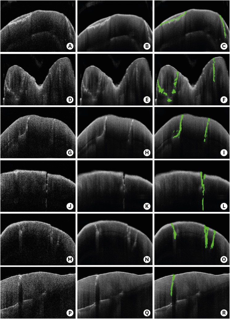
Methods: We evaluated SS-OCT with a near-infrared wavelength centered at 1,310 nm over a spectral bandwidth of 100 nm at a rate of 50 kHz as a new diagnostic tool for the detection of tooth cracks. The reliability of the SS-OCT images was verified by comparing the crack lines with those detected using conventional methods. After performing preprocessing of the obtained SS-OCT images to emphasize cracks, an algorithm was developed and verified to detect tooth cracks automatically.
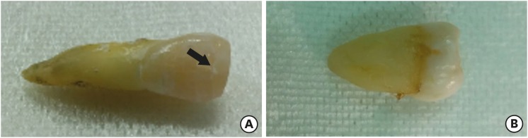
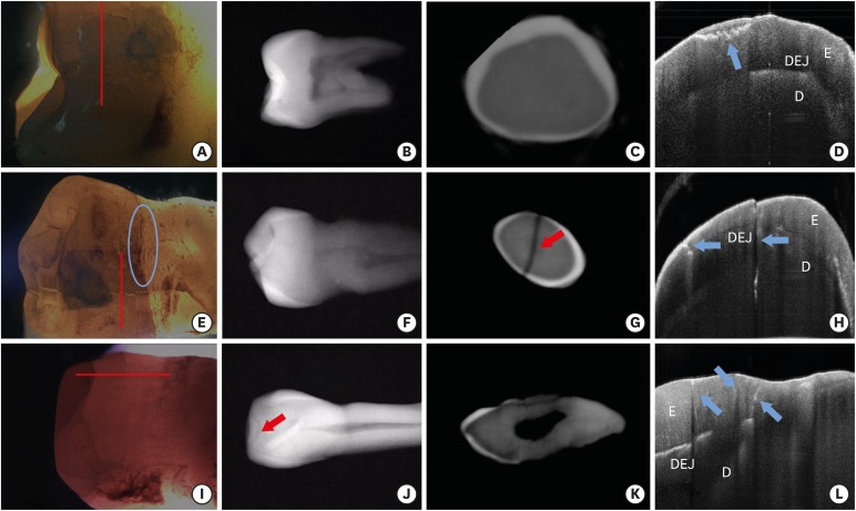
Results: The detection capability of SS-OCT was superior or comparable to that of trans-illumination, which did not discriminate among the cracks according to depth. Other conventional methods for the detection of tooth cracks did not sense initial cracks with a width of less than 100 μm. However, SS-OCT detected cracks of all sizes, ranging from craze lines to split teeth, and the crack lines were automatically detected in images using the Hough transform.
Conclusions: We were able to distinguish structural cracks, craze lines, and split lines in tooth cracks using SS-OCT images, and to automatically detect the position of various cracks in the OCT images. Therefore, the detection capability of SS-OCT images provides a useful diagnostic tool for cracked tooth syndrome.
Keywords: Computer-assisted image interpretation; Cracked tooth syndrome; Optical coherence tomography; Tooth fractures.
Conflict of interest statement
Conflict of Interest: No potential conflict of interest relevant to this article was reported.
Figures
Similar articles
-
Recent Advances in the Diagnosis of Enamel Cracks: A Narrative Review.Diagnostics (Basel). 2022 Aug 22;12(8):2027. doi: 10.3390/diagnostics12082027. Diagnostics (Basel). 2022. PMID: 36010379 Free PMC article. Review.
-
Tooth cracks detection and gingival sulcus depth measurement using optical coherence tomography.Annu Int Conf IEEE Eng Med Biol Soc. 2017 Jul;2017:4403-4406. doi: 10.1109/EMBC.2017.8037832. Annu Int Conf IEEE Eng Med Biol Soc. 2017. PMID: 29060873
-
Dental optical coherence tomography: new potential diagnostic system for cracked-tooth syndrome.Surg Radiol Anat. 2016 Jan;38(1):49-54. doi: 10.1007/s00276-015-1514-8. Epub 2015 Jul 14. Surg Radiol Anat. 2016. PMID: 26168856
-
Noninvasive cross-sectional visualization of enamel cracks by optical coherence tomography in vitro.J Endod. 2012 Sep;38(9):1269-74. doi: 10.1016/j.joen.2012.05.008. Epub 2012 Jul 20. J Endod. 2012. PMID: 22892749
-
Evaluation of dental caries, tooth crack, and age-related changes in tooth structure using optical coherence tomography.Jpn Dent Sci Rev. 2020 Nov;56(1):109-118. doi: 10.1016/j.jdsr.2020.08.001. Epub 2020 Oct 2. Jpn Dent Sci Rev. 2020. PMID: 33033549 Free PMC article. Review.
Cited by
-
Three Visual-Diagnostic Methods for the Detection of Enamel Cracks: An In Vitro Study.J Clin Med. 2023 Jan 27;12(3):973. doi: 10.3390/jcm12030973. J Clin Med. 2023. PMID: 36769621 Free PMC article.
-
A method of crack detection based on digital image correlation for simulated cracked tooth.BMC Oral Health. 2021 Oct 19;21(1):539. doi: 10.1186/s12903-021-01897-2. BMC Oral Health. 2021. PMID: 34666731 Free PMC article.
-
Recent Advances in the Diagnosis of Enamel Cracks: A Narrative Review.Diagnostics (Basel). 2022 Aug 22;12(8):2027. doi: 10.3390/diagnostics12082027. Diagnostics (Basel). 2022. PMID: 36010379 Free PMC article. Review.
-
The Use of Optical Coherence Tomography in Dental Diagnostics: A State-of-the-Art Review.J Healthc Eng. 2017;2017:7560645. doi: 10.1155/2017/7560645. Epub 2017 Jul 16. J Healthc Eng. 2017. PMID: 29065642 Free PMC article. Review.
-
Review of Cracked Tooth Syndrome: Etiology, Diagnosis, Management, and Prevention.Pain Res Manag. 2021 Dec 15;2021:3788660. doi: 10.1155/2021/3788660. eCollection 2021. Pain Res Manag. 2021. PMID: 34956432 Free PMC article. Review.
References
-
- Ehrmann EH, Tyas MJ. Cracked tooth syndrome: diagnosis, treatment and correlation between symptoms and post-extraction findings. Aust Dent J. 1990;35:105–112. - PubMed
-
- Lynch CD, McConnell RJ. The cracked tooth syndrome. J Can Dent Assoc. 2002;68:470–475. - PubMed
-
- Kahler W. The cracked tooth conundrum: terminology, classification, diagnosis, and management. Am J Dent. 2008;21:275–282. - PubMed
-
- Hayashi M, Kinomoto Y, Miura M, Sato I, Takeshige F, Ebisu S. Short-term evaluation of intentional replantation of vertically fractured roots reconstructed with dentin-bonded resin. J Endod. 2002;28:120–124. - PubMed
-
- Lin CC, Tsai YL, Li UM, Chang YC, Lin CP, Jeng JH. Horizontal/oblique root fractures in the palatal root of maxillary molars with associated periodontal destruction: case reports. Int Endod J. 2008;41:442–447. - PubMed
LinkOut - more resources
Full Text Sources
Other Literature Sources
