

Temporomandibular Joint Anatomy Assessed by CBCT Images
- PMID: 28261607
- PMCID: PMC5312052
- DOI: 10.1155/2017/2916953
Temporomandibular Joint Anatomy Assessed by CBCT Images
Abstract
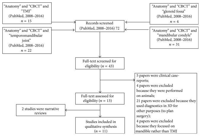
Aim. Since cone beam computed tomography (CBCT) has been used for the study of craniofacial morphology, the attention of orthodontists has also focused on the mandibular condyle. The purpose of this brief review is to summarize the recent 3D CBCT images of mandibular condyle. Material and Methods. The eligibility criteria for the studies are (a) studies aimed at evaluating the anatomy of the temporomandibular joint; (b) studies performed with CBCT images; (c) studies on human subjects; (d) studies that were not clinical case-reports and clinical series; (e) studies reporting data on children, adolescents, or young adults (data from individuals with age ≤ 30 years). Sources included PubMed from June 2008 to June 2016. Results. 43 full-text articles were initially screened for eligibility. 13 full-text articles were assessed for eligibility. 11 articles were finally included in qualitative synthesis. The main topics treated in the studies are the volume and surface of the mandibular condyle, the bone changes on cortical surface, the facial asymmetry, and the optimum position of the condyle in the glenoid fossa. Conclusion. Additional studies will be necessary in the future, constructed with longitudinal methodology, especially in growing subjects. The limits of CBCT acquisitions are also highlighted.
Conflict of interest statement
The authors declare that they have no competing interests.
References
-
- Chen J., Sorensen K. P., Gupta T., Kilts T., Young M., Wadhwa S. Altered functional loading causes differential effects in the subchondral bone and condylar cartilage in the temporomandibular joint from young mice. Osteoarthritis and Cartilage. 2009;17(3):354–361. doi: 10.1016/j.joca.2008.05.021. - DOI - PMC - PubMed
Publication types
MeSH terms
LinkOut - more resources
Full Text Sources
Other Literature Sources
