. 2017:2017:3427319.
doi: 10.1155/2017/3427319. Epub 2017 Feb 5.

Correlation between miR-148 Expression in Vitreous and Severity of Rhegmatogenous Retinal Detachment

Affiliations

Correlation between miR-148 Expression in Vitreous and Severity of Rhegmatogenous Retinal Detachment

Taichi Tsunekawa et al. Biomed Res Int. 2017.

Abstract

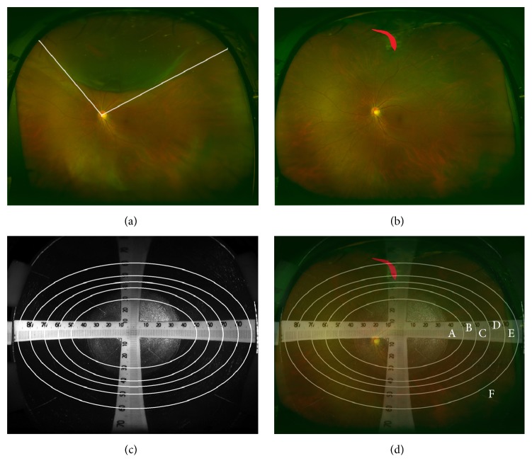
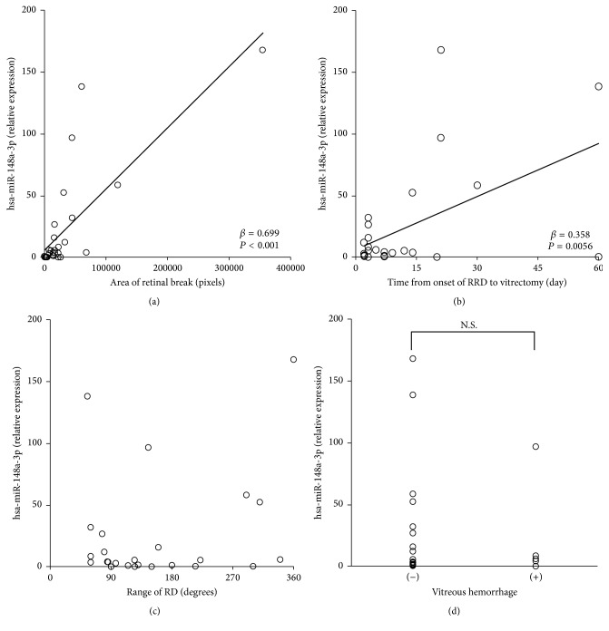
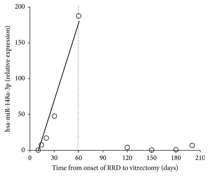
Purpose. We had earlier reported positive hsa-miR-148a-3p expression in eyes with rhegmatogenous retinal detachment (RRD) and its involvement in the epithelial-mesenchymal transition of retinal pigment epithelium in vitro. Here we investigated the association of hsa-miR-148a-3p expression levels in the vitreous fluid of patients with RRD with severity of RRD. Methods. The hsa-miR-148a-3p expression levels in the vitreous fluid, range (degree) of retinal detachment (RD), and pixels of retinal break were measured in 27 eyes with RRD. The association of hsa-miR-148a-3p expression levels with other factors was evaluated by multiple regression analysis. Results. The hsa-miR-148a-3p expression levels, time from onset of RRD to vitrectomy, range of RD, and pixels of retinal breaks were 23.68 ± 43.00, 12.07 ± 15.36 days, 155.85 ± 86.67 degrees, and 37000 ± 67100 pixels, respectively. Five eyes with RRD had vitreous hemorrhage preoperatively. The hsa-miR-148a-3p expression levels were significantly associated with pixels of retinal breaks (β = 0.699) and the time from onset of RRD to vitrectomy (β = 0.358) but not with the range of RD or presence of vitreous hemorrhage. Conclusion. The hsa-miR-148a-3p expression levels in the vitreous fluid were significantly associated with the size of retinal break and disease duration.


Conflict of interest statement

The authors declare that they have no competing interests.

Figures

Figure 1
Figure 1
Measurement of the pixels in retinal break and the range of RD. (a) Representative preoperative fundus image from 55-year-old male patient with RRD obtained with ultra-wide-field scanning ophthalmoscope. The angle of RD was measured by the two lines starting from the optic disc. (b) The postoperative fundus image from the same patient captured by ultra-wide-field scanning ophthalmoscope. The retinal break is filled in red. (c) The fundus image of the simulated eye with scale obtained with ultra-wide-field scanning ophthalmoscope. The circles delineating the areas with the same size were designed based on the scale. (d) Merged image of (b) and (c). Total pixels of the retinal break (filled in red) were corrected based on the ratio of each area (A–F). RRD, rhegmatogenous retinal detachment; RD, retinal detachment.
Figure 2
Figure 2
Relationship between hsa-miR-148a-3p expression and clinical parameters of RD. Open circles represent the values in each case (a, b, c, and d). The expression level of hsa-miR-148a-3p showed significant correlations with the area of the retinal break and the time from onset of RRD to vitrectomy. (c) The expression levels of hsa-miR-148a-3p did not show significant correlations with the range of the RD (P = 0.985). (d) The existence of the vitreous hemorrhage did not show significant difference in hsa-miR-148a-3p expression level (P = 0.661). RRD, rhegmatogenous retinal detachment; RD, retinal detachment; N.S., not significant.
Figure 3
Figure 3
Expression levels of hsa-miR-148a-3p in proliferative vitreoretinopathy (PVR) and time from onset of RRD to vitrectomy. In patients with PVR that underwent vitrectomy within 2 months after the onset of RRD (based on patient declaration), the expression levels of hsa-miR-148a-3p showed a significant correlation with time from onset of RRD to vitrectomy (r = 1.0, P < 0.01). Low expression levels of hsa-miR-148a-3p were observed in patients who underwent surgery ≥ 60 days after the onset of RRD. RRD, rhegmatogenous retinal detachment.

Similar articles

Cited by

References

    1. Bourne R. R. A., Stevens G. A., White R. A., et al. Causes of vision loss worldwide, 1990–2010: a systematic analysis. The Lancet Global Health. 2013;1(6):e339–e349. doi: 10.1016/s2214-109x(13)70113-x. - DOI - PubMed
    1. Mitry D., Charteris D. G., Fleck B. W., Campbell H., Singh J. The epidemiology of rhegmatogenous retinal detachment: geographical variation and clinical associations. British Journal of Ophthalmology. 2010;94(6):678–684. doi: 10.1136/bjo.2009.157727. - DOI - PubMed
    1. Colucciello M. Rhegmatogenous retinal detachment. Physician and Sportsmedicine. 2009;37(2):59–65. doi: 10.3810/psm.2009.06.1710. - DOI - PubMed
    1. Kreissig I. Surgical techniques for repair of primary retinal detachment: part I. Review of their development during the last 80 years. Folia Medica. 2009;51(4):5–11. - PubMed
    1. Sodhi A., Leung L.-S., Do D. V., Gower E. W., Schein O. D., Handa J. T. Recent trends in the management of rhegmatogenous retinal detachment. Survey of Ophthalmology. 2008;53(1):50–67. doi: 10.1016/j.survophthal.2007.10.007. - DOI - PubMed

MeSH terms

LinkOut - more resources