

Cutaneous CD8+ Cytotoxic T-Cell Lymphoma Infiltrates: Clinicopathological Correlation and Outcome of 35 Cases
- PMID: 28261650
- PMCID: PMC5315091
- DOI: 10.1007/s40487-016-0026-y
Cutaneous CD8+ Cytotoxic T-Cell Lymphoma Infiltrates: Clinicopathological Correlation and Outcome of 35 Cases
Abstract
Introduction: Cytotoxic CD8+ T-cell lymphomas are only rarely encountered and thus remain only poorly characterized. Our aim was to collect and correlate clinical and histological data of CD8+ skin lymphoma infiltrates to obtain a proper subtype assignment of CD8+ skin lymphoma infiltrates and to derive putative prognostic markers thereof.
Methods: Formalin-fixed and paraffin-embedded (FFPE) tissue of 35 patients with CD8+ cytotoxic cutaneous T-cell lymphoma infiltrates was retrieved from the archives of the Institute of Pathology and the Department of Dermatology, University Hospital Wuerzburg, dating back from 1998 until 2015. Cytological, histological, immunohistochemical and molecular genetic features were assessed and correlated with respective clinical data.
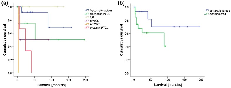
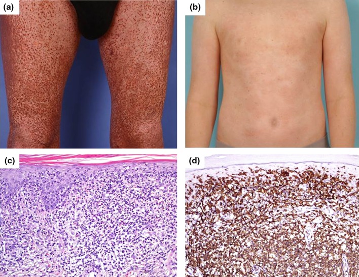
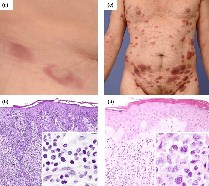
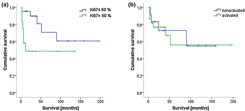
Results: The identified cases of CD8+ cytotoxic atypical lymphoproliferative infiltrates of the skin (n = 35) comprised 13 cases of mycosis fungoides (MF)/Sézary syndrome (SS), 4 cases of subcutaneous panniculitis-like T-cell lymphoma (SPTCL), 5 cases of primary cutaneous acral CD8+ lymphoma [formerly indolent CD8+ lymphoid proliferation (ILP)] and 1 case of aggressive epidermotropic primary cutaneous T-cell lymphoma (AECTCL). Moreover, nine cases were classified as primary cutaneous peripheral T-cell lymphoma, not otherwise specified (PTCL-NOS) and three cases as systemic PTCL-NOS. Multiple skin lesions, a high proliferative index and especially a final subtype attribution to AECTCL or systemic PTCL-NOS were associated with a worse survival. Coexpression of CD68 by tumor cells was exclusively observed in indolent acral CD8+ T-cell lymphoma and thus indicated an invariably benign clinical course. No further distinctive markers could be derived from our analysis.
Conclusion: Cutaneous infiltrates of CD8+ cytotoxic T-cell lymphoma comprise clinically and histologically heterogeneous entities of either primary cutaneous T-cell lymphomas or secondary infiltrates of otherwise systemic peripheral T-cell lymphomas. A thorough clinicopathological correlation with respective staging examinations remains the mainstay for correct subtype assignment and proper prognostication as long as no better markers have been defined.
Keywords: Cutaneous lymphomas; Cytotoxic; Histology; Prognosis.
Figures
Similar articles
-
Dermatopathology, Cutaneous Lymphomas.2023 Feb 16. In: StatPearls [Internet]. Treasure Island (FL): StatPearls Publishing; 2025 Jan–. 2023 Feb 16. In: StatPearls [Internet]. Treasure Island (FL): StatPearls Publishing; 2025 Jan–. PMID: 36944007 Free Books & Documents.
-
CD68 expression is a discriminative feature of indolent cutaneous CD8-positive lymphoid proliferation and distinguishes this lymphoma subtype from other CD8-positive cutaneous lymphomas.Br J Dermatol. 2015 Jun;172(6):1573-1580. doi: 10.1111/bjd.13628. Epub 2015 Apr 15. Br J Dermatol. 2015. PMID: 25524664
-
Primary cutaneous T-cell lymphomas other than mycosis fungoides and Sézary syndrome. Part II: Prognosis and management.J Am Acad Dermatol. 2021 Nov;85(5):1093-1106. doi: 10.1016/j.jaad.2021.04.081. Epub 2021 May 1. J Am Acad Dermatol. 2021. PMID: 33945836 Review.
-
Clinical and Histologic Variants of CD8+ Cutaneous T-Cell Lymphomas.Cancers (Basel). 2024 Sep 5;16(17):3087. doi: 10.3390/cancers16173087. Cancers (Basel). 2024. PMID: 39272944 Free PMC article. Review.
-
Primary cutaneous T-cell lymphomas other than mycosis fungoides and Sézary syndrome. Part I: Clinical and histologic features and diagnosis.J Am Acad Dermatol. 2021 Nov;85(5):1073-1090. doi: 10.1016/j.jaad.2021.04.080. Epub 2021 Apr 30. J Am Acad Dermatol. 2021. PMID: 33940098 Review.
Cited by
-
Tumor Immune Microenvironment in Lymphoma: Focus on Epigenetics.Cancers (Basel). 2022 Mar 13;14(6):1469. doi: 10.3390/cancers14061469. Cancers (Basel). 2022. PMID: 35326620 Free PMC article. Review.
-
An accurate diagnosis of dermal CD8+ lymphoproliferative disorders requires clinicopathological and immunophenotypic correlation.Br J Dermatol. 2022 May;186(5):769-771. doi: 10.1111/bjd.21299. Br J Dermatol. 2022. PMID: 35501942 Free PMC article.
-
C-C chemokine receptor 4 expression in CD8+ cutaneous T-cell lymphomas and lymphoproliferative disorders, and its implications for diagnosis and treatment.Histopathology. 2020 Jan;76(2):222-232. doi: 10.1111/his.13960. Epub 2019 Nov 13. Histopathology. 2020. PMID: 31355940 Free PMC article.
-
B Cells versus T Cells in the Tumor Microenvironment of Malignant Lymphomas. Are the Lymphocytes Playing the Roles of Muhammad Ali versus George Foreman in Zaire 1974?J Clin Med. 2020 Oct 24;9(11):3412. doi: 10.3390/jcm9113412. J Clin Med. 2020. PMID: 33114418 Free PMC article. Review.
References
LinkOut - more resources
Full Text Sources
Other Literature Sources
Research Materials
