

Spectroscopic and Imaging Characteristics of Pigmented Non-Melanoma Skin Cancer and Melanoma in Patients with Skin Phototypes III and IV
- PMID: 28261658
- PMCID: PMC5315082
- DOI: 10.1007/s40487-016-0036-9
Spectroscopic and Imaging Characteristics of Pigmented Non-Melanoma Skin Cancer and Melanoma in Patients with Skin Phototypes III and IV
Abstract
Introduction: Non-melanoma skin cancer is the most common malignancy worldwide. Differentiating between malignant and benign skin tumors, however, can be challenging. As a result, various auxiliary tools have been developed to aid in the diagnosis of cutaneous neoplasms. Here, skin tumors were investigated through analysis of their digital image histograms and spectroscopic response under ultraviolet (UV) and white light-emitting diodes (LEDs).
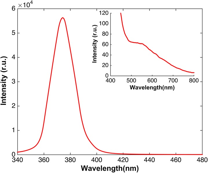
Methods: Fifty tumoral lesions were spectroscopically and histologically studied. For optical studies, UV at 375 nm and white LEDs were used to illuminate the lesions. Commercial cameras were used for imaging, and a miniature spectrometer with a bifurcated optical fiber was used for spectroscopic measurements.
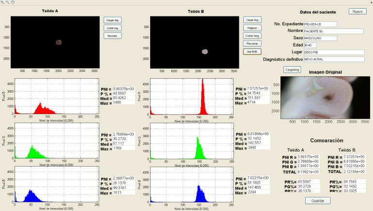
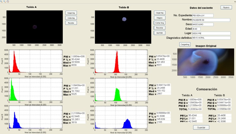
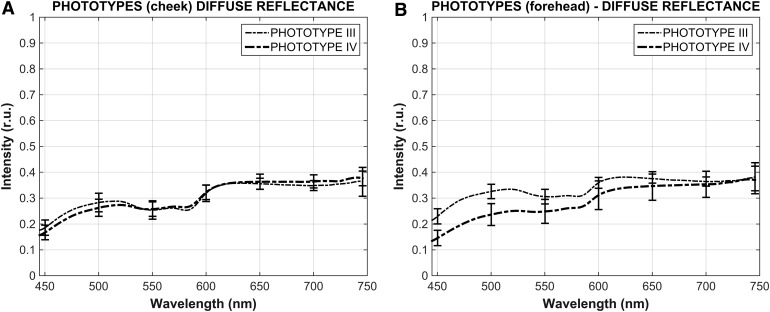
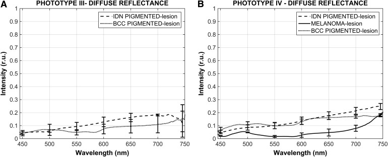
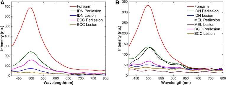
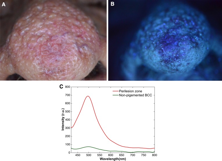
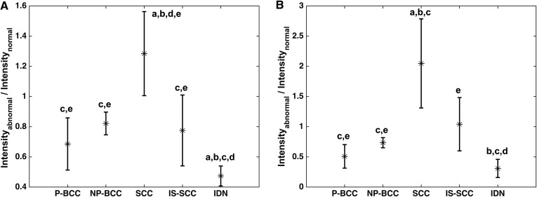
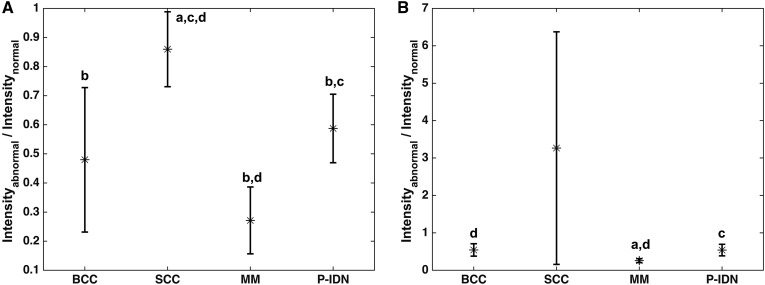
Results: In this study, the intensity histograms of the images taken under white and UV illumination and the spectroscopic response under white light showed clear differences between pigmented basal cell carcinoma (BCC), intradermal melanocytic nevus (IDN), and melanoma lesions for skin phototypes III and IV. However, there was little difference in their spectroscopic response to the UV LED.
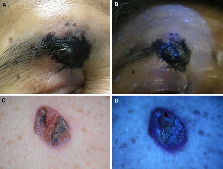
Conclusion: We found differences in the intensity and shape of diffuse reflectance spectra of pigmented BCC, IDN, and melanoma lesions in patients with skin phototypes III and IV. Also, images taken under UV and white light were helpful for differentiation of these pigmented lesions. Additional research is needed to ascertain the clinical utility of these tools for skin cancer diagnosis.
Keywords: Digital images; Melanoma; Skin cancer; Spectroscopy.
Figures
Similar articles
-
Role of In Vivo Reflectance Confocal Microscopy in the Analysis of Melanocytic Lesions.Acta Dermatovenerol Croat. 2018 Apr;26(1):64-67. Acta Dermatovenerol Croat. 2018. PMID: 29782304 Review.
-
High-resolution ultrasound reflex transmission imaging and digital photography: potential tools for the quantitative assessment of pigmented lesions.Skin Res Technol. 2006 Feb;12(1):50-9. doi: 10.1111/j.0909-725X.2006.00136.x. Skin Res Technol. 2006. PMID: 16420539 Clinical Trial.
-
Raman Spectroscopic Characterization of Melanoma and Benign Melanocytic Lesions Suspected of Melanoma Using High-Wavenumber Raman Spectroscopy.Anal Chem. 2016 Aug 2;88(15):7683-8. doi: 10.1021/acs.analchem.6b01592. Epub 2016 Jul 22. Anal Chem. 2016. PMID: 27382927
-
Assessment of pigmented skin lesions in terms of blood perfusion estimates.Skin Res Technol. 2004 Feb;10(1):43-9. doi: 10.1111/j.1600-0846.2004.00052.x. Skin Res Technol. 2004. PMID: 14731248 Clinical Trial.
-
Black and Brown Oro-facial Mucocutaneous Neoplasms.Head Neck Pathol. 2019 Mar;13(1):56-70. doi: 10.1007/s12105-019-01008-2. Epub 2019 Jan 29. Head Neck Pathol. 2019. PMID: 30693458 Free PMC article. Review.
Cited by
-
Polarimetric imaging combining optical parameters for classification of mice non-melanoma skin cancer tissue using machine learning.Heliyon. 2023 Nov 7;9(11):e22081. doi: 10.1016/j.heliyon.2023.e22081. eCollection 2023 Nov. Heliyon. 2023. PMID: 38034801 Free PMC article.
-
Characterization of Mueller matrix elements for classifying human skin cancer utilizing random forest algorithm.J Biomed Opt. 2021 Jul;26(7):075001. doi: 10.1117/1.JBO.26.7.075001. J Biomed Opt. 2021. PMID: 34227277 Free PMC article.
References
-
- Díaz González JM, Peniche Castellanos A, Fierro Arias JM, Ponce Olivera RM. Cáncer de piel en pacientes menores de 40 años. Experiencia de cuatro años en el Hospital General de México. Gac Méd Méx. 2011;147:17–21. - PubMed
-
- Gutierrez RM. Cáncer de piel. Rev Fac Med UNAM. 2003;46(4):166–171.
-
- Jurado-Santa Cruz F, Medina-Bojórquez A, Gutiérrez-Vidrio RM, Ruiz-Rosillo JM. Prevalencia del cáncer de piel en tres ciudades de México. Rev Med Ins Mex Seguro Soc. 2011;49(3):253–258. - PubMed
LinkOut - more resources
Full Text Sources
Other Literature Sources
Research Materials
