Immunohistochemical analysis of estrogen receptors in prostate and clinical correlation in men with benign prostatic hyperplasia
- PMID: 28261681
- PMCID: PMC5330371
- DOI: 10.4111/icu.2017.58.2.117
Immunohistochemical analysis of estrogen receptors in prostate and clinical correlation in men with benign prostatic hyperplasia
Abstract
Purpose: Estrogens act through interaction with 2 receptor subtypes, ER alpha (ERα) and ER beta (ERβ), in human prostate. The aim of the present study was to semiquantitatively assess the differential expression of ER subtypes in human benign prostatic hyperplasia (BPH) by use of immunocytochemistry (IHC) methods and to explore their relationship with various measures of BPH.
Materials and methods: A total of 45 patients with BPH undergoing transurethral resection of the prostate and 22 patients with bladder cancer with normal prostate undergoing surveillance cystoscopy were studied as cases and controls, respectively. Quantitative immunolabeling of ER subtypes was scored by use of a semiquantitative scale. Also, correlations were assessed between ER levels in prostate and various measures of BPH.
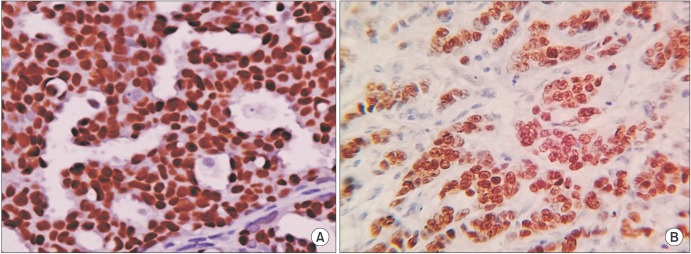
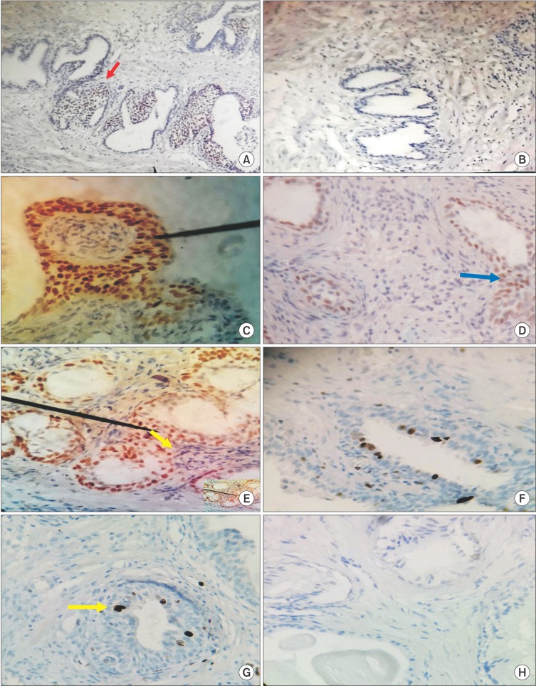
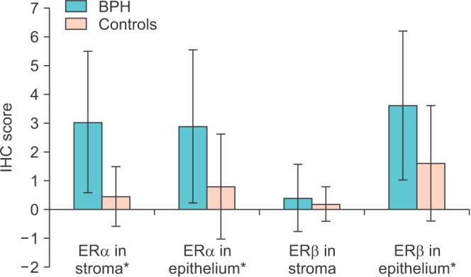
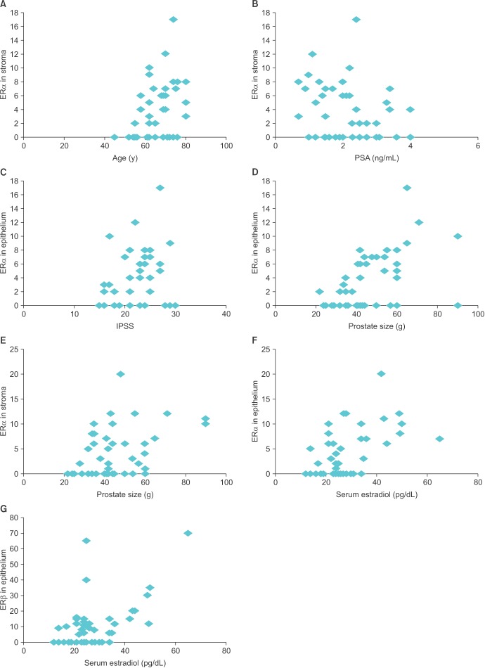
Results: Overall, we found strong immunostaining for ERα in stroma and for ERβ in epithelium, respectively. The IHC score for ERα differed significantly between BPH patients and controls in both stroma (p≤0.001) and epithelium (p=0.008), respectively. The ERβ IHC score was also significantly higher in the epithelium of BPH patients (p=0.01). Also, we found a significant correlation between prostatic ER levels and various clinical measures of BPH.
Conclusions: ERs may play an important role in the pathogenesis of BPH.
Keywords: Antibodies; Estrogens; Immunohistochemistry; Prostate.
Conflict of interest statement
CONFLICTS OF INTEREST: The authors have nothing to disclose.
Figures
Similar articles
-
Sex steroid receptor expression and localization in benign prostatic hyperplasia varies with tissue compartment.Differentiation. 2013 Apr-Jun;85(4-5):140-9. doi: 10.1016/j.diff.2013.02.006. Epub 2013 Jun 20. Differentiation. 2013. PMID: 23792768 Free PMC article.
-
Role of oestrogen receptor-α and -β in bladder tissue of patients with a clinical diagnosis of benign prostatic hyperplasia.BJU Int. 2018 Jan;121(1):130-138. doi: 10.1111/bju.14022. Epub 2017 Oct 18. BJU Int. 2018. PMID: 28941035
-
Expression of estrogen receptor-B ( ER-B ) in bengin and malignant prostatic epithelial cells and its correlation with the clinico-pathological features.J Egypt Natl Canc Inst. 2007 Dec;19(4):239-48. J Egypt Natl Canc Inst. 2007. PMID: 19672287
-
The role of estrogens and estrogen receptors in normal prostate growth and disease.Steroids. 2008 Mar;73(3):233-44. doi: 10.1016/j.steroids.2007.10.013. Epub 2007 Nov 12. Steroids. 2008. PMID: 18093629 Free PMC article. Review.
-
[Androgen and estrogen metabolism in human benign prostatic hyperplasia (BPH)].Verh Dtsch Ges Pathol. 1993;77:19-24. Verh Dtsch Ges Pathol. 1993. PMID: 7511279 Review. German.
Cited by
-
The Role of Cancer-Associated Fibroblasts in Prostate Cancer Tumorigenesis.Cancers (Basel). 2020 Jul 13;12(7):1887. doi: 10.3390/cancers12071887. Cancers (Basel). 2020. PMID: 32668821 Free PMC article. Review.
-
Concentrations of canine prostate specific esterase, CPSE, at baseline are associated with the relative size of the prostate at three-year follow-up.BMC Vet Res. 2021 Apr 26;17(1):173. doi: 10.1186/s12917-021-02874-1. BMC Vet Res. 2021. PMID: 33902583 Free PMC article.
-
The Effects of Caloric Restriction on Inflammatory Targets in the Prostates of Aged Rats.Int J Mol Sci. 2024 May 11;25(10):5236. doi: 10.3390/ijms25105236. Int J Mol Sci. 2024. PMID: 38791274 Free PMC article.
-
SMC1A is associated with radioresistance in prostate cancer and acts by regulating epithelial-mesenchymal transition and cancer stem-like properties.Mol Carcinog. 2019 Jan;58(1):113-125. doi: 10.1002/mc.22913. Epub 2018 Oct 5. Mol Carcinog. 2019. PMID: 30242889 Free PMC article.
-
Estrogen regulates the proliferation and inflammatory expression of primary stromal cell in benign prostatic hyperplasia.Transl Androl Urol. 2020 Apr;9(2):322-331. doi: 10.21037/tau.2020.02.08. Transl Androl Urol. 2020. PMID: 32420138 Free PMC article.
References
-
- Barry MJ. Epidemiology and natural history of benign prostatic hyperplasia. Urol Clin North Am. 1990;17:495–507. - PubMed
-
- Castagnetta LA, Carruba G. Human prostate cancer: a direct role for oestrogens. In: Bock GR, Goode JA, editors. Non-reproductive actions of sex steroids. Chichester: John Wiley Sons; 1995. pp. 269–289.
-
- Risbridger GP, Bianco JJ, Ellem SJ, McPherson SJ. Oestrogens and prostate cancer. Endocr Relat Cancer. 2003;10:187–191. - PubMed
-
- Greene GL, Gilna P, Waterfield M, Baker A, Hort Y, Shine J. Sequence and expression of human estrogen receptor complementary DNA. Science. 1986;231:1150–1154. - PubMed
MeSH terms
Substances
LinkOut - more resources
Full Text Sources
Other Literature Sources
Medical
Miscellaneous