Review
. 2018 Jun;175(11):1789-1804.
doi: 10.1111/bph.13770. Epub 2017 Apr 8.

Nicotinic acetylcholine receptors at the single-channel level

Affiliations
Review

Nicotinic acetylcholine receptors at the single-channel level

Cecilia Bouzat et al. Br J Pharmacol. 2018 Jun.

Abstract

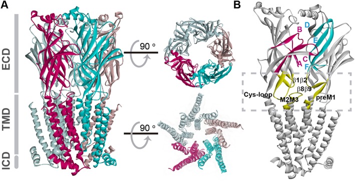
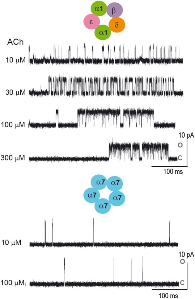
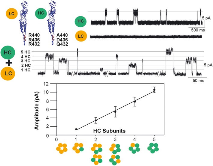
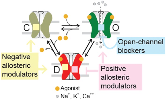
Over the past four decades, the patch clamp technique and nicotinic ACh (nACh) receptors have established an enduring partnership. Like all good partnerships, each partner has proven significant in its own right, while their union has spurred innumerable advances in life science research. A member and prototype of the superfamily of pentameric ligand-gated ion channels, the nACh receptor is a chemo-electric transducer, binding ACh released from nerves and rapidly opening its channel to cation flow to elicit cellular excitation. A subject of a Nobel Prize in Physiology or Medicine, the patch clamp technique provides unprecedented resolution of currents through single ion channels in their native cellular environments. Here, focusing on muscle and α7 nACh receptors, we describe the extraordinary contribution of the patch clamp technique towards understanding how they activate in response to neurotransmitter, how subtle structural and mechanistic differences among nACh receptor subtypes translate into significant physiological differences, and how nACh receptors are being exploited as therapeutic drug targets.

Linked articles: This article is part of a themed section on Nicotinic Acetylcholine Receptors. To view the other articles in this section visit http://onlinelibrary.wiley.com/doi/10.1111/bph.v175.11/issuetoc/.


Figures

Figure 1
Figure 1
Structural model of nACh receptor. The structure corresponds to the α4β2 plus nicotine (PDB 5KXI) (Morales‐Perez et al., 2016). (A) View parallel to the plasma membrane with colouring to highlight α (purple) and β subunits (cyan). Functional domains include the extracellular domain (ECD), which carries the binding sites at subunit interfaces; the TMD, which contains the ion pore and the gate and is formed by four α‐helices from each monomer (M1–M4); and the intracellular domain (ICD) between M3 and M4 domains, that contains determinants of channel conductance and sites for regulation. Most of the ICD is not shown since it was removed to obtain well‐diffracting crystals (Morales‐Perez et al., 2016). Views of the ECD and the TMD perpendicular to the membrane are shown on the right, upper and lower parts respectively. The TMD contains intra‐subunit and inter‐subunit transmembrane cavities involved in allosteric modulation. (B) Adjacent subunits showing the loops contributing to the binding site and the coupling region (squared). Each nACh receptor ECD monomer consists of an N‐terminal α‐helix and a core of ten β‐strands that form a β‐sandwich structure. Each agonist‐binding site is found at an interface between two adjacent subunits. The principal face, provided by the α subunit, contributes three loops that span β strands and harbour predominantly key aromatic residues, named as Loop A (β4β5 loop), Loop B (β7β8 loop) and Loop C (β9β10 loop). The adjacent subunit, which forms the complementary face, contributes three β strands with residues clustered in segments called Loops D–F. The interface between the ECD and TMD, named as the coupling region, is important for coupling agonist binding to channel opening as well as for determining open channel lifetime and rate of desensitization (Bouzat et al., 2004; Lee and Sine, 2005; Bouzat et al., 2008; Bartos et al., 2009; Yan et al., 2015). The main loops are shown in yellow.
Figure 2
Figure 2
Single‐channel recordings as a function of ACh concentration for muscle and α7 nACh receptors. For the muscle nACh receptor, single‐channel activity appears as bursts separated from each other by relatively long silent periods in which the receptor is desensitized. Bursts are composed of successive opening events corresponding to the same individual channel. The main closed time within a burst reflects the transitions between unliganded closed and diliganded open states and becomes progressively briefer with increasing ACh concentration. In contrast, the temporal pattern of α7 channel currents does not show any concentration dependence due to the fact that desensitization determines the rate of channel closing. Channel activity appears as brief (~0.1 ms) isolated events. Openings are shown as upward deflections. C and O correspond to closed and open states respectively. Membrane potential: −70 mV. Filter: 9 kHz.
Figure 3
Figure 3
Electrical fingerprinting strategy to determine receptor stoichiometry. To determine receptor stoichiometry, a subunit with a reporter mutation that alters unitary conductance is generated, and mutant and non‐mutant subunits are co‐expressed. Although receptors with a range of different subunit compositions are produced, patch clamp recordings reveal that the amplitude of each single channel opening event reports the number of mutant subunits in the receptor that originated that event. Due to its low conductance, single‐channel currents of the α7‐5HT3A chimeric receptor (LC) are not resolved from cell‐attached patches. The triple mutation increases the conductance (HC) and single‐channel openings appear as a uniform population of 10 pA at −120 mV. Recordings from cells expressing HC and LC subunits show discrete classes of channel amplitudes, each one corresponding to a receptor containing a given number of HC subunits. Thus, the amplitude of the opening event is the signature of the stoichiometry of the receptor that originated the event.
Figure 4
Figure 4
Schematic allosteric scheme for activation and drug modulation. The resting (C), open (O) and desensitized (D) states can be positively or negatively modulated by allosteric compounds. Open‐channel blockers bind to the ion channel and inhibit the flux of ions. NAMs stabilize resting or desensitized states. PAMs stabilize open states or decrease desensitization.
Figure 5
Figure 5
Effects of open‐channel blockers on nACh receptor activity as a function of the unblocking rate. Single‐channel currents for muscle nACh receptor activated by 100 μM ACh were simulated on the basis of the linear open‐channel blocking scheme, including a closed (C), an open (O) and a blocked state (OB). A desensitized state was also included to allow the termination of the bursts in the simulations. Rate constants are β = 50 000 s−1 (opening rate), d + = 100 s−1 (desensitization rate), d‐ = 1 s−1 (recovery from desensitization), k+b = 1 × 107 M−1 s−1, [B] = 5 mM, k‐b = 700, 7000 or 70 000 s−1.
Figure 6
Figure 6
Potentiation of human α7 receptors by type I and type II PAMs as a function of temperature. Single‐channel currents of human α7 receptors activated by 100 μM ACh in the absence and presence of potentiators. The increase of temperature reduces the long clusters induced by PNU‐120596 but does not affect significantly the duration of bursts induced by 5‐HI. Channel openings are shown as upward deflections. Membrane potential: −70 mV. ACh: 100 μM, PNU‐120596: 1 μM, 5‐HI: 2 mM.

Similar articles

Cited by

References

    1. Akk G, Steinbach JH (2005). Galantamine activates muscle‐type nicotinic acetylcholine receptors without binding to the acetylcholine‐binding site. J Neurosci 25: 1992–2001. - PMC - PubMed
    1. Alexander SPH, Peters JA, Kelly E, Marrion N, Benson HE, Faccenda E et al. (2015). The Concise Guide to PHARMACOLOGY 2015/16: Ligand‐gated ion channels. Br J Pharmacol 172: 5870–5903. - PMC - PubMed
    1. Amador M, Dani JA (1991). MK‐801 inhibition of nicotinic acetylcholine receptor channels. Synapse 7: 207–215. - PubMed
    1. Andersen N, Corradi J, Bartos M, Sine SM, Bouzat C (2011). Functional relationships between agonist binding sites and coupling regions of homomeric Cys‐loop receptors. J Neurosci 31: 3662–3669. - PMC - PubMed
    1. Andersen N, Corradi J, Sine SM, Bouzat C (2013). Stoichiometry for activation of neuronal α7 nicotinic receptors. Proc Natl Acad Sci U S A 110: 20819–20824. - PMC - PubMed

Publication types

LinkOut - more resources