. 2017 Mar 9;168(6):1135-1148.e12.
doi: 10.1016/j.cell.2017.02.009. Epub 2017 Mar 2.

An Intestinal Organ Culture System Uncovers a Role for the Nervous System in Microbe-Immune Crosstalk

Affiliations

An Intestinal Organ Culture System Uncovers a Role for the Nervous System in Microbe-Immune Crosstalk

Nissan Yissachar et al. Cell. .

Abstract

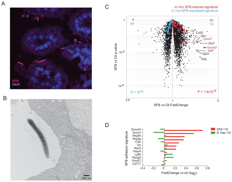
Investigation of host-environment interactions in the gut would benefit from a culture system that maintained tissue architecture yet allowed tight experimental control. We devised a microfabricated organ culture system that viably preserves the normal multicellular composition of the mouse intestine, with luminal flow to control perturbations (e.g., microbes, drugs). It enables studying short-term responses of diverse gut components (immune, neuronal, etc.). We focused on the early response to bacteria that induce either Th17 or RORg+ T-regulatory (Treg) cells in vivo. Transcriptional responses partially reproduced in vivo signatures, but these microbes elicited diametrically opposite changes in expression of a neuronal-specific gene set, notably nociceptive neuropeptides. We demonstrated activation of sensory neurons by microbes, correlating with RORg+ Treg induction. Colonic RORg+ Treg frequencies increased in mice lacking TAC1 neuropeptide precursor and decreased in capsaicin-diet fed mice. Thus, differential engagement of the enteric nervous system may partake in bifurcating pro- or anti-inflammatory responses to microbes.

Keywords: enteric nervous system; gut microbiota; neuropeptides; regulatory T cells; substance P.


Figures

Figure 1
Figure 1. Development of an intestinal organ culture system
(A) Schematics of the system design. Intact intestinal tissue is connected to input and output ports of the chamber (top), pumps controlling medium flow inside the lumen and in the external medium chamber. The entire device (bottom) contains six such chambers. (B) Histological analysis of colon tissue structure (H&E staining), and mucus production (PAS staining), of colon fragments cultured for different times. Data representative of >3 independent experiments. (C–D) Confocal imaging of colon segments cultured for 12h, immunostained for several intestinal differentiation or proliferation (Ki-67) markers. Representative of >3 independent experiments (additional time points in Fig. S3). (E) Whole-mount staining of the enteric nervous system in colon segments cultured for 8h (additional time points in Fig. S3 and MovieS2).
Figure 2
Figure 2. Intestinal immunocytes and microbiota are maintained in culture
(A) Representative flow cytometry profiles of colonic LP immunocytes isolated from colon segments from 12–14 days old SPF mice, quantitated below for 4–6 segments at each time point from two independent experiments. (B) Confocal imaging of colon cultures immunostained for the myeloid marker CX3CR1 (see MovieS5 for real time immunocyte dynamics) (C) Bacterial concentration (aerobic and anaerobic) in colon segments (as in A) cultured for different times (gray shading is the range observed in normal colon of SPF mice). (D) Bacterial concentration in cultured colon segments originating from GF mice. (E) C. ramosum bacterial concentration in SPF segments infused with C. ramosum.
Figure 3
Figure 3. Mucosal association and rapid transcriptional triggering of typical signature by SFB
(A–B) SFB associates with the intestinal epithelium after 2 hrs in cultured SI segment visualized by (A) FISH with an SFB-specific probe (red) or (B) electron microscopy. (C–D) Induction of intestinal gene expression by SFB. SI organ cultures were infused with bacteria-containing supernatant of feces from SFB- or B. fragilis-monocolonized mice or GF controls, and cultured for 2 hrs before microarray profiling of gene expression in the entire tissue. (C) Changes in gene expression on a “volcano plot” comparing SFB or GF infused cultures. Transcripts up- or down-regulated by SFB in whole SI in vivo are highlighted (red and blue, respectively). (D) Bar plot comparing the induction by SFB or B. fragilis (log2 FoldChange) of adhesion-mediated IEC activation signature genes (from (Atarashi et al., 2015)).
Figure 4
Figure 4. Early colonic response to Treg inducing microbes is enriched in neural genes
(A) Time course heatmap of changes in whole-tissue gene expression in response to C. ramosum in SPF colon cultures (log2 of FoldChange relative to the mean of medium-infused controls; each column an individual sample). Squares: genes observed as responding to SFB in vivo [Tan T. ms submitted and (Atarashi et al., 2015)] or to long-term colonization with C. ramosum in vivo (from N. Geva-Zatorsky et al, in revision). (B–E) Gene expression was profiled in colon segments from GF mice cultured and infused with different microbes. (B) Comparison of the 2 hr response to C. ramosum or P. magnus (FoldChange relative to medium control cultures, mean of triplicates). Common responses are labeled in black, those unique to C. ramosum in red or blue. (C) Correlation between transcriptional changes elicited in vitro by a panel of microbes and their ability to induce RORg+ Tregs in vivo. Top left: frequency of colonic RORg+ Tregs in monocolonized mice (data from (Sefik et al., 2015)); top right: ranked Pearson correlation coefficients, highlighting the 300 transcripts most positively or negatively correlated with colonic RORg+ Tregs (orange and green, respectively); bottom: heatmap representation of the row-normalized expression of these most-correlated transcripts (D) Volcano plot comparing C. ramosum-infused to control colon cultures (top) or SFB-infused to control SI cultures (bottom; data from Fig. 3), both highlighted with the 300 transcripts most positively or negatively correlated to RORg+ Treg frequencies, as defined in C (E) GeneOntology (biological process) enriched in these most correlated transcripts.
Figure 5
Figure 5. Treg-inducing microbes modulate colonic expression of neuropeptides and their receptors
Further analysis of gene expression in microbe-infused colon segments from Fig. 5. (A) Frequency of RORg+ Tregs in monocolonized mice (x-axis) vs mean expression of Tac1 (top) and TacR1 (bottom; encode Substance P and its receptor, respectively) in GF colon segments after 2 hr infusion with the same symbionts (y axis). (B–D) Ranked correlation to RORg+ Treg frequency (per Fig. 4C) highlighted for transcripts encoding neuropeptide receptors (B), neuropeptides (C) or neuropeptides most expressed in nociceptor neurons (D); Kolmogorov-Smirnoff test p-value and D-factor. (E) Log2 of FoldChanges vs control for C. ramosum-infused colon (red) or SFB-infused SI (blue) for neuropeptides most enriched in nociceptor neurons.
Figure 6
Figure 6. Differential activation of sensory neurons by RORg+ Treg inducing microbes compared to non-inducers via soluble mediators
(A–B) Multi-electrode array recordings of cultured dorsal root ganglia (T7-L6 DRG) sensory neurons. (A) Individual raster plots of recorded action potentials over time for C. ramosum or P. magnus; average action potential waveform for each electrode is shown at right. (B) Average spike rate comparison following stimulation by C. ramosum or P. magnus across MEA plates (n=6 per strain; two-way ANOVA p.value). (C–H) Mouse dorsal root ganglia (T7-L6 DRG) neurons were analyzed by Fura-2 ratiometric imaging for bacterial responses. Microbes were applied to cultured neurons (107–108 CFU/mL), followed by capsaicin (Cap) to mark nociceptor neurons, and KCl to activate all sensory neurons in the imaging field. (C) Representative fields showing calcium flux in neurons responding to C. ramosum (white arrows) but none to P. magnus. (D) Individual Fura-2 ratiometric traces of neurons showing calcium influx following application of bacteria, capsaicin, and KCl. Right: quantitation of the frequency of neurons responding to the two bacterial strains (response scored if the fold-change increase vs. basal state was >=1.2; student’s t-test p value<10−4). (E) Venn diagram of neuronal subsets responding to C. ramosum, capsaicin, or both. (F–H) Representative Fura-2 ratiometric traces of neurons (F, left) and quantitation of the frequency of neurons responding (F, right) to 6 different cultured microbes or to fecal suspensions from GF, SFB or C.ramosum mono-colonized mice (G). (H) Neurons respond to bacterial washoff and supernatant from C. ramosum but not from P. magnus. Bacterial cultures were washed into neuronal imaging medium for preparation of unconditioned supernatant (left) or incubated for 30min for conditioned supernatant (right).
Figure 7
Figure 7. Tgac1 deficiency and neuronal activation by capsaicin feeding alters colonic RORg+ Tregs levels in-vivo
(A) Frequency of RORg+ Tregs among colonic FoxP3+ Tregs in Tac1-deficient mice or control littermates (paired Student’s t-test); middle: KO/WT ratio of RORg+ Treg frequencies in each littermate pair; right: frequency of total colonic FoxP3+ Tregs. (B) Frequency of RORg+ Tregs among colonic FoxP3+ Tregs (left) and total colonic FoxP3+ Tregs (right) in mice fed with a capsaicin enriched diet vs. control diet (unpaired Student’s t-test).

Comment in

References

    1. Atarashi K, Tanoue T, Ando M, Kamada N, Nagano Y, Narushima S, Suda W, Imaoka A, Setoyama H, Nagamori T, et al. Th17 Cell Induction by Adhesion of Microbes to Intestinal Epithelial Cells. Cell. 2015;163:367–380. - PMC - PubMed
    1. Baral P, Mills K, Pinho-Ribeiro FA, Chiu IM. Pain and Itch: Beneficial or Harmful to Antimicrobial Defense? Cell Host Microbe. 2016;19:755–759. - PubMed
    1. Cao YQ, Mantyh PW, Carlson EJ, Gillespie AM, Epstein CJ, Basbaum AI. Primary afferent tachykinins are required to experience moderate to intense pain. Nature. 1998;392:390–394. - PubMed
    1. Caterina MJ, Schumacher MA, Tominaga M, Rosen TA, Levine JD, Julius D. The capsaicin receptor: a heat-activated ion channel in the pain pathway. Nature. 1997;389:816–824. - PubMed
    1. Chiu IM, Barrett LB, Williams EK, Strochlic DE, Lee S, Weyer AD, Lou S, Bryman GS, Roberson DP, Ghasemlou N, et al. Transcriptional profiling at whole population and single cell levels reveals somatosensory neuron molecular diversity. Elife. 2014:3. - PMC - PubMed

Publication types