

Influence of refractive error on pupillary dynamics in the normal and mild traumatic brain injury (mTBI) populations
- PMID: 28262507
- PMCID: PMC5904777
- DOI: 10.1016/j.optom.2016.12.005
Influence of refractive error on pupillary dynamics in the normal and mild traumatic brain injury (mTBI) populations
Abstract
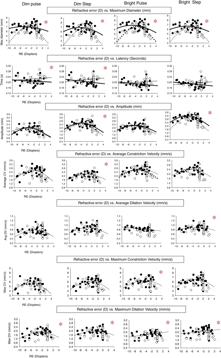
Purpose: There have been several studies investigating static, baseline pupil diameter in visually-normal individuals across refractive error. However, none have assessed the dynamic pupillary light reflex (PLR). In the present study, both static and dynamic pupillary parameters of the PLR were assessed in both the visually-normal (VN) and the mild traumatic brain injury (mTBI) populations and compared as a function of refractive error.
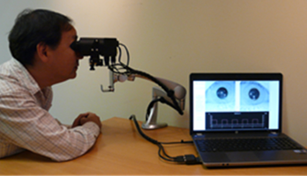
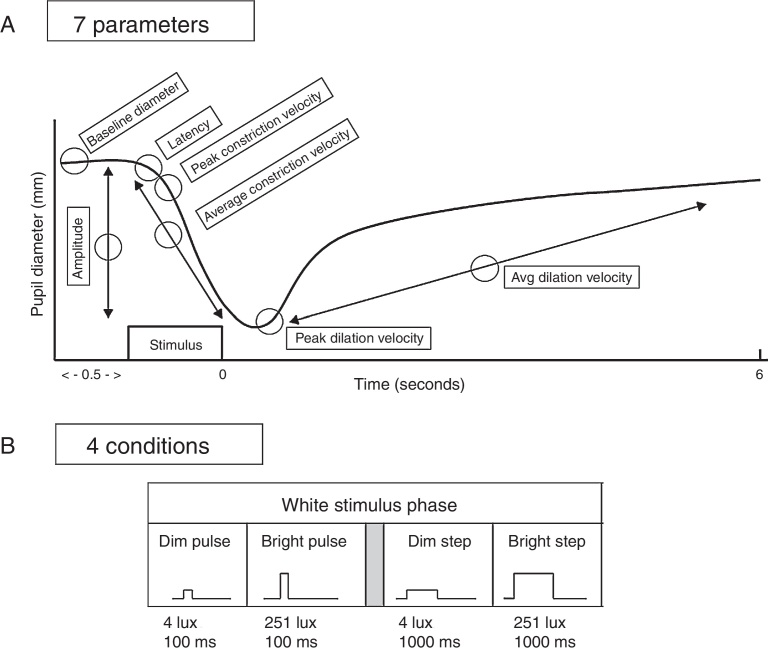
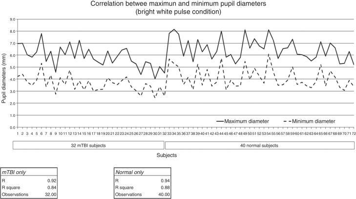
Methods: The VN population comprised 40 adults (22-56 years of age), while the mTBI population comprised 32 adults (21-60 years of age) over a range of refractive errors (-9.00D to +1.25D). Seven pupillary parameters (baseline static diameter, latency, amplitude, and peak and average constriction and dilation velocities) were assessed and compared under four white-light stimulus conditions (dim pulse, dim step, bright pulse, and bright step). The Neuroptics, infrared, DP-2000 binocular pupillometer (30Hz sampling rate; 0.05mm resolution) was used in the monocular (right eye) stimulation mode.
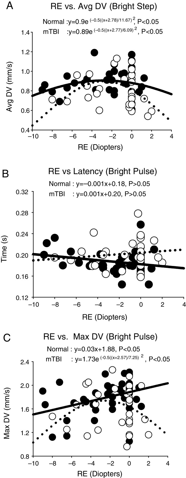
Results: For the majority of pupillary parameters and stimulus conditions, a Gaussian distribution best fit the data, with the apex centered in the low myopic range (-2.3 to -4.9D). Responsivity was reduced to either side of the apex.
Conclusions: Over a range of dynamic and static pupillary parameters, the PLR was influenced by refractive error in both populations. In cases of high refractive error, the PLR parameters may need to be compensated for this factor for proper categorization and diagnosis.
Objetivo: Existen diversos estudios que han investigado el diámetro pupilar estático y basal en individuos con visión normal en todo el espectro de errores refractivos. Sin embargo, ninguno de ellos ha evaluado el reflejo dinámico pupilar a la luz (RPL). En el presente estudio, se evaluaron tanto los parámetros pupilares estáticos como los dinámicos en poblaciones con visión normal (VN) y en las afectadas de lesiones cerebrales traumáticas leves (mTBI), comparándolos en función del error refractivo.
Métodos: La población VN incluyó a 40 adultos (de 22 a 56 años de edad), mientras que el grupo de mTBI incluyó a 32 adultos (de 21 a 60 años de edad) para un rango de errores refractivos (de -9D a + 1,25D). Se valoraron siete parámetros pupilares (diámetro estático basal, latencia, amplitud, constricción máxima y media, y velocidades de dilatación), comparándose bajo cuatro situaciones de estímulo con luz blanca (pulso tenue, punto tenue, pulso brillante, y punto brillante). Se utilizó el pupilómetro binocular con infrarrojos DP-200 de Neuroptics (30 Hz de muestreo; 0,05 mm de resolución) en el modo de estimulación monocular (ojo derecho).
Resultados: Para la mayoría de los parámetros pupilares y situaciones de estímulo, los datos se ajustaron a una distribución gausiana, centrándose el ápex en el rango miópico bajo (−2,3 to −4,9D). La respuesta se redujo a ambos extremos del ápex.
Conclusiones: Para un rango de parámetros pupilares dinámicos y estáticos, el RPL se vio influenciado por el error refractivo en ambas poblaciones. En casos de error refractivo elevado, los parámetros de RPL pueden necesitar compensarse por este factor, para su debida categorización y diagnóstico.
Keywords: Error refractivo; Infrared pupillometry; Lesión cerebral traumática leve (mTBI); Mild traumatic brain injury (mTBI); Miopía; Myopia; Pupil light reflex (PLR); Pupilometría por infrarrojos; Reflejo pupilar a la luz (RPL); Refractive error.
Copyright © 2017 Spanish General Council of Optometry. Published by Elsevier España, S.L.U. All rights reserved.
Figures
Similar articles
-
Understanding the effects of mild traumatic brain injury on the pupillary light reflex.Concussion. 2017 Aug 3;2(3):CNC36. doi: 10.2217/cnc-2016-0029. eCollection 2017 Nov. Concussion. 2017. PMID: 30202579 Free PMC article. Review.
-
Comparison of pupillary dynamics to light in the mild traumatic brain injury (mTBI) and normal populations.Brain Inj. 2016;30(11):1378-1389. doi: 10.1080/02699052.2016.1195922. Epub 2016 Aug 19. Brain Inj. 2016. PMID: 27541745
-
Quantifying pupillary asymmetry through objective binocular pupillometry in the normal and mild traumatic brain injury (mTBI) populations.Brain Inj. 2016;30(11):1372-1377. doi: 10.1080/02699052.2016.1192220. Epub 2016 Aug 11. Brain Inj. 2016. PMID: 27712127
-
Pupillary responses to light in chronic non-blast-induced mTBI.Brain Inj. 2015;29(12):1420-5. doi: 10.3109/02699052.2015.1045029. Epub 2015 Jul 16. Brain Inj. 2015. PMID: 26182230
-
Eyeing up the Future of the Pupillary Light Reflex in Neurodiagnostics.Diagnostics (Basel). 2018 Mar 13;8(1):19. doi: 10.3390/diagnostics8010019. Diagnostics (Basel). 2018. PMID: 29534018 Free PMC article. Review.
Cited by
-
Eye Movements in Mild Traumatic Brain Injury: Ocular Biomarkers.J Eye Mov Res. 2022 Jun 16;15(2):10.16910/jemr.15.2.4. doi: 10.16910/jemr.15.2.4. eCollection 2022. J Eye Mov Res. 2022. PMID: 36439911 Free PMC article.
-
Effects of low and moderate refractive errors on chromatic pupillometry.Sci Rep. 2019 Mar 20;9(1):4945. doi: 10.1038/s41598-019-41296-w. Sci Rep. 2019. PMID: 30894608 Free PMC article.
-
Understanding the effects of mild traumatic brain injury on the pupillary light reflex.Concussion. 2017 Aug 3;2(3):CNC36. doi: 10.2217/cnc-2016-0029. eCollection 2017 Nov. Concussion. 2017. PMID: 30202579 Free PMC article. Review.
-
iPhone-based Pupillometry: A Novel Approach for Assessing the Pupillary Light Reflex.Optom Vis Sci. 2018 Oct;95(10):953-958. doi: 10.1097/OPX.0000000000001289. Optom Vis Sci. 2018. PMID: 30234829 Free PMC article.
-
Autonomic nervous system dysfunction in pediatric sport-related concussion: a systematic review.J Can Chiropr Assoc. 2023 Dec;67(3):246-268. J Can Chiropr Assoc. 2023. PMID: 38283159 Free PMC article.
References
-
- Truong J.Q. 2016. Mild traumatic brain injury (mTBI) and photosensitivity: objective pupillometric findings [Doctoral dissertation] Retrieved from https://dspace.sunyconnect.suny.edu/handle/1951/67743 [accessed July 20]
-
- Truong J.Q., Ciuffreda K.J. Comparison of pupillary dynamics to light in the mild traumatic brain injury (mTBI) and normal populations. Brain Injury. 2016;30:1378–1389. - PubMed
-
- Gracitelli C.P., Duque-Chica G.L., Moura A.L. A positive association between intrinsically photosensitive retinal ganglion cells and retinal nerve fiber layer thinning in glaucoma pupillary response and glaucoma damage. Invest Ophthalmol Vis Sci. 2014;55:7997–8005. - PubMed
-
- Law C.L., Siu M., Modica P., Backus B. Stimulus characteristics affect assessment of pupil defects in amblyopia. Optom Vis Sci. 2015;92:551–558. - PubMed
MeSH terms
LinkOut - more resources
Full Text Sources
Other Literature Sources
Medical
Miscellaneous
