. 2017 Mar 14;8(3):548-560.
doi: 10.1016/j.stemcr.2017.01.025. Epub 2017 Mar 2.

Notch Inhibition Enhances Cardiac Reprogramming by Increasing MEF2C Transcriptional Activity

Affiliations

Notch Inhibition Enhances Cardiac Reprogramming by Increasing MEF2C Transcriptional Activity

Maria Abad et al. Stem Cell Reports. .

Abstract

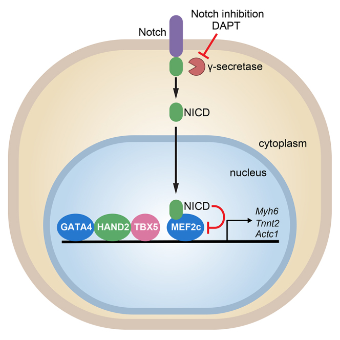
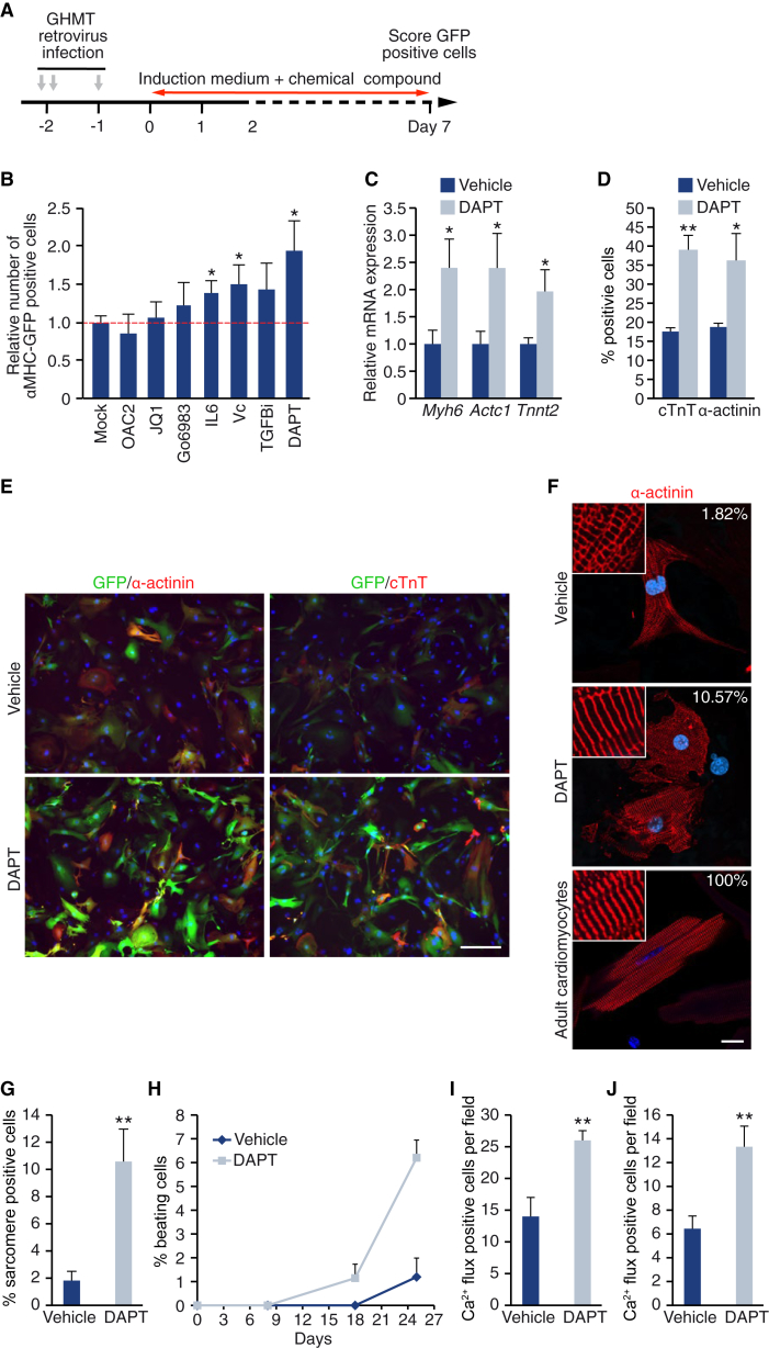
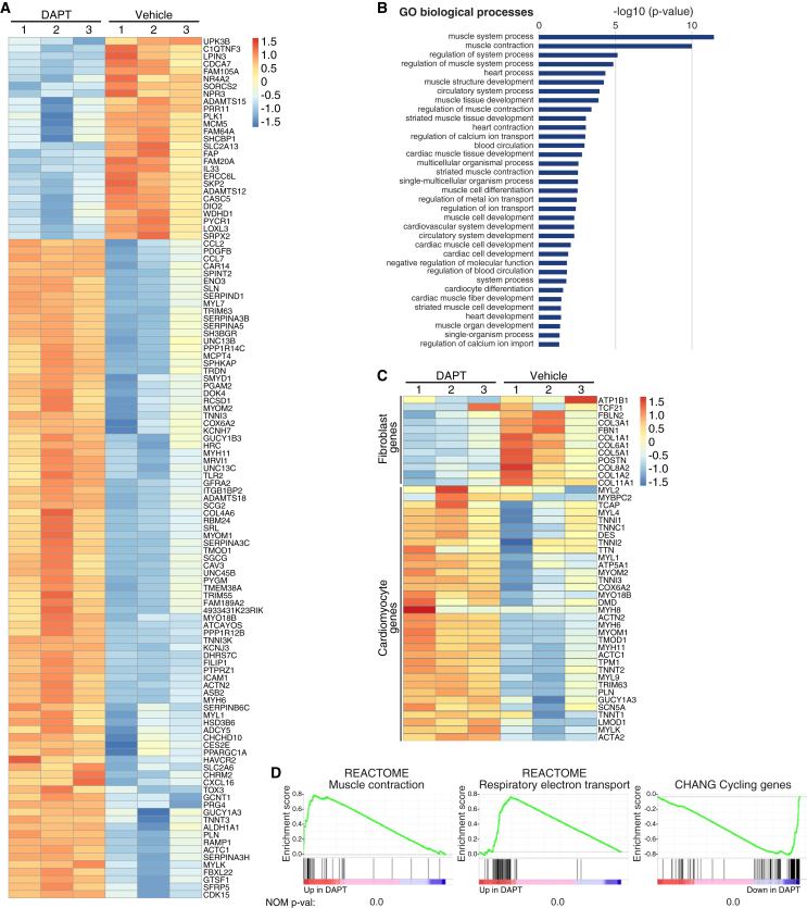
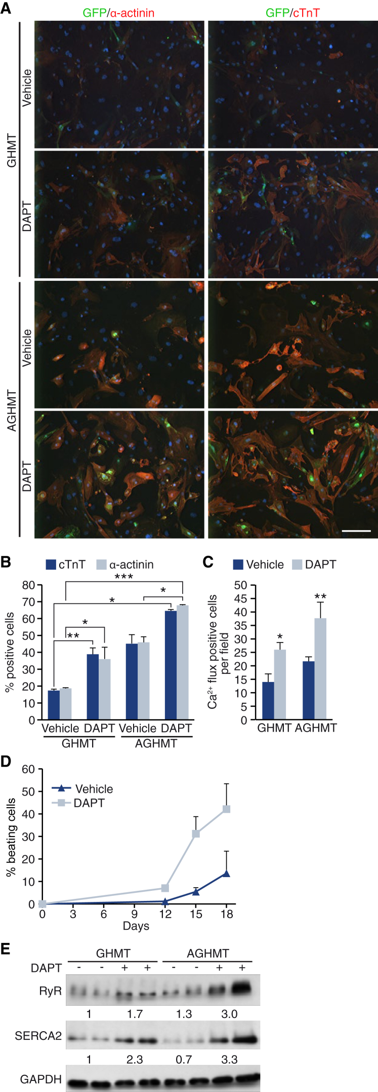
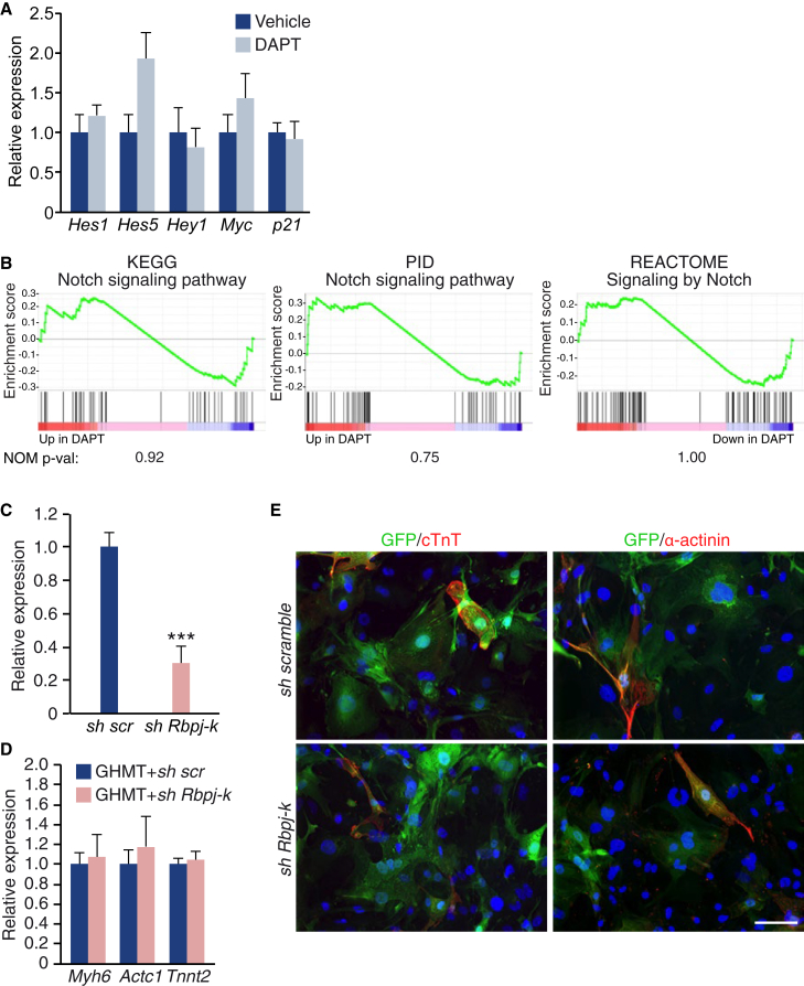
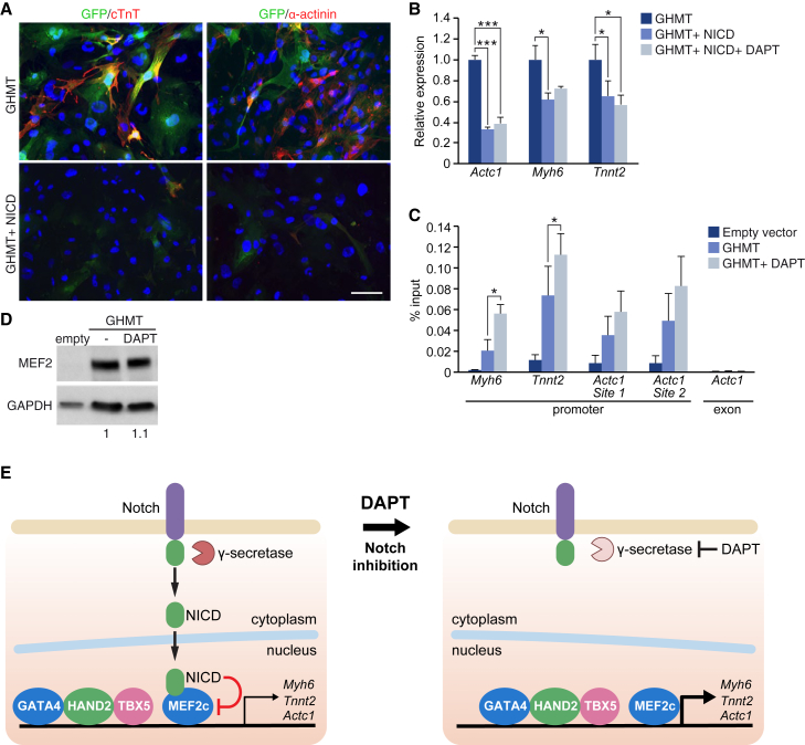
Conversion of fibroblasts into functional cardiomyocytes represents a potential means of restoring cardiac function after myocardial infarction, but so far this process remains inefficient and little is known about its molecular mechanisms. Here we show that DAPT, a classical Notch inhibitor, enhances the conversion of mouse fibroblasts into induced cardiac-like myocytes by the transcription factors GATA4, HAND2, MEF2C, and TBX5. DAPT cooperates with AKT kinase to further augment this process, resulting in up to 70% conversion efficiency. Moreover, DAPT promotes the acquisition of specific cardiomyocyte features, substantially increasing calcium flux, sarcomere structure, and the number of spontaneously beating cells. Transcriptome analysis shows that DAPT induces genetic programs related to muscle development, differentiation, and excitation-contraction coupling. Mechanistically, DAPT increases binding of the transcription factor MEF2C to the promoter regions of cardiac structural genes. These findings provide mechanistic insights into the reprogramming process and may have important implications for cardiac regeneration therapies.

Keywords: DAPT; Notch signaling; cardiomyocytes; cell-fate conversion; direct cellular reprogramming; heart regeneration; regenerative medicine; transdifferentiation.


Figures

None
Graphical abstract
Figure 1
Figure 1
DAPT Increases Calcium Flux, Sarcomere Structure, and Spontaneous Beating in iCLMs (A) Experimental strategy for drug screening to enhance cardiac reprogramming. (B) Quantification of the number of αMHC-GFP-positive MEFs after 7 days of reprogramming by GHMT and treatment with the indicated chemicals. Values are shown as relative to the mock control; n = 3 biological replicates. (C) αMHC-GFP MEFs were infected with GHMT, and treated with DMSO or DAPT for 2 weeks. Analysis by qPCR of the mRNA expression of the indicated genes, relative to expression in DMSO-treated cells; n = 3 biological replicates. (D) Quantification of the number of cells positive for cTnT and α-actinin by immunostaining; n = 3 biological replicates. (E) Representative image of the immunostaining quantified in (D). Scale bar, 200 μm. (F) Representative confocal images of immunostaining against α-actinin showing sarcomere structure in iCLMs (treated with DMSO or DAPT) and mouse adult cardiomyocytes. The percentage of cells presenting a sarcomere structure is shown in the upper-right corner of every panel. Scale bar, 20 μm. (G) Quantification of the percentage of α-actinin-positive cells with sarcomeric structure. The average of three different experiments is shown, with a total n = 471 in vehicle-treated cells and n = 570 in DAPT-treated cells. (H) Percentage of beating cells, relative to the number of input cells at indicated times; n = 3 biological replicates. (I) Quantification of Ca2+ flux-positive GCaMP3 cells after 2 weeks of reprogramming; n = 3 biological replicates. (J) Quantification of Ca2+ flux-positive MEFs treated with 4-Fluo AM dye after 2 weeks of reprogramming; n = 3 biological replicates. Data are presented as mean ± SD. p < 0.05, ∗∗p < 0.01. See also Figures S1–S3 and Table S1.
Figure 2
Figure 2
DAPT Treatment Induces Genetic Programs Related to Muscle Development, Differentiation, and Excitation-Contraction Coupling RNA-seq was performed on MEFs infected with GHMT and treated for 15 days with DMSO (vehicle) or DAPT. (A) Heatmap generated with differentially expressed genes. (B) Graphical representation of gene ontology analysis using PANTHER Overrepresentation Test. (C) Hierarchical clustering and heatmap using cardiomyocyte- and fibroblast-specific genes. (D) Enrichment plots of the indicated gene sets and their nominal p values. See also Figure S4 and Tables S2–S4.
Figure 3
Figure 3
Notch Inhibition Cooperates with AKT1 to Enhance Cardiac Reprogramming αMHC-GFP MEFs (or GCaMP MEFs in C) were infected with GHMT or AGHMT, and treated with DMSO (vehicle) or DAPT. (A) Representative immunostaining images of GFP, α-actinin, and cTnT at day 15 of reprogramming. Scale bar, 200 μm. (B) Quantification of cells positive for α-actinin and cTnT as determined by immunostaining; n = 3 biological replicates. (C) Quantification of Ca2+ flux-positive cells in GCaMP MEFs at day 15; n = 3 biological replicates. (D) Percentage of beating cells, relative to the number of input cells; n = 3 biological replicates. (E) Immunoblot against the Ca2+ handling proteins ryanodine receptor (RyR) and SERCA2 at day 15 of reprogramming. Densitometric quantification is shown as the average of every replicate, relative to GAPDH. Data are presented as mean ± SD. p < 0.05, ∗∗p < 0.01, ∗∗∗p < 0.001.
Figure 4
Figure 4
Inhibition of the Canonical Notch Pathway Does Not Enhance Reprogramming (A) qPCR analysis of Notch target genes in MEFs reprogrammed by GHMT, at day 15; n = 3 biological replicates. (B) Enrichment plots of Notch pathways gene sets, and its nominal p value. (C–E) αMHC-GFP MEFs were infected with GHMT and an shRNA scramble (scr) or an shRNA against Rbpj-k. At day 15, Rbpj-k mRNA expression was analyzed by qPCR (C), and different cardiac markers were analyzed by qPCR (D) or by immunostaining (E); n = 3 biological replicates. Scale bar, 100 μm. Data are presented as means ± SD. ∗∗∗p < 0.001. See also Figure S5.
Figure 5
Figure 5
NICD Inhibition Increases the Transcriptional Activity of MEF2C at Cardiac Gene Promoters (A and B) αMHC-GFP MEFs were infected with GHMT, or GHMT + NICD, and treated or untreated with DAPT. At day 15, different cardiac markers were analyzed (A) by immunostaining or (B) by qPCR; n = 3 biological replicates. Scale bar, 100 μm. (C and D) MEFs were infected with empty vector or GHMT, and treated with vehicle (DMSO) or with DAPT for 15 days. (C) Chromatin immunoprecipitation of MEF2C followed by qPCR of the indicated genomic regions. An exonic region of Actc1 was used as a control; n=3, three independent immuoprecipitations. (D) Immunoblot using MEF2C antibody. Densitometric quantification, relative to GAPDH, is shown. (E) Model illustrating proposed molecular mechanism by which Notch inhibition enhances cardiac reprogramming. Data are presented as means ± SD. p < 0.05, ∗∗∗p < 0.001.

References

    1. Addis R.C., Ifkovits J.L., Pinto F., Kellam L.D., Esteso P., Rentschler S., Christoforou N., Epstein J.A., Gearhart J.D. Optimization of direct fibroblast reprogramming to cardiomyocytes using calcium activity as a functional measure of success. J. Mol. Cell Cardiol. 2013;60:97–106. - PMC - PubMed
    1. Andersen P., Uosaki H., Shenje L.T., Kwon C. Non-canonical Notch signaling: emerging role and mechanism. Trends Cell Biol. 2012;22:257–265. - PMC - PubMed
    1. Andersson E.R., Sandberg R., Lendahl U. Notch signaling: simplicity in design, versatility in function. Development. 2011;138:3593–3612. - PubMed
    1. Batty J.A., Lima J.A., Jr., Kunadian V. Direct cellular reprogramming for cardiac repair and regeneration. Eur. J. Heart Fail. 2016;18:145–156. - PubMed
    1. Beltrami A.P., Urbanek K., Kajstura J., Yan S.M., Finato N., Bussani R., Nadal-Ginard B., Silvestri F., Leri A., Beltrami C.A. Evidence that human cardiac myocytes divide after myocardial infarction. N. Engl. J. Med. 2001;344:1750–1757. - PubMed

Publication types

MeSH terms