. 2017 Mar 6:8:14539.
doi: 10.1038/ncomms14539.

Rapid self-assembly of complex biomolecular architectures during mussel byssus biofabrication

Affiliations

Rapid self-assembly of complex biomolecular architectures during mussel byssus biofabrication

Tobias Priemel et al. Nat Commun. .

Abstract

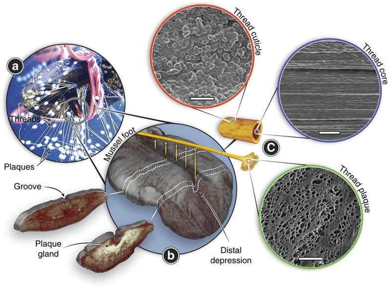
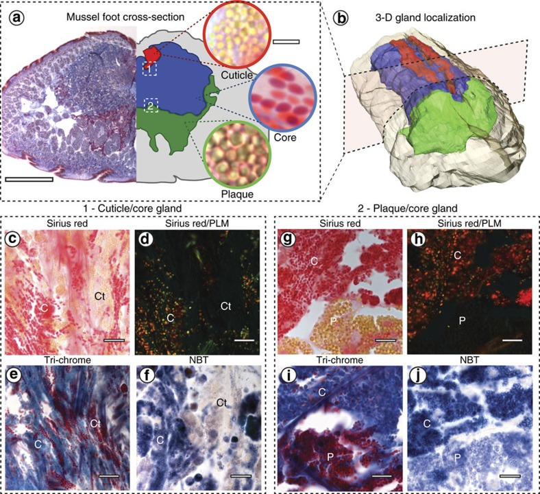
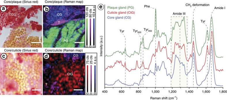
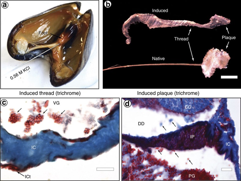
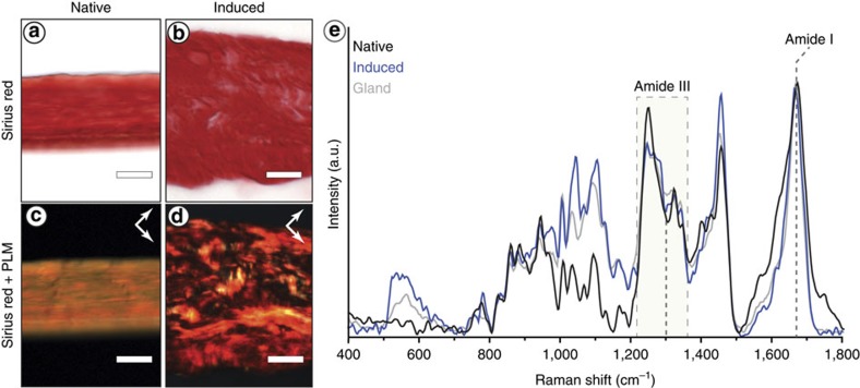
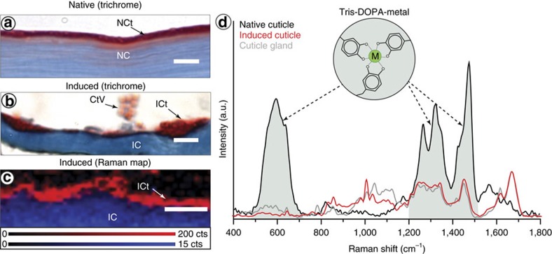
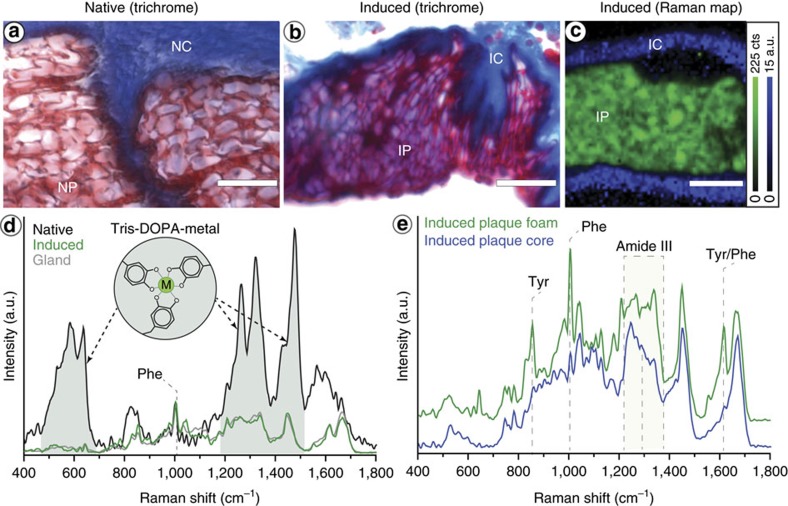
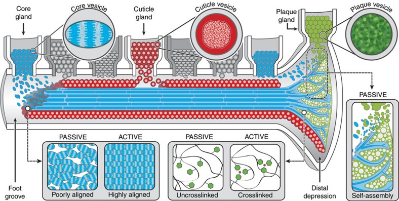
Protein-based biogenic materials provide important inspiration for the development of high-performance polymers. The fibrous mussel byssus, for instance, exhibits exceptional wet adhesion, abrasion resistance, toughness and self-healing capacity-properties that arise from an intricate hierarchical organization formed in minutes from a fluid secretion of over 10 different protein precursors. However, a poor understanding of this dynamic biofabrication process has hindered effective translation of byssus design principles into synthetic materials. Here, we explore mussel byssus assembly in Mytilus edulis using a synergistic combination of histological staining and confocal Raman microspectroscopy, enabling in situ tracking of specific proteins during induced thread formation from soluble precursors to solid fibres. Our findings reveal critical insights into this complex biological manufacturing process, showing that protein precursors spontaneously self-assemble into complex architectures, while maturation proceeds in subsequent regulated steps. Beyond their biological importance, these findings may guide development of advanced materials with biomedical and industrial relevance.


Conflict of interest statement

The authors declare no competing financial interests.

Figures

Figure 1
Figure 1. Morphology and fabrication of the mussel byssus.
(a) Photo of a mussel in the process of extruding threads onto a plexiglass surface with its foot. Note the many threads already deposited. (b) μ-CT image of the ventral surface of an iodine-stained mussel foot with two sections cut out to show the inner gland and groove structure in the middle of the foot and near the distal depression. Byssal threads are synthesized one at a time, by secreting protein building blocks into the groove on the ventral side of the mussel foot. (c) Schematic of a single byssal thread, with scanning electron microscopy images highlighting the complex micron-scale morphologies of the protective cuticle (scale bar, 3 μm), fibrous core (scale bar, 5 μm) and the adhesive plaque (scale bar, 50 μm).
Figure 2
Figure 2. Histological investigation of mussel foot secretory glands.
(a) Trichrome stained transverse cross-section of the mussel foot tissue (left; scale bar, 1 mm) and accompanying colour-coded illustration showing localization of the three secretory glands (right). High magnification light microscopy images of the vesicles in each gland are shown on the far right (scale bar, 3 μm for all three images). (b) 3D reconstruction of a mussel foot in the resting state made from serial trichrome stained transverse foot sections showing gland distribution using the same colour scheme shown in a. The colour scheme is arbitrary and is used throughout the paper to identify the specific glands and their contents. (cj) Microscopy images of histologically stained mussel foot sections from regions approximately indicated by the numbered white dashed boxes in a. Panels cf show regions from the interface between the cuticle (Ct) and core (C) glands (box 1), while panels gj show regions from interface of the plaque (P) and core (C) glands (box 2). (c,g) Sirius red stains collagen bright red. (d,h) PLM imaging of the same region in c and g, respectively, showing the birefringence of the vesicles in the core gland indicating collagen alignment. (e,i) Masson's trichrome stains the core gland vesicles blue indicating presence of collagen, whereas the cuticle and plaque vesicles are stained red. (f,j) Positive, purple-blue coloration with NBT-glycinate staining indicates the presence of oxidized DOPA groups (that is, DOPA-quinone). Scale bars for cj are 10 μm.
Figure 3
Figure 3. Raman confocal microspectroscopy of mussel foot glands.
(a) Sirius red staining and (b) composite Raman image of an analogous region of the core/plaque gland interface. The composite Raman confocal spectroscopic image was integrated over three spectral regions, which were found to correspond to the core gland (CG) (Blue; ratio of integration of 1,200–1,292 cm−1 to 1,292–1,377 cm−1), the plaque gland (PG; green; 1,600–1,631 cm−1) and surrounding tissue (T; violet; 1,550–1,600 cm−1). (c) Sirius red staining and (d) composite Raman image of an analogous region of the core/cuticle gland interface The composite Raman confocal spectroscopic image was integrated over three spectral regions corresponding to the cuticle gland (CtG; red; 1,600–1,631 cm−1), core gland (CG; blue; ratio of integration of 1,200–1,292 cm−1 to 1,292–1,377 cm−1) and surrounding tissue (T; violet; 1,550–1,600 cm−1). Scale bar is 8 μm for panels a and b, and 3 μm for c and d. (e) Average spectra from each of the secretory glands showing a distinctive spectral fingerprint and thus, different protein composition.
Figure 4
Figure 4. Induced thread formation.
(a) Injection of KCl into the foot induces secretion of byssal thread secretory vesicles. (b) Induced thread morphology is impaired compared to native threads (scale bar, 1 mm). (c) Trichrome-stained section of an induced foot within the ventral groove (VG) showing the self-assembly of the induced thread core (IC) and cuticle (ICt). Small arrows indicate vesicles from the cuticle gland in the ventral groove prior to assembly on the core surface (scale bar, 10 μm). (d) Trichrome-stained section of an induced foot within the distal depression (DD) showing the self-assembly of the induced thread plaque foam (IP) and core (IC) with cross-sectional morphology similar to that of a native thread. Small arrows indicate secretory vesicles from the core gland (CG) and plaque gland (PG) in the distal depression as they move towards the tissue assembly point (scale bar, 10 μm).
Figure 5
Figure 5. Histological and Raman characterization of induced thread core.
(a,b) Sirius red-stained longitudinal cryo-section of a (a) native thread and (b) induced thread. (c,d) PLM images of the same region in (a) and (b) showing differences in birefringence and thus, protein alignment. Arrows show the orientation of the crossed polarizers. (e) Raman spectra from the core gland vesicles, induced core and native core, highlighting the differences in native thread spectra. Scale bar is 20 μm for panels ad.
Figure 6
Figure 6. Histological and Raman characterization of induced thread cuticle.
(a) Trichrome-stained native thread section with blue-staining core (NC) and red-staining granular cuticle (NCt). Scale bar is 4 μm. (b) Trichrome-stained section of induced thread core and cuticle with blue-staining core (IC) and red-staining cuticle (ICt). Clustered cuticle vesicles (CtV) can be seen in the ventral groove before assembly. Scale bar is 4 μm. (c) Composite Raman confocal spectroscopic image of an induced thread integrated over two spectral regions corresponding to the induced core (blue; 1,226–1,299 cm−1) and the induced cuticle (red; 1,600–1,630 cm−1). Scale bar is 3 μm. (d) Comparison of Raman spectra from native cuticle, induced cuticle and cuticle gland. The intensities of the gland and induced spectra are normalized to the Amide I band height; however, the native cuticle spectra is not normalized due to the extremely high intensity of the DOPA-metal resonance bands.
Figure 7
Figure 7. Histological and Raman characterization of induced thread plaque.
(a) Trichrome-stained native plaque showing the interface between native plaque foam (NP) and native core fibres (NC). (b) Trichrome-stained induced plaque showing the interface between induced plaque foam (IP) and induced core fibres (IC). Native and induced plaques show a very similar microscale structure. (c) Raman confocal spectroscopic image of an induced plaque integrated over two spectral regions corresponding to the induced plaque foam (green; 1,600–1,630 cm−1) and the induced core fibres (Blue; ratio of integration of 1,200–1,292 to 1,292–1,377 cm−1). Clear separation of the core and plaque proteins is observed, confirming that precursors are stored and assemble as a dense immiscible phase. (d) Comparison of Raman spectra from native plaque, induced plaque and plaque gland. The intensities of the three spectra are normalized to the Phe peak height at 1,006 cm−1, emphasizing the much larger intensity of the DOPA resonance Raman peaks. (e) Raman spectra extracted from the plaque foam and core fibre regions from panel c. Scale bars are 10 μm for panels ac.
Figure 8
Figure 8. Illustrative model of passive and active aspects of byssus assembly.
Byssus assembly proceeds in a manner similar to polymer injection moudling in which proteins organized in specific secretory vesicles are released into foot groove where they coalesce and spontaneously organize into native-like structures. The preCol proteins in the core vesicles are pre-organized into a liquid crystal phase, which facilitates the local organization of proteins during assembly; however, larger scale alignment of the semi-crystalline core structure likely proceeds through biologically regulated mechanical drawing. Plaque vesicle proteins are stored as a dense fluid phase that spontaneously acquires the native foam-like structure as vesicles coalesce and envelops the fibrous core, creating a root-like structure. Likewise, cuticle proteins spontaneously coalesce and spread over the core surface, creating a granular coating. Although, the cuticle and plaque require DOPA-based metal coordination for mechanical integrity, they acquire native-like structure spontaneously in the absence of metal coordination, suggesting that metal ions are likely infiltrated into the structure in a subsequent biologically regulated processing step.

References

    1. Cranford S. W., de Boer J., van Blitterswijk C. & Buehler M. J. Materiomics: an-omics approach to biomaterials research. Adv. Mater. 25, 802–824 (2013). - PubMed
    1. Fratzl P. & Weinkamer R. Nature's hierarchical materials. Prog. Mater. Sci. 52, 1263–1334 (2007).
    1. Keten S., Xu Z., Ihle B. & Buehler M. J. Nanoconfinement controls stiffness, strength and mechanical toughness of beta-sheet crystals in silk. Nat. Mater. 9, 359–367 (2010). - PubMed
    1. Denny M. W. & Gaylord B. Marine ecomechanics. Ann. Rev. Mar. Sci. 2, 89–114 (2010). - PubMed
    1. Tamarin A., Lewis P. & Askey J. Structure and formation of byssus attachment plaque in Mytilus. J. Morphol. 149, 199–221 (1976). - PubMed

Publication types