. 2017 Mar 6:7:43578.
doi: 10.1038/srep43578.

Exosomes function in antigen presentation during an in vivo Mycobacterium tuberculosis infection

Affiliations

Exosomes function in antigen presentation during an in vivo Mycobacterium tuberculosis infection

Victoria L Smith et al. Sci Rep. .

Abstract

Mycobacterium tuberculosis-infected macrophages and dendritic cells are limited in their ability to present antigen to CD4+ T cells suggesting that other mechanism of antigen presentation are driving the robust T cell response observed during an M. tuberculosis infection. These mechanisms could include antigens present in apoptotic bodies, necrotic debris, exosomes or even release of non-vesicular antigen from infected cells. However, there is limited data to support any of these mechanisms as important in driving T cell activation in vivo. In the present study we use Rab27a-deficient mice which show diminished trafficking of mycobacterial components to exosomes as well as M. tuberculosis strains that express recombinant proteins which traffic or fail to traffic to exosomes. We observed that exosomes released during a mouse M. tuberculosis infection contribute significantly to its T cell response. These finding imply that exosomes function to promote T cell immunity during a bacterial infection and are an important source of extracellular antigen.


Conflict of interest statement

The authors declare no competing financial interests.

Figures

Figure 1
Figure 1. Quantitative and qualitative changes in exosomes released from Mtb-infected Rab27a-deficient BMMs.
Exosomes were isolated from the cell culture supernatant of C57BL/6 and Rab27a-deficient BMMs infected either with Mtb at a 3:1 MOI or left uninfected. Purified exosomes were (A) quantified for protein concentration by BCA and (B) for vesicle number by Nanosight analysis. Shown is a representative Nanosight profile for exosomes released from infected or uninfected wild-type or Rab27a-deficient macrophages. (C) 10 μg of exosomes were probed for the presence of exosomal markers, Lamp-1, CD63, CD81 and Tsg-101 by western blot. (D) 20 μg of exosomes from Mtb infected BMM were assayed for mycobacterial proteins using an antibody that recognizes multiple Mtb culture filtrate proteins (CFP) and an antibody against the Mtb 19 kDa lipoprotein. The BCA data is the average protein concentration across 3 independent experiments +/−SD and statistical analysis was performed comparing Rab27a-deficient to wild-type infected macrophages (*p < 0.05). The NanoSight and western blot data are representative of three independent experiments. Uncropped western blots are shown in Supplementary Figure 1.
Figure 2
Figure 2. Rab27a-deficient mice infected with Mtb show decreased serum exosome concentration, increased bacterial load and diminished T cell response compared to WT infected mice.
Bacterial load in the spleen (A) and serum exosome concentration (B) were defined at different times post-infection. (C) Cells isolated from lung and spleens of infected mice at the times indicated were stimulated ex vivo with Mtb whole cell lysate and the amount of secreted IFN-γ quantified by ELISA. The results are expressed as the IFN-ɣ concentration after stimulation of 1 × 106 cells with the Mtb cell lysate. (D) Splenocytes were isolated from wild-type C57BL/6 and Rab27a-deficient mice at different times post-infection with Mtb. The cells were stained with PE-conjugated anti-mouse CD69 and CD4 or with an isotype control antibody. CD4+ T cells were analyzed for CD69 surface expression with a Beckman Coulter flow cytometer, and the percentage of CD69 positive relative to total CD4+ cells was calculated. Results are defined for each mouse with +/−SD and statistical analysis was performed comparing Rab27a-deficient mice to wild-type infected mice (*p < 0.05). The data is representative of three independent experiments for a total of 9 mice per mouse strain per time point.
Figure 3
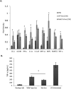
Figure 3. Exosomes purified from the serum of Mtb-infected wild-type mice were more pro-inflammatory compared to exosomes from infected Rab27a-deficient mice.
Serum exosomes from Mtb-infected wild-type and Rab27a-deficient mice (3 mice/group) were purified and used at 250 μg/ml to treat BMMs for 16 hours. (A) The supernatants were harvested and assayed for specific proteins using a mouse cytokine array. The pixel densities for each spot of the array were calculated using ImageJ software and plotted. Results are defined for each individual mouse +/− SD and statistical analysis was performed comparing Rab27a-deficient mice to WT infected mice (*p < 0.05). (B) The culture supernatants were also analyzed for TNF-α by ELISA. The results are the mean of three separate experiments (9 mice total) with SD shown. Statistical analysis was performed comparing Rab27a-deficient to wild-type infected mice (*p < 0.05). Exo; exosomes. Rab; Rab27a.
Figure 4
Figure 4. Increased trafficking of DsRed to exosomes in RAW264.7 cells infected with M. bovis BCG expressing the Ag85A-DsRed fusion protein.
(A) Fluorescence microscopic and (B) western blot analysis of recombinant M. bovis BCG expressing DsRed or Ag85A-DsRed. (C) Western blot analysis of exosomes and RAW264.7 whole cell lysate (WCL) following a 72 hour infection with the recombinant M. bovis strains or from unifected cells. Tubulin and Lamp-1 were used as loading control for WCL and exosomes respectively. (D) Fluorescence microscopic analysis of Ds-Red positive exosomes captured on protein G Sepharose beads coupled to anti-CD63. (E) Quantitation of the DsRed-positive Sepharose beads observed in (D). Uncropped western blots are shown in Supplementary Figure 1.
Figure 5
Figure 5. DsRed-specific T cell response is elevated in mice infected with M. bovis BCG expressing Ag85A-DsRed compared to bacilli expressing DsRed only.
(A) Wild-type mice were intranasally infected with M. bovis BCG expressing DsRed or Ag85A-DsRed and the lung and splenic cells were harvested 2 weeks post-infection and stimulated ex-vivo with DsRed. The number of IFN-ɣ producing T cells responding to the DsRed antigen was measured by ELISPOT. (B) Wild-type C57BL/6 or Rab27a-deficient mice were intranasally infected with M. bovis BCG expressing either DsRed or Ag85A-DsRed. The number of IFN-ɣ producing T cells responding to the antigen DsRed was determined by ELISPOT. Results are defined for each mouse (4 mice/group) +/−SD and statistical analysis was performed between BCG expressing DsRed or Ag85A-DsRed (A) or between infected WT and Rab27-deficient mice (B) (*p < 0.05). The data is representative of two independent experiments.
Figure 6
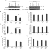
Figure 6. Increased trafficked of HspX to exosomes during a Mtb mouse infection enhances an HspX-specific T cell response.
(A) RAW264.7 cells were infected with ∆HspX H37Rv or ∆HspX H37Rv expressing either His-tagged wild type HspX or K85R HspX and 72 hours post-infection exosomes were purified from the culture media. Raw264.7 cell lysates were also obtained. A separate well of Raw264.7 cells were treated with 40 μg/ml of purified His-tagged HspX (rHspX) and 24 hours post-treatment culture media and cell lysates were obtained. 10 μg of purified exosomes or cell lysates was analyzed for the His-tagged HspX using an anti-His antibody. Blots were also probed for Lamp1 as a loading control. (B–E) Splenocytes were harvested from wild-type C57BL/6 mice 10 days (B,C) or 15 days (D,E) post infection or from Rab27a-deficient mice 10 days post-infection (F,G). Mice (3 to 4 mice/group) were infected with either 106 CFU of wild-type H37Rv, ∆HspX H37Rv or ∆HspX H37Rv expressing either wild-type HspX or K85R HspX. The splenocytes were analyzed 20 h after ex vivo stimulation with 5 μg/ml HspX (B,D,F) or 10 μg/ml Mtb CFP (C,E,G). The number of T cells producing IFN-ɣ upon HspX antigen stimulation was determined by ELISPOT and the number of positive cells counted +/−SD between individual mouse infections. Significance between samples is indicated (*p < 0.05). Data is representative of three independent experiments. Uncropped western blots are shown in Supplementary Figure 1.

Similar articles

Cited by

References

    1. Cooper A. M. T cells in mycobacterial infection and disease. Current opinion in immunology 21, 378–384 (2009). - PMC - PubMed
    1. Srivastava S. & Ernst J. D. Cell-to-cell transfer of M. tuberculosis antigens optimizes CD4 T cell priming. Cell host & microbe 15, 741–752, doi: 10.1016/j.chom.2014.05.007 (2014). - DOI - PMC - PubMed
    1. Cheng Y. & Schorey J. S. Exosomes carrying mycobacterial antigens can protect mice against Mycobacterium tuberculosis infection. Eur J Immunol 43, 279–290. (2013). - PMC - PubMed
    1. Giri P. K., Kruh N. A., Dobos K. M. & Schorey J. S. Proteomic analysis identifies highly antigenic proteins in exosomes from M. tuberculosis-infected and culture filtrate protein-treated macrophages. Proteomics 10, 3190–3202 (2010). - PMC - PubMed
    1. Schorey J. S. & Bhatnagar S. Exosome function: from tumor immunology to pathogen biology. Traffic 9, 871–881 (2008). - PMC - PubMed

Publication types

MeSH terms