. 2017 Apr 3;127(4):1475-1484.
doi: 10.1172/JCI90193. Epub 2017 Mar 6.

Loss of DDRGK1 modulates SOX9 ubiquitination in spondyloepimetaphyseal dysplasia

Loss of DDRGK1 modulates SOX9 ubiquitination in spondyloepimetaphyseal dysplasia

Adetutu T Egunsola et al. J Clin Invest. .

Abstract

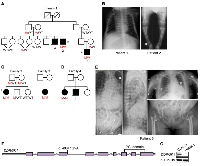
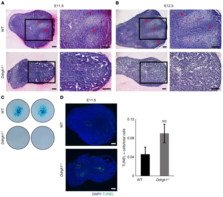
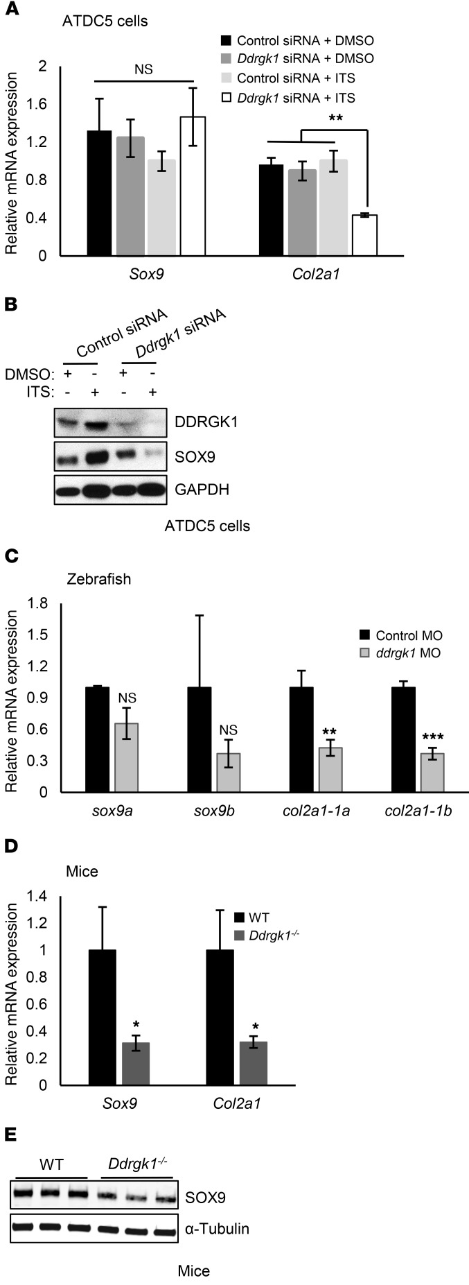
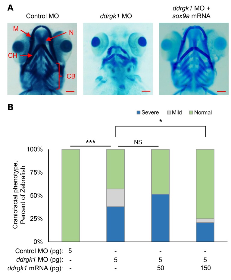
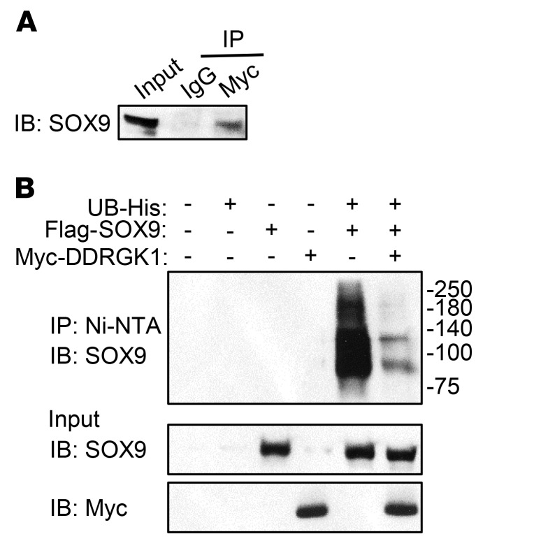
Shohat-type spondyloepimetaphyseal dysplasia (SEMD) is a skeletal dysplasia that affects cartilage development. Similar skeletal disorders, such as spondyloepiphyseal dysplasias, are linked to mutations in type II collagen (COL2A1), but the causative gene in SEMD is not known. Here, we have performed whole-exome sequencing to identify a recurrent homozygous c.408+1G>A donor splice site loss-of-function mutation in DDRGK domain containing 1 (DDRGK1) in 4 families affected by SEMD. In zebrafish, ddrgk1 deficiency disrupted craniofacial cartilage development and led to decreased levels of the chondrogenic master transcription factor sox9 and its downstream target, col2a1. Overexpression of sox9 rescued the zebrafish chondrogenic and craniofacial phenotype generated by ddrgk1 knockdown, thus identifying DDRGK1 as a regulator of SOX9. Consistent with these results, Ddrgk1-/- mice displayed delayed limb bud chondrogenic condensation, decreased SOX9 protein expression and Col2a1 transcript levels, and increased apoptosis. Furthermore, we determined that DDRGK1 can directly bind to SOX9 to inhibit its ubiquitination and proteasomal degradation. Taken together, these data indicate that loss of DDRGK1 decreases SOX9 expression and causes a human skeletal dysplasia, identifying a mechanism that regulates chondrogenesis via modulation of SOX9 ubiquitination.


Conflict of interest statement

Conflict of interest: The authors have declared that no conflict of interest exists.

Figures

Figure 1
Figure 1. Homozygous DDRGK1 loss-of-function mutation identified in Iraqi Jewish families with Shohat-type SEMD.
(A) Pedigree of family 1. M represents mutant allele. Asterisks indicate individuals who were selected for WES. (B) Radiographs from family 1 reveal platyspondyly and hypomineralized epiphyses and metaphyses in patients 1 and 2, respectively. (C and D) Pedigrees of families 2, 3, and 4. (E) Radiographs show severe scoliosis, vertebral compression factures, platyspondyly, broaden hypomineralized metaphyses, and smaller than average hypomineralized epiphyses in patient 5 from family 4. (F) Schematic of the c.408+1G>A mutation in the DDRGK1 gene. (G) Western analysis of whole cell lysates reveals absence of DDRGK1 expression in patient LCLs.
Figure 2
Figure 2. Overexpression of ddrgk1 mRNA rescues the ddrgk1 knockdown cartilage phenotype in zebrafish.
(A) Ventral view at 120 h.p.f. of Alcian blue–stained WT embryos and embryos injected with either control MO or ddrgk1 MO with or without exogenous ddrgk1 mRNA. M, Meckel’s cartilage; CH, ceratohyal cartilage; N, neurocranial cartilage; CB, ceratobranchial cartilage. Scale bars: 100 μM. (B) Quantification of the craniofacial phenotype in the embryos. Embryos with normal craniofacial features had properly formed Meckel’s cartilage, ceratohyal cartilage, neurocranial cartilage, and ceratobranchial cartilage. Embryos lacking ceratobranchial cartilage showed mild craniofacial defects, while embryos with several craniofacial defects had no neurocranial or ceratobranchial cartilage and poorly developed Meckel’s cartilage and ceratohyal cartilage. WT, n = 44; 5 pg control MO, n = 36; 3 pg ddrgk1 MO, n = 50; 4 pg ddrgk1 MO, n = 47; 5 pg ddrgk1 MO, n = 43; 5 pg ddrgk1 MO + 25 pg ddrgk1 mRNA, n = 45; 5 pg ddrgk1 MO + 50 pg ddrgk1 mRNA, n = 38. *P < 0.05; ***P < 0.001; Kruskal-Wallis rank-sum test followed by Wilcoxon’s rank-sum test with continuity correction.
Figure 3
Figure 3. Deletion of Ddrgk1 delays chondrogenic mesenchymal condensation and increases apoptosis in mouse limb buds.
(A and B) Mesenchymal condensation is detected in WT, but not in Ddrgk1–/–, limb buds stained with H&E at (A) E11.5 and (B) E12.5. Red asterisks indicate regions undergoing mesenchymal condensation. Images on the right are higher magnification depictions of the boxed regions of the images to the left. (C) After 7 days, micromass cultures of dissociated Ddrgk1–/– (n = 2) mesenchymal cells produced fewer Alcian blue–positive cartilaginous nodules than WT (n = 2) mesenchymal cells. Images are of 2 independent experiments. (D) TUNEL staining reveals Ddrgk1–/– limb buds (n = 3) have increased apoptosis compared with WT limb buds (n = 4) at E11.5, trending toward significance. Values are represented as mean ± SEM. Two-tailed t test. Scale bars: 100 μM.
Figure 4
Figure 4. Ddrgk1 deficiency decreases SOX9 protein and Col2a1 mRNA expression.
(A and B) ATDC5 cells were transiently transfected with control or Ddrgk1 siRNA and treated with either DMSO or ITS to induce differentiation 24 hours later. The cells were harvested 7 days after treatment. (A) RT-PCR of total RNA from ATDC5 cells treated with control siRNA + DMSO (n = 3), Ddrgk1 siRNA + DMSO (n = 3), control siRNA + ITS (n = 3), and Ddrgk1 siRNA + ITS (n = 3). Values are represented as mean ± SEM. **P < 0.01, 2-way ANOVA followed by Tukey’s post-hoc test. (B) Immunoblots of total cell lysates from ATDC5 cells. The immunoblots are representative of 3 independent experiments. (C) ddrgk1 morphants have less mRNA expression of col2a1, but not of sox9, than control morphants. Zebrafish embryos were injected with 5 pg control MO (n = 3) or 5 pg ddrgk1 MO (n = 3), and total RNA was collected for RT-PCR 72 h.p.f. later. Values are represented as mean ± SEM. **P < 0.01; ***P < 0.001, 2-tailed t test. (D and E) Deletion of Ddrgk1 decreases transcript levels of Col2a1 via SOX9 protein reduction in E11.5 limb buds. (D) RT-PCR of total RNA from E11.5 WT (n = 3) and Ddrgk1–/– (n = 3) limb buds. Values are represented as mean ± SEM. *P < 0.05, 2-tailed t test. (E) Immunoblots of total cell lysates from E11.5 WT (n = 3) and Ddrgk1–/– (n = 3) limb buds.
Figure 5
Figure 5. Expression of sox9a mRNA rescues the ddrgk1 knockdown cartilage phenotype in zebrafish.
(A) Ventral view at 120 h.p.f. of Alcian blue–stained embryos injected with either control MO or ddrgk1 MO with or without sox9a mRNA. Scale bars: 100 μM. (B) Quantification of the craniofacial phenotype in the embryos. The categories of craniofacial features are described in Figure 2. Control MO, n = 36; 5 pg ddrgk1 MO, n = 42; 5 pg ddrgk1 MO + 50 pg ddrgk1 mRNA, n = 31; 5 pg ddrgk1 MO + 150 pg ddrgk1 mRNA, n = 41. *P < 0.05; ***P < 0.001, Kruskal-Wallis rank-sum test followed by Wilcoxon’s rank-sum test with continuity correction.
Figure 6
Figure 6. DDRGK1 forms a complex with SOX9 and inhibits SOX9 ubiquitination.
(A) SOX9 forms a complex with DDRGK1. 293T cells were transiently transfected with plasmids expressing Myc-tagged Ddrgk1 and FLAG-tagged Sox9. After 48 hours, SOX9 was detected by immunoblotting with a FLAG-specific antibody after immunoprecipitation of cell lysates with anti-Myc or IgG antibody. As a control, the input was immunoblotted with the FLAG-specific antibody. The immunoblot is representative of 3 independent experiments. (B) Ddrgk1 overexpression decreases SOX9 ubiquitination in 293T cells. 293T cells were transiently transfected with plasmids expressing His-tagged UB, FLAG-tagged SOX9, and Myc-tagged DDRGK1. Twenty-four hours later, the cells were treated with 20 μM of proteasome inhibitor MG132 for 6 hours. Afterwards, we pulled down His-tagged ubiquitinated proteins using Ni-NTA beads, and ubiquitinated SOX9 was probed using anti-SOX9 antibody. As controls, the input was immunoblotted with anti-SOX9 and anti-Myc antibodies. The immunoblots are representative of 3 independent experiments.

References

    1. Spranger J, Winterpacht A, Zabel B. The type II collagenopathies: a spectrum of chondrodysplasias. Eur J Pediatr. 1994;153(2):56–65. - PubMed
    1. Kuivaniemi H, Tromp G, Prockop DJ. Mutations in fibrillar collagens (types I, II, III, and XI), fibril-associated collagen (type IX), and network-forming collagen (type X) cause a spectrum of diseases of bone, cartilage, and blood vessels. Hum Mutat. 1997;9(4):300–315. - PubMed
    1. Wirth T. [Spondyloepiphyseal and metaphyseal dysplasia] Der Orthopäde. 2008;37(1):8–16. doi: 10.1007/s00132-007-1179-z. - DOI - PubMed
    1. Cormier-Daire V. Spondylo-epi-metaphyseal dysplasia. Best Pract Res Clin Rheumatol. 2008;22(1):33–44. doi: 10.1016/j.berh.2007.12.009. - DOI - PubMed
    1. Figuera LE, Ramírez-Dueñas ML, Gallegos-Arreola MP, Cantú JM. Spondyloepimetaphyseal dysplasia (SEMD) Shohat type. Am J Med Genet. 1994;51(3):213–215. doi: 10.1002/ajmg.1320510307. - DOI - PubMed

MeSH terms

Supplementary concepts