. 2017 Apr;23(4):424-428.
doi: 10.1038/nm.4298. Epub 2017 Mar 6.

Glucocorticoid hormone-induced chromatin remodeling enhances human hematopoietic stem cell homing and engraftment

Affiliations

Glucocorticoid hormone-induced chromatin remodeling enhances human hematopoietic stem cell homing and engraftment

Bin Guo et al. Nat Med. 2017 Apr.

Abstract

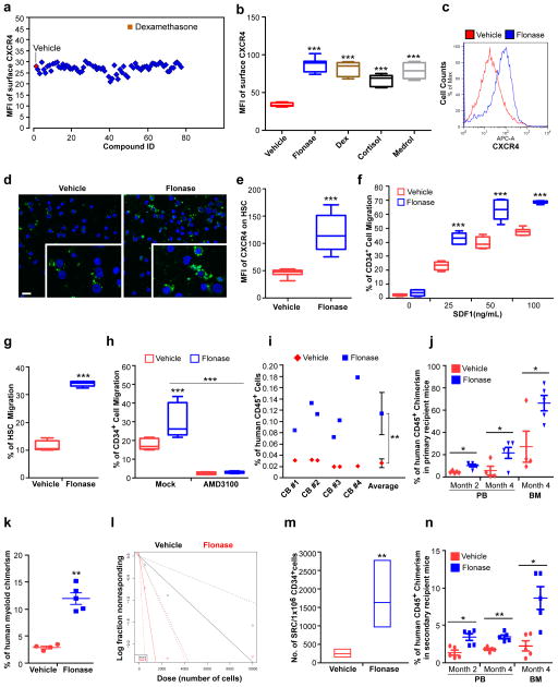
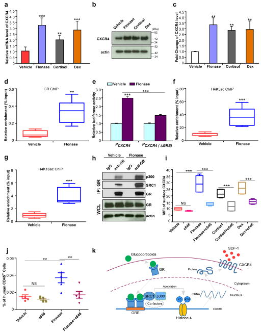
Efficient hematopoietic stem cell (HSC) homing is important for hematopoietic cell transplantation (HCT), especially when HSC numbers are limited, as in the use of cord blood (CB). In a screen of small-molecule compounds, we identified glucocorticoid (GC) hormone signaling as an activator of CXCR4 expression in human CB HSCs and hematopoietic progenitor cells (HPCs). Short-term GC pretreatment of human CB HSCs and HPCs promoted SDF-1-CXCR4-axis-mediated chemotaxis, homing, and long-term engraftment when these cells were transplanted into primary- and secondary-recipient NSG mice. Mechanistically, activated glucocorticoid receptor binds directly to a glucocorticoid response element in the CXCR4 promoter and recruits the SRC-1-p300 complex to promote H4K5 and H4K16 histone acetylation, facilitating transcription of CXCR4. These results suggest a new and readily available means to enhance the clinical efficacy of CB HCT.


Conflict of interest statement

COMPETING FINANCIAL INTERESTS

Dr. Broxmeyer is a member of the Medical Scientific Advisory Board of CordUse, a cord blood banking company based in Orlando, Florida.

Figures

Figure 1
Figure 1. Glucocorticoids increase surface expression of CXCR4 and promote SDF-1/CXCR4 axis mediated chemotaxis, homing and long term engraftment of human hematopoietic stem and progenitor cells
(a) Mean fluorescence intensity (MFI) of surface CXCR4 of human cord blood (CB) CD34+ cells after treatment of the cells for 16 hours with compounds from a nuclear receptor ligand library. The concentration of all compounds used in this study was 1 μM unless otherwise stated. (b) Quantification of mean fluorescence intensity (MFI) of surface CXCR4 of human CB CD34+ cells treated with vehicle, Flonase, dexamethasone (Dex), cortisol or methylprednisolone (Medrol). Data pooled from three independent experiments are shown (n=9 cultures per group, one-way ANOVA). (c) Histogram of surface CXCR4 expression of human CB CD34+ cells treated with vehicle or Flonase. Representative histograms from three independent experiments are shown. (d) Confocal imaging analysis of surface CXCR4 expression of human CB CD34+ cells treated with vehicle or Flonase. FITC (green) indicates CXCR4 expression; DAPI (blue) labels the cell nucleus. Representative images from two independent experiments are shown (the inset shows the amplified part of the image). Scale bar: 20 μm. (e) Quantification of mean fluorescence intensity (MFI) of surface CXCR4 of human CB HSCs (CD34+CD38CD45RACD90+CD49f+) treated with vehicle or Flonase (n=9 cultures per group). Data pooled from three independent experiments are shown. (f) Migration of human CB CD34+ cells towards human recombinant SDF-1α, as quantified by flow cytometry. The cells were cultured in the presence of vehicle or Flonase for 16 hours and then allowed to migrate towards the indicated concentrations of SDF-1α for 4 hours. Data pooled from two independent experiments are shown (n=6 cultures per group, two-way ANOVA). (g) Migration of human phenotypic HSCs in CB CD34+ cells towards human recombinant SDF-1α (50 ng/mL), as quantified by flow cytometry. The migration percentage of HSCs was calculated as the HSC (CD34+CD38CD45RACD90+CD49f+) frequency among migrated CD34+ cells. Data pooled from two independent experiments are shown (n=6 cultures per group). (h) Migration of vehicle or Flonase treated human CB CD34+ cells in the presence of the CXCR4 antagonist, AMD3100 (5 μg/mL), as quantified by flow cytometry. Data pooled from three independent experiments are shown (n=9 cultures per group, one-way ANOVA). (i) The percentage of human CD45+ cells in the bone marrow of NSG mice 24 h after transplantion with 500,000 CB CD34+ cells that had been treated with vehicle or Flonase. CD34+ cells from four cord blood samples (CB#1-4) were tested. (n=6 mice per group). (j) The percentage of human CD45+ cells in peripheral blood (PB) and BM of NSG mice at the indicated time points after transplantation with 10,000 CB CD34+ cells that had been treated with vehicle or Flonase. (n=4 mice in vehicle group and n=5 mice in Flonase group). (k) The percentage of human CD33+ myeloid cells in BM of NSG mice 4 months after transplantation with 10,000 CB CD34+ cells that had been treated with vehicle or Flonase (n=4 mice in vehicle group and n=5 mice in Flonase group). (l,m) The frequency of human SRCs in CB CD34+ cells treated with vehicle or Flonase, as determined by transplantations of graded doses of treated cells into NSG mice and determination of human CD45+ cell chimerism 3 months after transplantation (n=4–5 mice per group, see Supplementary Table 2). (l) Poisson statistical analysis of data from Supplementary Table 2. Shapes (circle or triangle in the plot) represent the percentage of negative mice for each dose of cells. The inverted triangles indicate that all tested mice were positive in this group. Solid lines indicate the best-fit linear model for each data set. Group A (black line) indicates vehicle group, Group B (red line) indicates Flonase group. Dotted lines represent 95% confidence intervals. (m) HSC frequencies (line in the box) and 95% confidence intervals (box) presented as the number of SRCs in 1×10⁶ CD34+ cells. (n) Human CD45+ cell chimerism in the PB and BM of secondary recipient NSG mice, which had been transplanted with 5×10⁶ bone marrow cells from primary recipient NSG mice (n=5 mice per group). Data are shown as dot plots (mean±s.e.m.) in j, k, n, or as box-and-whisker plots (the lines indicate median values, the whiskers indicate minimum and maximum values, the boxes indicate interquartile range) in b and eh. *p<0.05. **p<0.01. ***p<0.001.
Figure 2
Figure 2. Glucocorticoids enhance H4K5 and H4K16 acetylation associated with the CXCR4 promoter, facilitate expression of CXCR4 and promote homing of human CB CD34+ cells
(a) CXCR4 mRNA levels in Flonase, cortisol or Dex treated human CB CD34+ cells, relative to vehicle-treated cells, as assessed by quantitative realtime-PCR. Data pooled from three independent experiments are shown (n=9 replicates per group, one-way ANOVA). (b,c) Total CXCR4 protein levels in Flonase, cortisol or Dex treated human CB CD34+ cells, relative to vehicle-treated cells, as assessed by western blotting. A representative blot is shown in b, and quantification of CXCR4 protein levels from three independent western blot assays is shown in c (n=3 experiments, one-way ANOVA). Actin was used as a loading control. Uncropped images of blots are shown in Supplementary Figure 4a. (d) GR levels at the CXCR4 promoter in vehicle or Flonase treated human CB CD34+ cells, as assessed by a chromatin immunoprecipitation (ChIP) assay. Data pooled from two independent experiments are shown (n=6 replicates per group). (e) Promoter activities of the full length CXCR4 promoter (PCXCR4) and a glucocorticoid response element (GRE) defective form of the CXCR4 promoter (PCXCR4 (ΔGRE)) treated by vehicle or Flonase, as determined by a dual-luciferase reporter assay system. The relative luciferase activity of vehicle treated full length CXCR4 promoter group was set to 1 (n=3 replicates per group, one-way ANOVA). Representative data from three independent experiments are shown. (f,g) Acetylated H4K5 (H4K5ac, f) and H4K16 (H4K16ac, g) levels at the CXCR4 promoter in vehicle or Flonase treated human CB CD34+ cells, as assessed by a ChIP assay. Data pooled from two independent experiments are shown (n=8 replicates per group). (h) Levels of SRC1 and p300 that are co-immunoprecipitated with receptor GR in vehicle or Flonase treated human CB CD34+ cells. Extracts of treated cells were immunoprecipitated with anti-GR antibody and the resulting precipitates were analyzed by western blot. GR and actin in the whole cell lysate (WCL) serve loading controls. Representative data from three independent experiments are shown. Uncropped images of blots are shown in Supplementary Figure 4b. (i) Mean fluorescence intensity of surface CXCR4 in vehicle, c646 (30 μM), Flonase (10 nM), Flonase+c646, cortisol (1μM), cortisol+c646, Dex (100 nM), or Dex+c646 treated human CB CD34+ cells, as assessed by flow cytometry. Data pooled from two independent experiments are shown (n=6 cultures per group, one-way ANOVA). (j) The percentage of human CD45+ cells in the bone marrow of NSG mice 24 h after transplantion with 500,000 CB CD34+ cells that had been treated with vehicle, c646 (30 μM), Flonase (10 nM) or Flonase+c646. (n=5 mice per group, one-way ANOVA). Data are shown as dot plots (mean±s.e.m.) in j, or as box-and-whisker plots (the lines indicate median values, the whiskers indicate minimum and maximum values, the boxes indicate interquartile range) in d, f, g and i. NS, not significant. **p<0.01. ***p<0.001. (k) Model for the role of glucocorticoids in regulating CXCR4 expression in human HSC/HPCs. Glucocorticoid binding to GR in the cytoplasm results in GR activation, translocation to the nucleus and dimerization. The GR homodimer recognizes and binds to a GRE in CXCR4 promoter. Activated GR recruits SRC1, p300 and other co-factors with histone acetyltransferase activity to enhance acetylation of histone 4 on lysine 5 and lysine 16. Acetylation of histone 4 facilitates chromatin remodeling and promotes expression of CXCR4, and thus enhances HSC/HPC homing and engraftment.

Similar articles

Cited by

References

    1. Doulatov S, Notta F, Laurenti E, Dick JE. Cell Stem Cell. 2012;3:120–136. - PubMed
    1. Ballen KK, Gluckman E, Broxmeyer HE. Blood. 2013;122:491–498. - PMC - PubMed
    1. Fares I, et al. Science. 2014;345:1509–1512. - PMC - PubMed
    1. Boitano AE, et al. Science. 2010;329:1345–1348. - PMC - PubMed
    1. Wagner JE, et al. Cell Stem Cell. 2016;18:144–155. - PMC - PubMed

MeSH terms