. 2017 Apr:105:59-69.
doi: 10.1016/j.yjmcc.2017.03.001. Epub 2017 Mar 3.

Neuregulin-1β induces proliferation, survival and paracrine signaling in normal human cardiac ventricular fibroblasts

Affiliations

Neuregulin-1β induces proliferation, survival and paracrine signaling in normal human cardiac ventricular fibroblasts

Annet Kirabo et al. J Mol Cell Cardiol. 2017 Apr.

Abstract

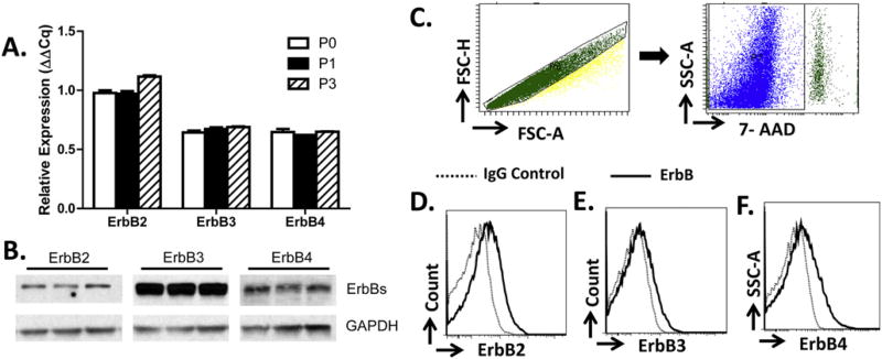
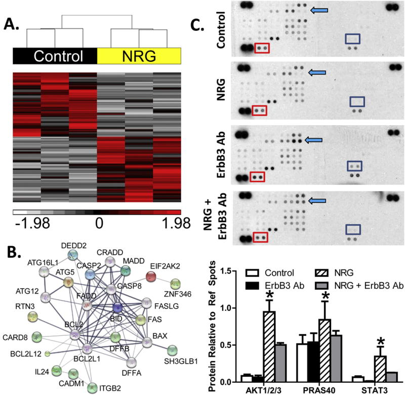
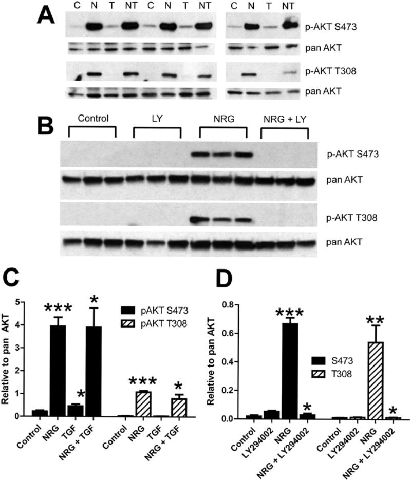
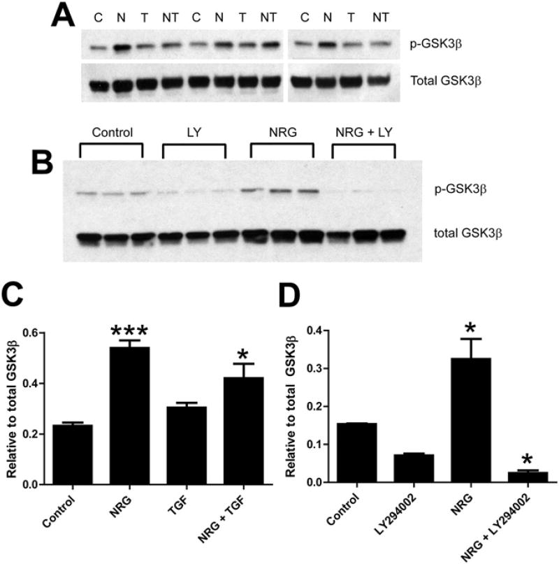
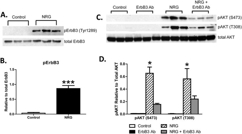
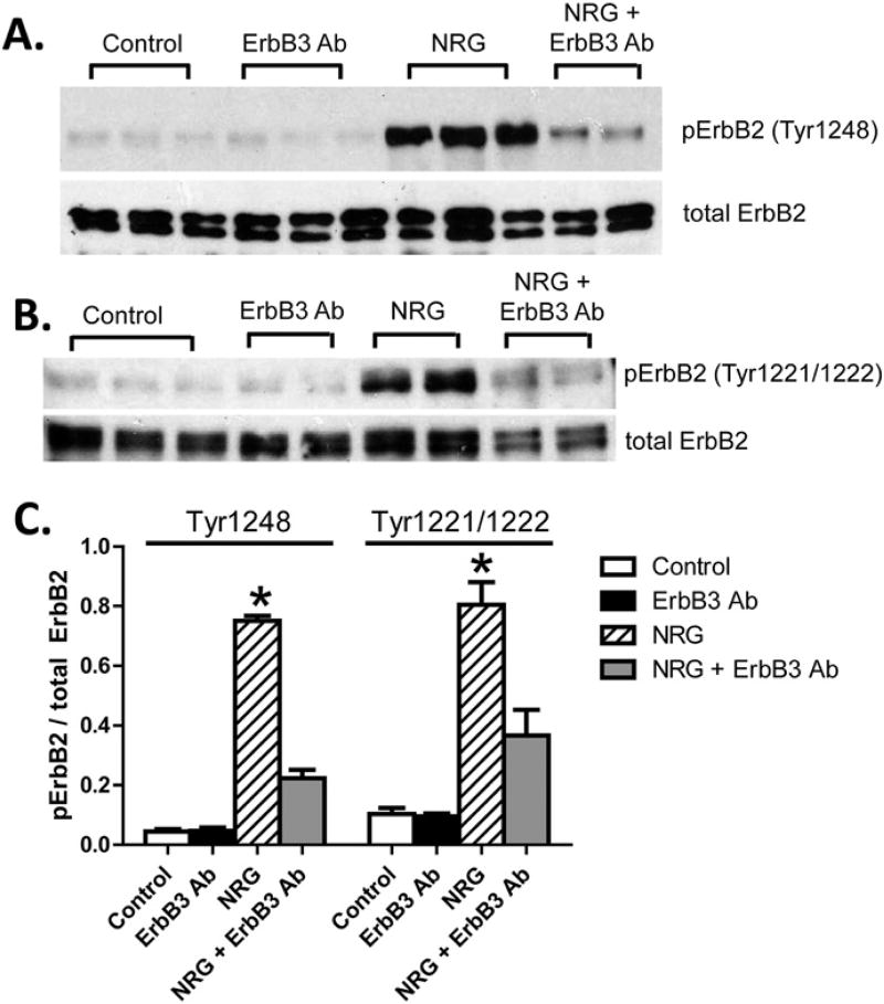
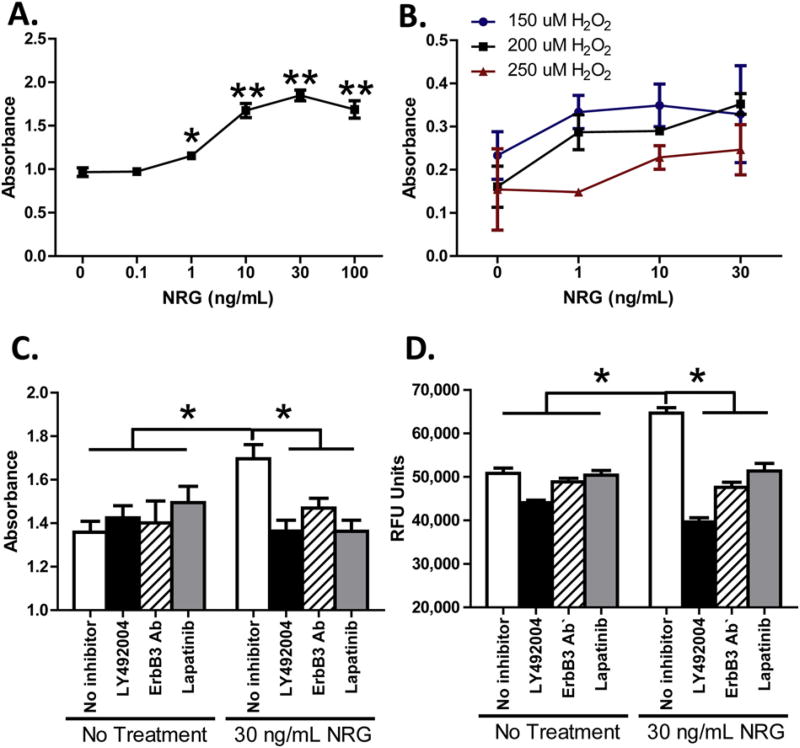
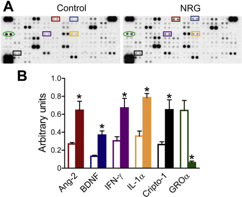
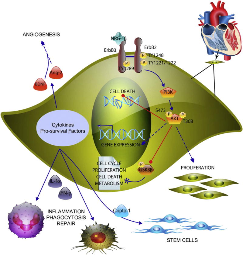
Neuregulin-1β (NRG-1β) is critical for cardiac development and repair, and recombinant forms are currently being assessed as possible therapeutics for systolic heart failure. We previously demonstrated that recombinant NRG-1β reduces cardiac fibrosis in an animal model of cardiac remodeling and heart failure, suggesting that there may be direct effects on cardiac fibroblasts. Here we show that NRG-1β receptors (ErbB2, ErbB3, and ErbB4) are expressed in normal human cardiac ventricular (NHCV) fibroblast cell lines. Treatment of NHCV fibroblasts with recombinant NRG-1β induced activation of the AKT pathway, which was phosphoinositide 3-kinase (PI3K)-dependent. Moreover, the NRG-1β-induced PI3K/AKT signaling in these cells required phosphorylation of both ErbB2 and ErbB3 receptors at tyrosine (Tyr)1248 and Tyr1289 respectively. RNASeq analysis of NRG-1β-treated cardiac fibroblasts obtained from three different individuals revealed a global gene expression signature consistent with cell growth and survival. We confirmed enhanced cellular proliferation and viability in NHCV fibroblasts in response to NRG-1β, which was abrogated by PI3K, ErbB2, and ErbB3 inhibitors. NRG-1β also induced production and secretion of cytokines (interleukin-1α and interferon-γ) and pro-reparative factors (angiopoietin-2, brain-derived neurotrophic factor, and crypto-1), suggesting a role in cardiac repair through the activation of paracrine signaling.

Keywords: Cardiac fibroblast; ErbB3; Neuregulin; RNASeq; Survival.


Figures

Fig. 1
Fig. 1
NHCV fibroblasts express ErbB Receptors. A, relative mRNA levels of ErbB2, ErbB3, and ErbB4 receptors in normal human ventricular cardiac (NHCV) fibroblasts after three passages is shown. B, Western blot analysis shows protein expression of ErbB2, ErbB3, and ErbB4 receptors in unstimulated NHCF cells. Anti-rabbit GAPDH served as a loading control. C, gating strategy to identify single live cells. Representative flow cytometry histograms showing isotype controls and surface expression of ErbB2 (D), ErbB3 (E), and ErbB4 (F) (n = 3 in 3 independent experiments).
Fig. 2
Fig. 2
RNA sequencing and protein array results for neuregulin-treated fibroblasts. A, hierarchical clustering of RNA transcript RPKM (Reads Per Kilobase per Million mapped reads) for differential genes between untreated and NRG-1β-treated NHCV fibroblasts. The cluster was produced using Partek Genomics Suite with average linkage and Euclidian distance measures. Bright red, white, and black indicate the highest, lowest, and median normalized reads, respectively. Vertical dendrograms represent the individual donor fibroblasts, of which there are three replicates for each group. B, differentially expressed genes associated with cell death formed a functional protein interaction network when analyzed using STRING software code and libraries, which are freely available online (string-db.org). C, representative immunoblots show differentially expressed phosphorylated AKT pathway proteins in NHCV fibroblasts that were untreated or treated for 30 min with NRG-1β, with and without pre-treatment with MAB3481, an ErbB3 blocking antibody. Each protein is represented on the array in duplicate, and each array includes three positive references (top left, top right, and bottom left) as well as negative reference spots (bottom right). Colored squares indicate significantly differential proteins. D, graphical representation of differentially expressed proteins identified using immunoblot arrays. Data includes densitometry analysis results for four replicate experiments (n = 4, *p < 0.05 for NRG-1β-treated cells, when compared to the various control treatments).
Fig. 3
Fig. 3
Neuregulin-1β treatment induces AKT activation in human cardiac fibroblasts. A, Western blot analysis of protein lysates of NHCV fibroblasts treated for 30 min with 30 ng/mL of NRG-1β. Bands shown are 60 kDa and were detected using antibodies that recognize phosphorylated AKT (pAKT) at serine position 473 (S473) or threonine position 308 (T308). Blots re-probed with pan AKT served as protein loading controls. Experimental conditions included untreated control cells, denoted by a “C”, and cells treated with NRG-1β (N), 1 ng/mL TGF-β (T), or both NRG-1β and TGF-1β (NT). Representative blots are shown. B, Western blots of protein lysates of NHCV fibroblasts probed with pAKT at S473 or T308 and re-probed with pan AKT. Treatments shown (in triplicate) include untreated control cells, and cells treated with the PI3K inhibitor LY294002 (LY), 30 ng/mL of NRG-1β (NRG), or both LY and NRG. C and D, Graphical representations of densitometry analysis results for the differentially expressed proteins shown in A and B, respectively (n = 3, *p < 0.05, **p < 0.01, and ***p < 0.001, respectively for NRG-1β-treated cells, when compared to appropriate control treatments).
Fig. 4
Fig. 4
Neuregulin-1β treatment induces phosphorylation of GSK-3β in human cardiac fibroblasts. A, Western blot analysis of protein lysates of NHCV fibroblasts treated for 30 min with 30 ng/mL of NRG-1β. Bands shown are 46 kDa and were detected using anti-rabbit GSK-3β and after re-probing with pan GSK-3β. Experimental conditions included untreated control cells, denoted by a “C”, and cells treated with NRG-1β (N), 1 ng/mL TGF-β (T), or both NRG-1β and TGF-1β (NT). Representative blots are shown. B, Western blots of protein lysates of NHCV fibroblasts probed first with pGSK-3β and then with pan GSK-3β (as a loading control). Treatments shown (in triplicate) include untreated control cells, and cells treated with the PI3K inhibitor LY294002 (LY), 30 ng/mL of NRG-1β (NRG), or both LY and NRG. C and D, Graphical representations of densitometry analysis results for the differentially expressed proteins shown in A and B, respectively (*p < 0.05 and ***p < 0.001, respectively for NRG-1β-treated cells, when compared to appropriate control treatments).
Fig. 5
Fig. 5
Neuregulin-1β treatment induces phosphorylation of ErbB3 receptor in human cardiac fibroblasts. A, Western blot analysis of protein lysates of NHCV fibroblasts treated for 30 min with 30 ng/mL of NRG-1β. Bands shown are 185 kDa and were detected using an anti-rabbit antibodies that recognize phosphorylated ErbB3 (pErbB) at tyrosine position 1289 (Tyr1289) and pan ErbB3. B, Graphical representation shows densitometry analysis result for blot shown in A. C, Western blots of protein lysates of NHCV fibroblasts probed with pAKT at S473 or T308 and re-probed with pan AKT. Treatments shown (in triplicate) include untreated control cells, and cells treated with an ErbB3 blocking antibody (MAB3481), 30 ng/mL of NRG-1β (NRG), or both ErbB3Ab and NRG. Graphical representations of densitometry analysis results for the differentially expressed proteins are shown in C (n=3, *p < 0.05, ***p < 0.001 for NRG-1β-treated cells compared to untreated fibroblasts).
Fig. 6
Fig. 6
Neuregulin-1β treatment induces phosphorylation of ErbB2 receptor in human cardiac fibroblasts. A, Western blot analysis of protein lysates of NHCV fibroblasts treated for 30 min with 30 ng/mL of NRG-1β. Bands shown are 185 kDa and were detected using an anti-rabbit antibodies that recognize phosphorylated ErbB2 (pErbB2) at tyrosine position 1289 (Tyr1248) and pan ErbB2. A representative blot is shown. B, Western blot analysis of protein lysates of NHCV fibroblasts treated for 30 min with 30 ng/mL of NRG-1β. Bands shown are 185 kDa and were detected using anti-rabbit antibodies that recognize phosphorylated ErbB2 (pErbB2) at tyrosine positions 1221 and 1222 (Tyr1221/1222), and pan ErbB2. C, Graphical representation showing densitometry analysis result for blots shown in A and B. Asterisks represent significance (n= 3, *p < 0.05 for NRG-1β-treated cells compared to controls).
Fig. 7
Fig. 7
NRG enhances proliferation and survival of human cardiac fibroblasts. A, graphical representation of BrdU incorporation in NHCV fibroblasts treated with increasing dosages of NRG (1–100 ng/mL), as shown on the abscissa. Cells were treated for 24 h, then treated with BrdU, incubated an additional 24 h, and the absorbance recorded as shown on the ordinate. B, graphical representation of cell viability in NHCV fibroblasts exposed to hydrogen peroxide (H2O2) and treated with NRG for 24 h. Absorbance at 590/600 nm is shown on the ordinate and concentration of NRG is displayed on the abscissa. Three concentrations of H2O2 are shown, as represented by blue (150 µm), black (200 µm), and red (250 µm) lines. C, results of cell proliferation assays for untreated and NRG-treated NHCV fibroblasts with and without pre-treatment with inhibitors for PI3K (LY294002, ErbB3 (MAB3481), or ErbB2 (Lapatinib). Treatments and BrdU incorporation were performed as described in A. D, results of viability assays for untreated and NRG-treated NHCV fibroblasts with and without pre-treatment with inhibitors for PI3K (LY294002), ErbB3 (MAB3481), or ErbB2 (Lapatinib). Untreated and treated cells were challenged with 200 µm for 24 h to induce cell death, as described in B (*p < 0.05 for NRG-1β-treated cells compared to controls).
Fig. 8
Fig. 8
Cytokines secreted from neuregulin-treated fibroblasts. A, representative immunoblots show differentially expressed cytokines in the supernatants collected from NHCV fibroblasts that were untreated or treated for 24 h with NRG-1β. Each cytokine is represented on the array in duplicate, and each array includes three positive references (top left, top right, and bottom left) as well as negative reference spots (bottom right). Colored squares indicate significantly differential cytokines. B, graphical representation of differentially expressed cytokines identified using cytokine profile arrays. Data includes densitometry analysis results for three replicate experiments conducted using NHCV fibroblast cell populations established from three separate individuals (n = 3, *p < 0.05 for NRG-1β-treated cells, when compared to the various control treatments). Open and solid bars represent untreated control cells and NRG-1β-treated cells, respectively. Bar colors correspond to colors of squares shown in A.
Fig. 9
Fig. 9
Model illustrating the intracellular signaling pathway leading to cell proliferation and survival in response to NRG-1β treatment. NRG-1β induces phosphorylation of the kinase-dead ErbB3 receptor, which leads to recruitment and phosphorylation of the kinase active ErbB2 receptor. This leads to activation of phosphoinositide-3 kinase (PI3K) with subsequent phosphorylation of protein kinase B (AKT) at serine (S)473 and threonine (T)308. AKT phosphorylates and inactivates GSK-3β, which participates in multiple signaling pathways, per the literature (paper icon).

Similar articles

Cited by

References

    1. Fan D, Takawale A, Lee J, Kassiri Z. Cardiac fibroblasts, fibrosis and extracellular matrix remodeling in heart disease. Fibrogenesis Tissue Repair. 2012;5:15. - PMC - PubMed
    1. Meyer D, Birchmeier C. Multiple essential functions of neuregulin in development. Nature. 1995;378:386–390. - PubMed
    1. Kramer R, Bucay N, Kane DJ, Martin LE, Tarpley JE, Theill LE. Neuregulins with an Ig-like domain are essential for mouse myocardial and neuronal development. Proc. Natl. Acad. Sci. U. S. A. 1996;93:4833–4838. - PMC - PubMed
    1. Odiete O, Hill MF, Sawyer DB. Neuregulin in cardiovascular development and disease. Circ. Res. 2012;111:1376–1385. - PMC - PubMed
    1. Fuller SJ, Sivarajah K, Sugden PH. ErbB receptors, their ligands, and the consequences of their activation and inhibition in the myocardium. J. Mol. Cell. Cardiol. 2008;44:831–854. - PubMed

Publication types