. 2017 Apr 15;424(2):221-235.
doi: 10.1016/j.ydbio.2017.02.006. Epub 2017 Mar 2.

SHH E176/E177-Zn2+ conformation is required for signaling at endogenous sites

Affiliations

SHH E176/E177-Zn2+ conformation is required for signaling at endogenous sites

Diana S Himmelstein et al. Dev Biol. .

Abstract

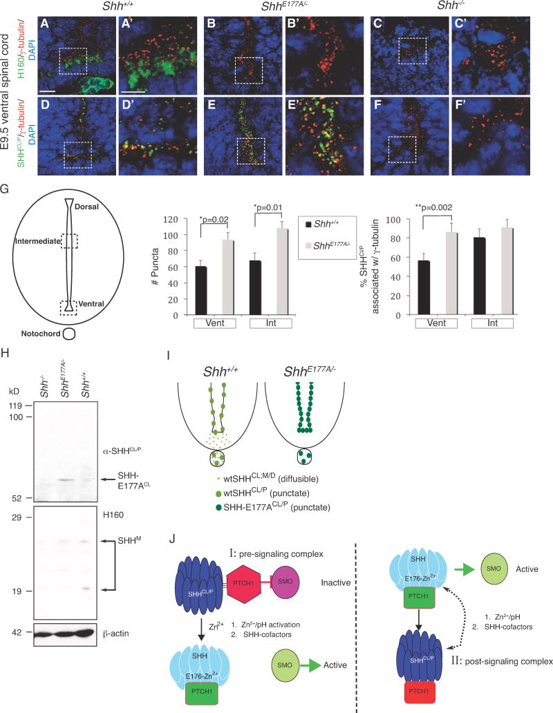
Sonic hedgehog (SHH) is a master developmental regulator. In 1995, the SHH crystal structure predicted that SHH-E176 (human)/E177 (mouse) regulates signaling through a Zn2+-dependent mechanism. While Zn2+ is known to be required for SHH protein stability, a regulatory role for SHH-E176 or Zn2+ has not been described. Here, we show that SHH-E176/177 modulates Zn2+-dependent cross-linking in vitro and is required for endogenous signaling, in vivo. While ectopically expressed SHH-E176A is highly active, mice expressing SHH-E177A at endogenous sites (ShhE177A/-) are morphologically indistinguishable from mice lacking SHH (Shh-/-), with patterning defects in both embryonic spinal cord and forebrain. SHH-E177A distribution along the embryonic spinal cord ventricle is unaltered, suggesting that E177 does not control long-range transport. While SHH-E177A association with cilia basal bodies increases in embryonic ventral spinal cord, diffusely distributed SHH-E177A is not detected. Together, these results reveal a novel role for E177-Zn2+ in regulating SHH signaling that may involve critical, cilia basal-body localized changes in cross-linking and/or conformation.


Figures

Figure 1
Figure 1. Conformation-specific antibody (α-SHHCl/P) recognizes cross-linked wtSHH in vitro and punctate, cilia BB-associated SHH in embryonic spinal cord ventricles
A. uSHHN (lipid lacking N terminal fragment), cross-linked wtSHH (wtSHHCL, N-and C-Lipid containing), and soluble monomeric wtSHH (wtSHH monomer, N-and C-lipid containing) are separated by SDS-PAGE and probed by Western blot analysis using α-SHHCl/P and H160 (α-SHH, Santa Cruz). α-SHHCl/P recognizes wtSHHCL, but not soluble monomeric wtSHH or monomeric uSHHN. H160 recognizes monomeric uSHHN and monomeric wtSHH, but not cross-linked wtSHHCL. Schematics for generating wtSHHCL,mono (N-terminal palmitate (Palm) and C-terminal cholesterol (Chol) containing) and uSHHN (lipid lacking) are shown on the right. B. Immunocytochemistry using α-SHHCl/P (green) and DAPI stain for nuclei (blue) detects SHH in C17 cell lines stably transfected with wtSHH, but not C24S-SHHN (N-and C-lipid lacking), and non-transfected control cells (C17). α-SHHCl/P detects wtSHH, but does not detect C24S-SHHN, as it is not expected to remain associated with the membrane. No staining is detected in non-transfected control cells, C17. C. Schematic of coronal section through the E9.5 spinal cord and notochord, with dashed boxes highlighting the regions shown in each subsequent image. D–L. Confocal analysis of immunofluorescent staining in E9.5 spinal cord using α-SHHCl/P (green; D-G′, I, L) or H160 (green; H, J, K) with γ-tubulin (cilia BBs, red; D–F, F′, H–L) or α-acetylated α-tubulin (axonemes, α-tubulin, red; G, G′). Notochord staining using γ-tubulin and α-SHHCl/P (I) or H160 (J). Shh−/− spinal cord staining using H160 (K) and α–SHHCl/P (L). DAPI (nuclei, blue). (F′–G′) Threedimensional reconstructions of ten confocal images taken sequentially along the z-axis from images F and G, respectively (AMIRA software). M. Quantification of the numbers of SHHCL/P puncta (green bars) or SHHCL/P/γ-tubulin double labeled puncta (yellow bars) comparing 2000μm2 regions in INT or VENT regions. *p<0.05, Student’s t-test. N. Schematic of the ventral spinal cord depicting the location of SHHCL/P detected by α-SHHCl/P (cilia BB-associated, black puncta) and H160 (non-BB associated, soluble, diffuse SHH, gray area). Scale bars = 5 μm (D–G, H–L) and 0.5 μm (F′-G′).
Figure 2
Figure 2. SHHCL/P localization in the embryonic ventral spinal cord of Ptc1−/−, ShhN/, and Ift172/Wimple mice
SHHCl/P co-localization with cilia BBs is determined using immunofluorescence microscopy in embryonic ventral spinal cord sections of mice lacking PTC1, Shh cholesterol modification, and IFT172 mutation (Wimple). E8.75 ventral spinal cord: Shh+/+ (A–C, A′–C′), Ptcl−/− (D–F, D′–F′). E9.5 ventral spinal cord, Shh+/+ (G, H, G′, H′), ShhN/−, lacking the C-terminal cholesterol modification, (I,J, I′,J′), Ift172/Wimple (K, L, K′, L′). White dotted lines outline ventral spinal cord ventricles and notocord. Anti-SHH antibodies (α-SHHCL;P and α-SHHCL;M/D) are green. Anti-γ-tubulin detects cilia BBs in red (A, B, D, E, A′, B′, D′, E′, G–L, G–L′, M)). Anti-a-tubulin detects cilia axonemes in red (C, C′, F, F′). Loss of PTC1 causes increased diffusion and aggregation of SHHCL/P (H, H′, I, I′), and diffusion of SHHCL;M/D (G, G′). α-SHHCL;M/D recognizes floorplate and notochord localized SHH, but not cilia BB associated SHH puncta. M. Anti-SHHCL;M/D (green) does not recognize targets in Shh−/− E9.5 ventral embryonic spinal cord, γ-tubulin (red). N. Western analysis using α-SHHCL;M/D recognizing an epitope shared by both wtSHHCL and uSHHN proteins. Scale Bars: 10μm (A–M), 5μm (A′–L′).
Figure 3
Figure 3. SHH-E176 controls Zn2+-dependent cross-linking and recognition by conformation-specific antibodies
Factors affecting the formation of SHH non-reducible cross-linked dimers (CLdimer) A. uSHHN (lanes 1–9), lanes 1–2, pH 6.5, lanes 3–4, pH 7.0, lanes 5–9, pH 7.5, lanes 2, 4, 6, 9, 1 mM Zn2+, lane 7, 1 mM Mg2+, lanes 8 and 9, 1 mM EDTA. Western analysis was performed using anti-SHH antibody H160. B. uSHHN (lanes 1–5), lane 1, Zn2+ addition 30 minutes after cross-linking reaction starts, lane 2, minus Zn2+, lane 3, Zn2+ addition from the beginning of the cross-linking reaction, lanes 4–5, 100ng heparin, lane 5, 1 mM Zn2+. Western analysis was performed using anti-SHH antibody H160. Solid arrows indicate CLdimer and monomer, and dashed arrow indicates CLintra. C. SHHN proteins affinity purified from secreted cell culture supernatants, affinity purified on a 5E1 column (5E1, monoclonal anti-SHH, Developmental Hybridoma Studies Bank), and assayed for cross-linking. SHHN (lanes 1–2, 1′–2′), SHHN-E176A (lanes 3–4, 3′–4′), wtSHH (lanes 5–6, 5′–6′), and SHH-E176A (lanes 7–8, 7′–8′) were incubated in the presence (lanes 2, 4, 6, 8) or absence (lanes 1, 3, 5, 7) of 1mM Zn2+. Western analysis was performed using α-SHHCL/P (lanes 1′–8′), stripped and reprobed with H160 (lanes 1–8). Arrows indicate CLdimer and monomer. D. Description of recombinant proteins used in A–D: *Full-length construct, produces N-and C-lipid modified SHH proteins. SHH proteins are affinity purified from culture supernatants of transfected C17 neural cells, as previously described for wtSHH (Feng et al., 2004), **N-terminal construct produces N-lipid containing, C-lipid lacking SHH proteins. SHH proteins are affinity purified from supernatants of transfected C17 neural cells, previously described for (Feng et al., 2004), §N-terminal construct lacks both N-and C-lipid modifications. uSHHN is produced in E.coli, Ni-Agarose affinity purified, histidine tag cleaved, as previously described in (Williams et al., 1999). + (antibody recognizes), − (antibody does not recognize), weak (antibody weakly recognizes), ND (not determined), +ç (recognizes wtSHH crosslinked form after storage at −80°C for 6 months). Note: Recombinant proteins (1–5) do not contain tags at the N-or C-termini. Proteins 1–4 were purified by affinity chromatography on a 5E1 anti-SHH-antibody column, while the 6Xhis-tag on uSHHN (protein 5) was removed prior to crosslinking assays.
Figure 4
Figure 4. SHH-E176A is highly active when ectopically expressed in the developing mouse forebrain
A. Diagram of retroviral backbone (pCLE) used to express the human SHH protein. Full length human cDNA sequence was inserted into cloning site (CS), producing N-and C-lipid modified SHH proteins. Placental alkaline phosphatase (PLAP) is bicistronic with the Shh cDNA, allowing detection of virally infected cells (Gaiano et al., 1999; Kohtz et al., 2001). B. Whole cell extracts of C17 cells infected with wtSHH and SHH-E176A expressing viruses are Western-blotted and probed with H160 antibody, showing that both viruses express monomeric, cell associated SHH proteins. C–K. Analysis of embryos injected with retroviruses at E9.5 and analyzed at E12.5 (C–E) uninfected littermates, (F–H) wtSHH, (I–K) Shh-E176A. Whole embryos (C, F, I), and sections (D, E, G, H, J, K) of E12.5 mouse brains 3 days after infection with SHH expressing. Infected viral clusters are visualized by alkaline phosphatase staining in the dorsal midline of the telencephalon (G, J). Uninfected littermate control does not contain alkaline phosphatase-expressing clusters of cells (D). In situ hybridization of adjacent sections probed for ectopic expression of Dlx2 (E, H, K). wtSHH, n=8/8, SHH-E176A, n=4/4 display gross morphological defects. Scale bar in D, (same for E, G, H, J, K) is 300 μm. Arrows indicate ventralization of dorsal telencephalon by SHH overexpression and ectopic expression of Dlx2.
Figure 5
Figure 5. E177 is required for Shh signaling in vivo
A. ShhE177A/− embryos display gross morphological defects compared to Shh+/+ at E12.5. Schematic showing homologous recombination in embryonic stem cells. The mouse bacterial artificial chromosome 429m20 contains a 16 kb fragment spanning the Shh genomic region. A glutamate to alanine point mutation at amino acid 177 was introduced in exon 2, and a conditional triple polyadenylation signal was inserted in the 5′ UTR. Schematic representation shows homologous recombination between the BAC targeting vector and the mouse Shh (mShh) genomic locus to generate the floxed transcription stop E177A targeted allele (ShhTS-E177A). ShhTS-E177A/+ mice were bred with EllaCre mice that express Cre recombinase under the control of the adenovirus Ella promoter to excise the floxed transcription stop. The resulting allele contains only one loxP site and the E177A mutation (ShhE177A). Black rectangles represent exons, lines represent introns. Correct targeting in ES cells is shown by Southern blotting. ApaLI and HindIII restriction sites are indicated. Stars represent the location of probes. B. RNA isolated from Shh+/−, ShhE177A/+ and ShhE177A/− embryos is reverse transcribed and cDNA is amplified by PCR. The E177A point mutation results in the generation of an MluI restriction enzyme site. MluI digest of an intron-spanning PCR on cDNA shows that the E177A mutation is expressed and the transcript is correctly spliced. Sequencing of cDNAs isolated from E9.5 Shh+/+ and ShhE177A/− embryos confirms introduction of E177A, and Mlu1 restriction site. C. X-galstaining for Ptc-lacZ expression in whole E9.5 embryos shows Ptc1 target gene expression is lost in ShhE177A/−PtclacZ/+ and Shh−/−PtclacZ/+ embryos. D–L. RNA in situ hybridization analysis using Ptc1 (D–F), Gli1 (G–I) and Shh (J–L) probes on E9.5 lumbar spinal cord sections in Shh+/+ (D,G,J), ShhE177A/− (E,H,K), and Shh−/− (F,I,L). M–a. Immunofluorescence staining of spinal cord progenitor markers HNF3β (M–O), Nkx2.2 (P–R), Nkx6.1 (S–U), Pax6 (V–X) and Pax7 (Y–a) on Shh+/−, ShhE177A/−, and Shh−/− E9.5 lumbar spinal cord sections, as indicated. (b–d) Nkx2.1 RNA situ hybridization of E12.5 forebrain sections from Shh+/+ (b), and anterior sections from ShhE177A/−(c) and Shh−/− (d). Scale bars: 500 μm (A,C) and 50 μm (D–a).
Figure 6
Figure 6. Increased SHHCl/P cilia BB association and SHH cross-linking in ShhE177A/− embryonic spinal cord
A–F′. Confocal analysis of immunofluorescence staining using H160 (A–C′, green) and α-SHHCL/P (D–F′, green), on E9.5 Shh+/+ (A, A′, D, D′), ShhE177A/− (B, B′, E, E′), and Shh−/− (C, C′, F, F′) lumbar spinal cord sections. Scale bars 10μm (A–F), 5μm (A′–F′). G. Schematic of the spinal cord highlighting the regions where SHHCl/P puncta are counted. Bar graph on the left shows the total number of puncta per 2000μm2 area; bar graph on the right shows the %SHHCL/P associated with γ-tubulin. Counting is performed in ventral (Vent) and intermediate (Int) regions of the Shh+/+ (black bars) and ShhE177A/− (gray bars) E9.5 lumbar spinal cord. In Shh+/+, the dorsal boundary of the intermediate region is 200 μm from the dorsal edge of the spinal cord; the ventral boundary of the intermediate region is 320 μm from the ventral edge of the spinal cord. A corresponding region is used for ShhE177A/− embryos. Data represented as mean ± SD, Student’s t-test, total number of puncta: *p=0.02.(ventral region) and *p= 0.01, (intermediate region), %SHHCL/P associated with γ-tubulin *p=0.002 (ventral region). n=3 embryos for each region. H. Western analysis of membrane extracts isolated from anterior region of Shh+/+, ShhE177A/−, and Shh−/− E10.5 embryos. The top of the blot was probed with α-SHHCL/P and bottom was probed with H160. The lower part of the blot was reprobed with anti-β-actin for a loading control. A long gel (14 cm) was run for better protein separation. α-SHHCl/P identifies SHH-E177ACL in ShhE177A/−, indicated by the arrow. H160 identifies SHH monomers migrating as multiple bands between 19–29 kD (SHHM). There is a shift from the 20kD monomer to the cross-linked form in Shh+/+ compared to ShhE177A/−. Shh−/− extracts are loaded as a negative control. I. Ventral spinal cord schematics show that ShhE177A/− mutants lack diffuse Shh staining (light green) in floorplate, with increased puncta (dark green) expression in the spinal cord ventricle. J. Models proposing possible roles of SHH-E177, Zn2+ and SHH/CL/P in SHH signaling. I. Pre-signaling E176/E177-Zn2+ activation model of SHH signaling at cilia BBs. II. Post-signaling model of SHHCL/P formation at cilia BBs. While the pre-signaling model is preferred based on the findings listed below, it is also possible that SHHCL/P can function in both pre and post-signaling. The model is based on the following findings:
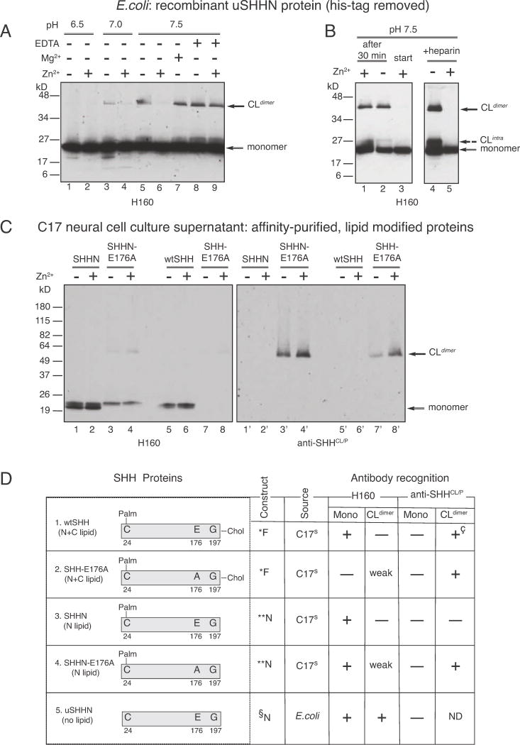
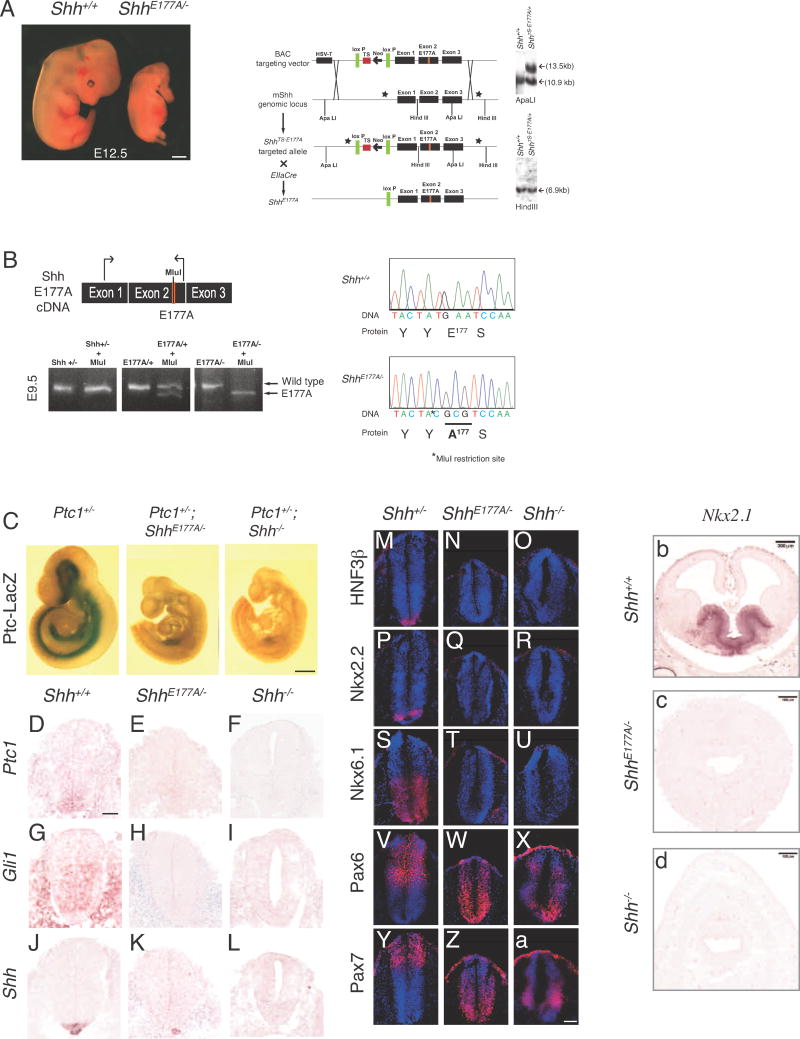
  1. E176A/E177A modulates Zn2+-mediated conformational change, detected by increased formation of cross-linked dimers.

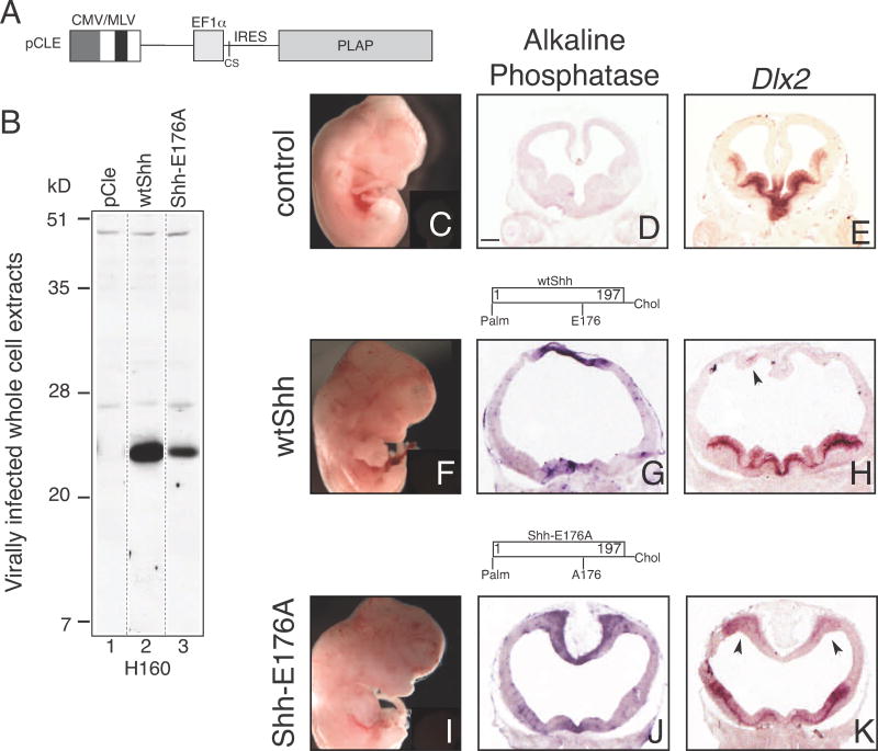
  2. E176A/E177A is active at ectopic, but not endogenous sites in vivo.

  3. In ShhE177A/− mutant spinal cord, increased accumulation of SHH-E177A occurs near cilia BBs.

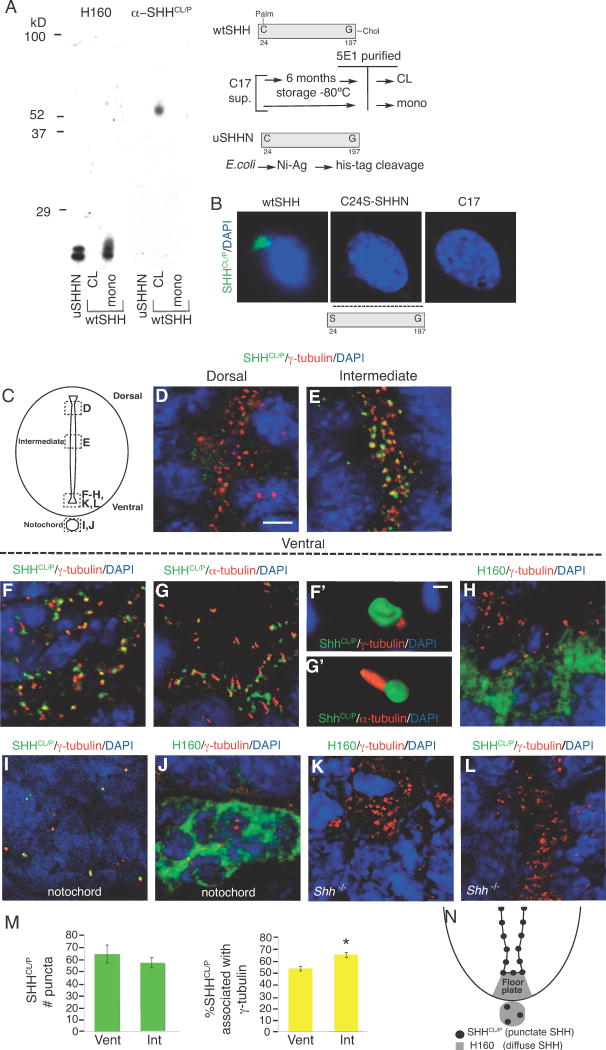
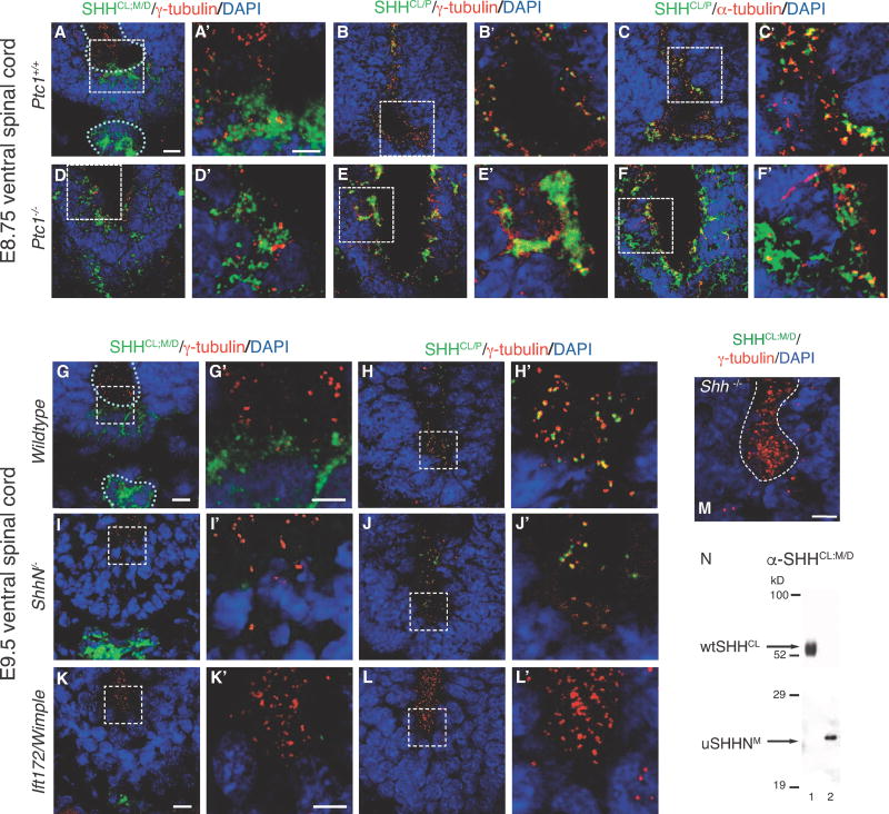
  4. SHHCL/P is still present in the ventral spinal cord even in the absence of the SHH receptor PTC1, supporting a pre-signaling role of SHHCL/P.

We propose that SHH signaling is regulated by a Zn2+-induced cross-linked/conformational change that occurs at cilia BBs. The E176/E177 site is required for this cross-linked/conformational change at cilia BBs, explaining why E177A mutation eliminates SHH signaling at endogenous sites. Co-factors that regulate the availability/concentration of Zn2+, pH, the C-terminal cholesterol modification, and PTC1 are likely to contribute to SHH signaling at endogenous sites, an environment that is not recapitulated in ectopic signaling events. The dotted arrow indicates the possibility of a dynamic state of conversion between inactive and active states. Proof of such conversions will rely on the development of tools to study these events in vivo.

Similar articles

Cited by

References

    1. Beachy PA, Hymowitz SG, Lazarus RA, Leahy DJ, Siebold C. Interactions between Hedgehog proteins and their binding partners come into view. Genes & development. 2010;24:2001–2012. - PMC - PubMed
    1. Briscoe J, Ericson J. Specification of neuronal fates in the ventral neural tube. Curr Opin Neurobiol. 2001;11:43–49. - PubMed
    1. Bumcrot DA, Takada R, McMahon AP. Proteolytic processing yields two secreted forms of sonic hedgehog. Molecular and cellular biology. 1995;15:2294–2303. - PMC - PubMed
    1. Chamberlain CE, Jeong J, Guo C, Allen BL, McMahon AP. Notochord-derived Shh concentrates in close association with the apically positioned basal body in neural target cells and forms a dynamic gradient during neural patterning. Development. 2008;135:1097–1106. - PubMed
    1. Chan JA, Balasubramanian S, Witt RM, Nazemi KJ, Choi Y, Pazyra-Murphy MF, Walsh CO, Thompson M, Segal RA. Proteoglycan interactions with Sonic Hedgehog specify mitogenic responses. Nature neuroscience. 2009;12:409–417. - PMC - PubMed

Publication types

MeSH terms

LinkOut - more resources