. 2017 Jul 27;36(30):4243-4252.
doi: 10.1038/onc.2017.8. Epub 2017 Mar 6.

GATA3 interacts with and stabilizes HIF-1α to enhance cancer cell invasiveness

Affiliations

GATA3 interacts with and stabilizes HIF-1α to enhance cancer cell invasiveness

M-C Lin et al. Oncogene. .

Erratum in

Abstract

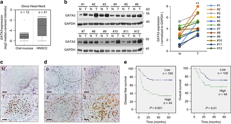
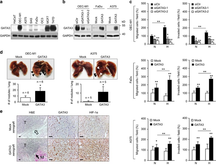
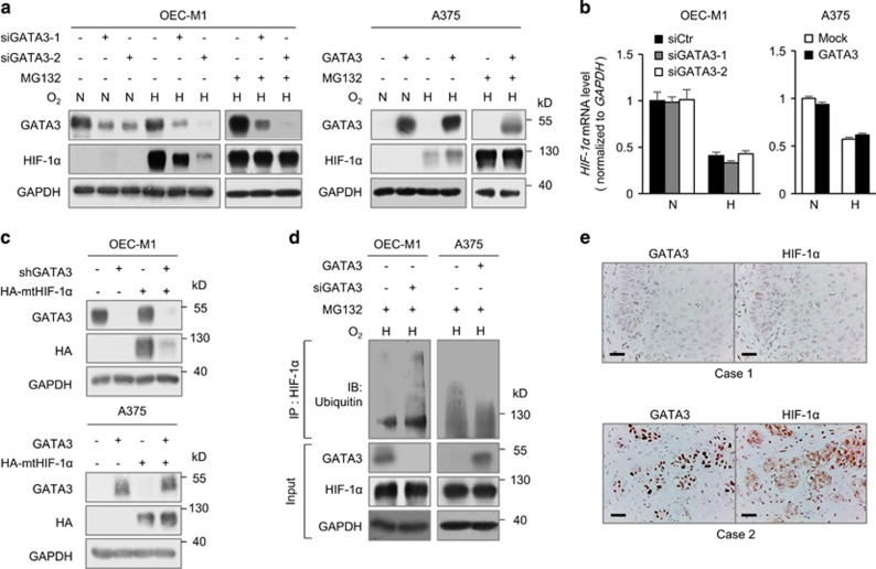
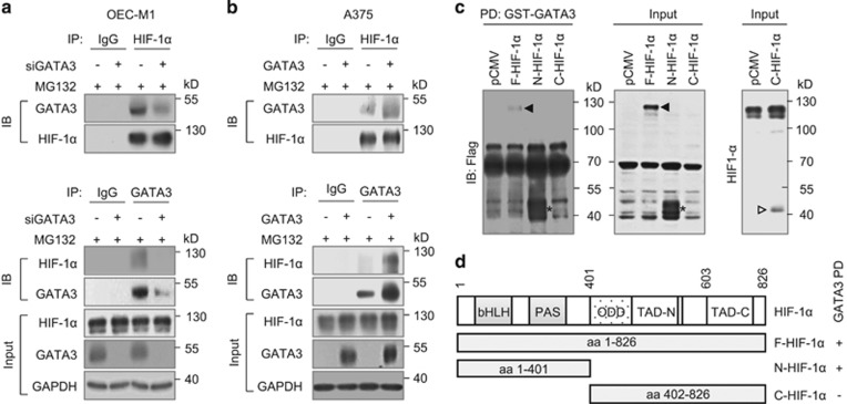
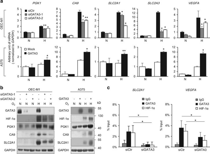
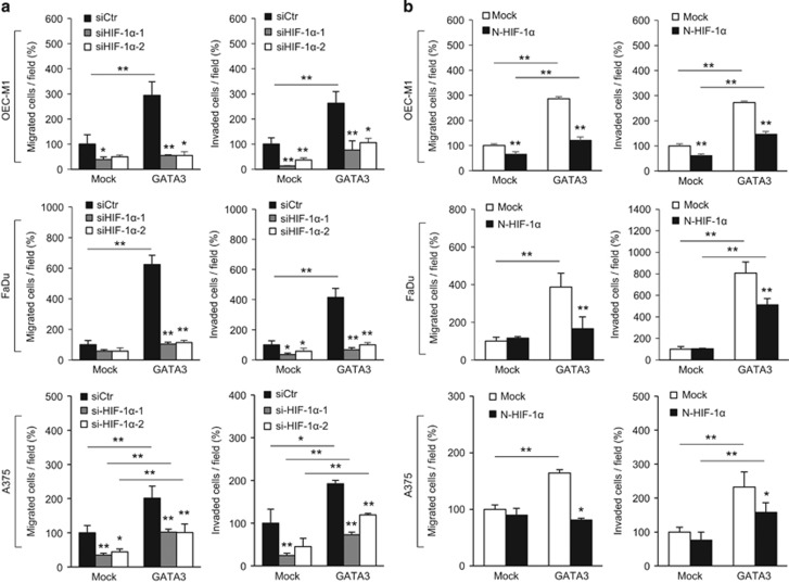
GATA binding protein 3 (GATA3) is indispensable in development of human organs. However, the role of GATA3 in cancers remains elusive. Hypoxia inducible factor (HIF)-1 plays an important role in pathogenesis of human cancers. Regulation of HIF-1α degradation is orchestrated through collaboration of its interacting proteins. In this study, we discover that GATA3 is upregulated in head and neck squamous cell carcinoma (HNSCC) and is an independent predictor for poor disease-free survival. GATA3 promotes invasive behaviours of HNSCC and melanoma cells in vitro and in immunodeficient mice. Mechanistically, GATA3 physically associates with HIF-1α under hypoxia to inhibit ubiquitination and proteasomal degradation of HIF-1α, which is independent of HIF-1α prolyl hydroxylation. Chromatin immunoprecipitation assays show that the GATA3/HIF-1α complex binds to and regulates HIF-1 target genes, which is also supported by the microarray analysis. Notably, the GATA3-mediated invasiveness can be significantly reversed by HIF-1α knockdown, suggesting a critical role of HIF-1α in the underlying mechanism of GATA3-mediated effects. Our findings suggest that GATA3 stabilizes HIF-1α to enhance cancer invasiveness under hypoxia and support the GATA3/HIF-1α axis as a potential therapeutic target for cancer treatment.


Conflict of interest statement

The authors declare no conflict of interest.

Figures

Figure 1
Figure 1
GATA3 is overexpressed in HNSCC and high GATA3 expression is associated with poor survivals. (a) GATA3 mRNA is overexpressed in HNSCC. GATA3 mRNA levels are higher in HNSCC (n=41) compared with normal oral mucosa (n=13). The data are retrieved from Ginos Head-Neck in the Oncomine database (https://www.oncomine.org). (b) Western blot analysis of GATA3 expression in paired HNSCC tumours (T) and adjacent non-tumour mucosa tissues (N) from 12 patients. Left: Western blots showing GATA3 expression and GAPDH control. Right: GATA3 expression was quantified and analysed by paired Student’s t-test. **P<0.01. (c) Representative images of immunohistochemistry of GATA3 in paired adjacent non-tumour mucosa (N) and tumour tissues (T) from HNSCC patients (n=15). Scale bars, 50 μm. (d) Scoring of immunohistochemistry of GATA3 in HNSCC tumour tissues. The intensity of GATA3 staining is scored from 0 to 3 and grouped into low (score=0, 1) and high (score=2, 3) expression. (e) Kaplan–Meier survival analysis. Patients with follow-up period over 18 months are included (n=144). Left and right panels indicate disease-free and overall survivals of patients with low and high GATA3 expression, respectively.
Figure 2
Figure 2
GATA3 promotes tumour invasiveness in vitro and in vivo. (a) Western blot analysis of GATA3 expression in various cell lines as indicated. GAPDH was an internal control. (b) Knockdown and overexpression of GATA3 were confirmed by western blot analysis. For knockdown, OEC-M1 cells were transfected with non-targeting siRNA (siCtr) or two independent siRNAs against GATA3 (siGATA3-1 and siGATA3-2). For overexpression, FaDu and A375 cells were transfected with empty pcDNA3.1 (Mock) or GATA3/pcDNA3.1 (GATA3). (c) Effects of GATA3 on cell migration and invasion analysed by transwell migration and Matrigel invasion assays, respectively. Cells were seeded after transfection for 48 h. Normoxia (N, O2=21%) or hypoxia (H, O2=1%) is as indicated. Results are represented as mean±s.d. from three independent experiments. *P<0.05, **P<0.01. (d) Effects of GATA3 on experimental metastasis in vivo. Upper panel, representative images of lungs and metastatic tumour nodules indicated by arrowheads. Lower panel, statistical analysis of tumour nodules. Mock or GATA3 overexpressing OEC-M1 and A375 stable transfectants (1 × 10⁶ cells / per injection) were injected through tail veins into NOD-SCID mice (n=6 and 5, respectively). Results are represented as mean±s.d. Data are analysed by Student’s t-test. *P<0.05. (e) Expression of GATA3 and HIF-1α in subcutaneous tumour xenografts. Left, hematoxylin and eosin staining. The hollow arrow indicates the intact capsule separating the tumour from surrounding tissues. The black arrow indicates the invasion of tumour into skeletal muscles. M, skeletal muscle. T, tumour. Middle and right, immunohistochemical staining of GATA3 and HIF-1α. Scale bars, 50 μm.
Figure 3
Figure 3
GATA3 expression increases HIF-1α protein levels. (a) Western blot analysis of GATA3 and HIF-1α under normoxia (N) or hypoxia (H) for overnight with or without a proteasome inhibitor, MG132 (20 μM), as indicated. GAPDH was an internal control. OEC-M1 cells were transfected with non-targeting siRNA or GATA3 siRNA (siGATA3-1 or siGATA3-2) and A375 cells were transfected with Mock or GATA3 plasmid. (b) Real-time RT-PCR analysis of HIF-1α mRNA expression. The total RNA was extracted from cells grown under normoxia or hypoxia for overnight. The results are represented as mean±s.d. from three independent experiments. (c) Western blot analysis of GATA3 and P402A/P564A HIF-1α mutant. GAPDH was an internal control. pLKO or shGATA3/pLKO (shGATA3) expressing OEC-M1 stable transfectants as well as Mock or GATA3 overexpressing A375 stable transfectants were transfected with empty vector or HA-P402A/P564A HIF-1α/pcDNA3.1 (HA-mtHIF-1α). (d) Effects of GATA3 on HIF-1α ubiquitination. OEC-M1 cells transfected with non-targeting siRNA or GATA3 siRNA (siGATA3-2) and A375 cells transfected with Mock or GATA3 plasmid were incubated with MG132 (20 μM) under hypoxia for 6 h. Cell lysates were immunoprecipitated with anti-HIF-1α antibody and then immunoblotted with anti-ubiquitin antibody. GATA3, HIF-1α and GAPDH in whole cell lysates (input) were shown. (e) Representative images of immunohistochemical staining of GATA3 and HIF-1α in serial sections of HNSCC tissues. Scale bars, 50 μm.
Figure 4
Figure 4
GATA3 physically interacts with HIF-1α. (a, b) Co-immunoprecipitation assays of GATA3 and HIF-1α in OEC-M1 and A375 cells. OEC-M1 cells were transfected with non-targeting siRNA or GATA3 siRNA (siGATA3-2) and A375 cells were transfected with Mock or GATA3 plasmid. Cells were incubated with MG132 (20 μM) and CoCl2 (800 μM) for 6 h. Upper panel, cell lysates were immunoprecipitated with IgG control antibody or anti-HIF-1α antibody and then immunoblotted with anti-GATA3 or anti-HIF-1α antibody. Lower panel, cell lysates were immunoprecipitated with IgG control antibody or anti-GATA3 antibody and then immunoblotted with anti-HIF-1α or anti-GATA3 antibody. HIF-1α, GATA3 and GAPDH in whole cell lysates (input) were shown. (c) GST pull-down assays. 293FT cells were transfected with F-, N- or C-HIF-1α/pCMV-Tag 4A (Flag-tagged) and incubated with CoCl2 (800 μM) for 4 h. Left panel, GST-GATA3 fusion protein pulled down F-HIF-1α and N-HIF-1α, but not C-HIF-1α. On the same membrane, C-HIF-1α could not be detected by the rabbit anti-HIF-1α polyclonal antibody (Cell Signaling Technology), which is known to recognize this region (Ser653, data not shown). The black arrowhead indicates F-HIF-1α. The asterisk indicates N-HIF-1α. The expression of F-HIF-1α and N-HIF-1α was confirmed by western blotting and shown in the middle panel. For unknown reason, C-HIF-1α could not be detected by anti-Flag antibody. We used rabbit anti-HIF-1α polyclonal antibody (Cell Signaling Technology) to confirm C-HIF-1α expression as shown in the right panel (hollow arrowhead). (d) The design of HIF-1α constructs is based on known functional domains and the pull-down assay results were summarized schematically. PAS, Per-ARNT-Sim.
Figure 5
Figure 5
GATA3 regulates expression of HIF-1 target genes under hypoxic conditions. (a) Real-time RT-PCR analysis of the expression of HIF-1α target genes, including PGK1, CA9, SLC2A1, SLC2A3 and VEGFA in OEC-M1 and A375 cells. (b) Western blot analysis of GATA3, HIF-1α, PGK1, CA9 and SLC2A1. GAPDH was an internal control. Total RNAs and cell lysates were extracted from cells grown under normoxia (N) or hypoxia (H), as indicated. (c) ChIP assays with anti-GATA3 or anti-HIF-1α antibody and SLC2A1 and VEGFA genes in OEC-M1 cells. Control or GATA3 knockdown cells were cultured under hypoxia for overnight and then harvested for ChIP assays. Isotype matched IgG was used as controls. The results are represented as mean±s.d. from three independent experiments. *P<0.05, **P<0.01.
Figure 6
Figure 6
GATA3-mediated cancer cell invasiveness is reversed by HIF-1α knockdown or N-HIF-1α expression. (a) Effects of HIF-1α knockdown on migration and invasion in OEC-M1, FaDu and A375 cells overexpressing GATA3. Mock or GATA3 overexpressing OEC-M1, FaDu and A375 stable transfectants were transiently transfected with non-targeting siRNA (siCtr) or two independent siRNAs against HIF-1α (siHIF-1α-1, siHIF-1α-2). Transwell migration and Matrigel invasion assays were carried out under hypoxia. (b) Effects of the ectopic N-HIF-1α expression on GATA3-mediated migration and invasion. Mock or GATA3 overexpressing OEC-M1, FaDu and A375 stable transfectants were transfected with empty vector (Mock) or N-HIF-1α/pCMV-Tag 4A (N-HIF-1α). Transwell migration and Matrigel invasion assays were carried out under hypoxia. The results are represented as mean±s.d. from three independent experiments. *P<0.05, **P<0.01.

Similar articles

Cited by

References

    1. Simon MC. Gotta have GATA. Nat Genet 1995; 11: 9–11. - PubMed
    1. Ho IC, Pai SY. GATA-3 – not just for Th2 cells anymore. Cell Mol Immunol 2007; 4: 15–29. - PubMed
    1. Hosoya T, Maillard I, Engel JD. From the cradle to the grave: activities of GATA-3 throughout T-cell development and differentiation. Immunol Rev 2010; 238: 110–125. - PMC - PubMed
    1. Asselin-Labat ML, Sutherland KD, Barker H, Thomas R, Shackleton M, Forrest NC et al. Gata-3 is an essential regulator of mammary-gland morphogenesis and luminal-cell differentiation. Nat Cell Biol 2007; 9: 201–209. - PubMed
    1. Labastie MC, Catala M, Gregoire JM, Peault B. The GATA-3 gene is expressed during human kidney embryogenesis. Kidney Int 1995; 47: 1597–1603. - PubMed

MeSH terms

Substances