. 2017 Mar 6;13(3):e1006265.
doi: 10.1371/journal.ppat.1006265. eCollection 2017 Mar.

Dengue virus genomic variation associated with mosquito adaptation defines the pattern of viral non-coding RNAs and fitness in human cells

Affiliations

Dengue virus genomic variation associated with mosquito adaptation defines the pattern of viral non-coding RNAs and fitness in human cells

Claudia V Filomatori et al. PLoS Pathog. .

Abstract

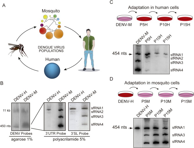
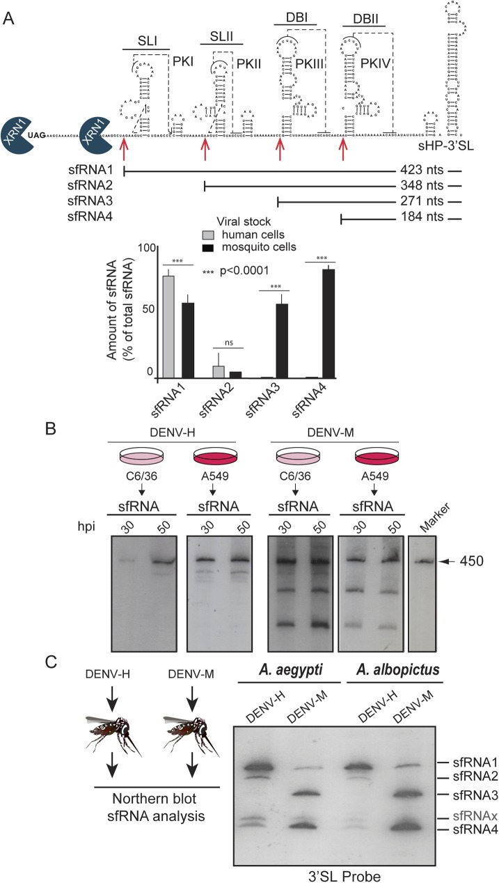
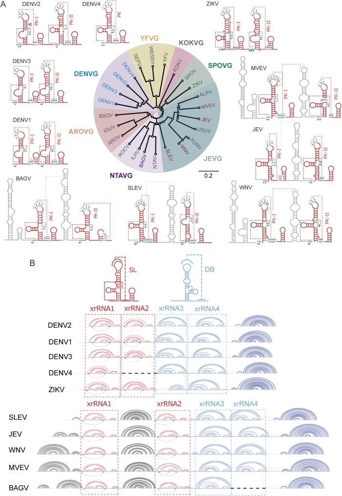
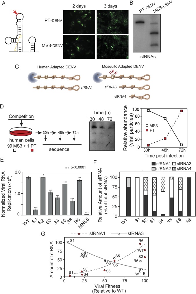
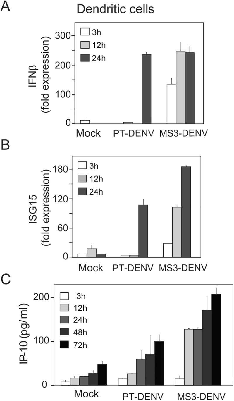
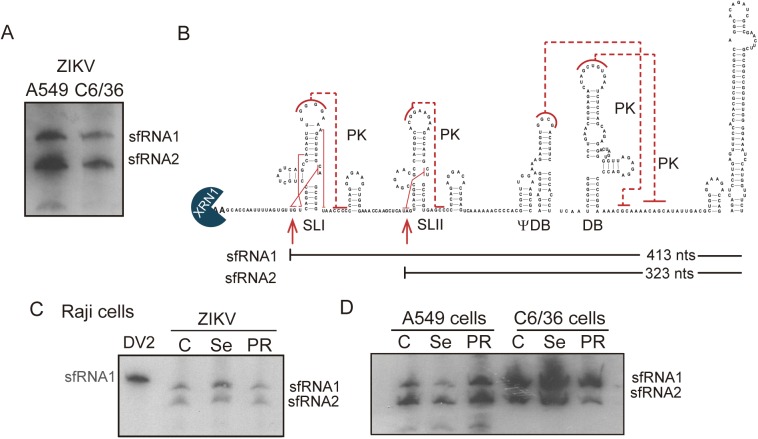
The Flavivirus genus includes a large number of medically relevant pathogens that cycle between humans and arthropods. This host alternation imposes a selective pressure on the viral population. Here, we found that dengue virus, the most important viral human pathogen transmitted by insects, evolved a mechanism to differentially regulate the production of viral non-coding RNAs in mosquitos and humans, with a significant impact on viral fitness in each host. Flavivirus infections accumulate non-coding RNAs derived from the viral 3'UTRs (known as sfRNAs), relevant in viral pathogenesis and immune evasion. We found that dengue virus host adaptation leads to the accumulation of different species of sfRNAs in vertebrate and invertebrate cells. This process does not depend on differences in the host machinery; but it was found to be dependent on the selection of specific mutations in the viral 3'UTR. Dissecting the viral population and studying phenotypes of cloned variants, the molecular determinants for the switch in the sfRNA pattern during host change were mapped to a single RNA structure. Point mutations selected in mosquito cells were sufficient to change the pattern of sfRNAs, induce higher type I interferon responses and reduce viral fitness in human cells, explaining the rapid clearance of certain viral variants after host change. In addition, using epidemic and pre-epidemic Zika viruses, similar patterns of sfRNAs were observed in mosquito and human infected cells, but they were different from those observed during dengue virus infections, indicating that distinct selective pressures act on the 3'UTR of these closely related viruses. In summary, we present a novel mechanism by which dengue virus evolved an RNA structure that is under strong selective pressure in the two hosts, as regulator of non-coding RNA accumulation and viral fitness. This work provides new ideas about the impact of host adaptation on the variability and evolution of flavivirus 3'UTRs with possible implications in virulence and viral transmission.


Conflict of interest statement

The authors have declared that no competing interests exist.

Figures

Fig 1
Fig 1
Different patterns of sfRNA produced during dengue virus host adaptation (A) Schematic representation of DENV 3’UTR diversification during host switch. Variants in viral populations obtained in mosquito or human cells are represented by circles. The size and color of circles represent the frequency and number of mutations of variants, respectively. The distance between variants is also shown by a fan dendrogram. (B) Genomic and subgenomic DENV RNAs produced in infections using viruses generated in human (DENV-H) or mosquito (DENV-M) cells. Cells were infected at MOI of 1 with DENV-H or DENV-M. Northern blots using a mix of radiolabeled probes that recognize the viral 3’UTR, capsid and NS5 (left), only the 3’UTR (middle) or only the 3’SL (right) are shown. The species of sfRNAs detected are indicated on the right. (C) Northern blot showing sfRNAs produced during the process of DENV adaptation to human cells. Specific probes complementary to the viral 3’UTR were used to hybridize RNA isolated from A549 cells infected with the indicated viruses. P5H, P10H and P15H indicate viral stocks passaged 5, 10 and 15 times in human cells, respectively. (D) Northern blot showing sfRNAs produced during the process of DENV adaptation to mosquito cells. Specific probes complementary to the viral 3’UTR were used to hybridize RNA isolated from C6/36 cells infected with the indicated viruses.
Fig 2
Fig 2. Mapping the sfRNAs generated in DENV infected human and mosquito cells.
(A) Secondary structure of DENV 3’UTR indicating the location and size of sfRNAs identified by sequencing analysis. Below, plots representing relative amounts of sfRNA species produced using human or mosquito infected cells. The amount of each sfRNA was estimated by ImageJ-quantitation and expressed as the mean +/- SD of the relative percentage from total sfRNAs (n = 3). (B) Northern blot hybridization using specific probes complementary to the viral 3’UTR employing RNA extracted at 30 and 50 hpi from C6/36 or A549 cells infected with either DENV-M or DENV-H stocks, as indicated. (C) DENV sfRNAs produced in infected Ae. albopictus and Ae. aegypi mosquitos. Northern blot hybridization using RNA extracted from mosquitos infected with DENV-M or DENV-H. A probe complementary to the viral 3’SL was used for detection.
Fig 3
Fig 3. Comparative analysis of predicted RNA structures of the 3’UTR of mosquito-borne flaviviruses (MBFV).
(A) Conserved stem loops of selected viruses from each subgroup of MBFV are shown in red. Predicted H-type pseudoknots are indicated with red dashed lines and pseudoknots including nucleotides present in the three-way junction are shown in black dashed lines. Information obtained from crystallographic studies using MVEV and ZIKV RNAs was included [25,39]. Group-specific RNA structures are indicated in grey. The distance tree was drawn using the neighbor joining method of all complete genome sequences for each virus available in GenBank. (B) Representation of the complete 3’UTR structure of different MBFVs showing the conserved elements involved in Xrn1 stalling. Conserved stem loop (SL) and dumbbell (DB) structures are shown in red and blue, respectively. Arc plots of RNA structures corresponding to the conserved xrRNA1, xrRNA2, xrRNA3 and xrRNA4 are shown for DENV1 to 4, ZIKV, WNV, MVEV, SLEV and BAGV. The conserved 3’SL structure is shown in dark blue.
Fig 4
Fig 4. Dissecting the molecular determinants for sfRNA accumulation during DENV host change.
(A) Schematic representation of mosquito-selected viral variants with mutations and deletions within the viral 3’UTR. DENV variants were found with deletions, variant S1 and S2, or point mutations variants S3 to S6. The location of the mutations is indicated. (B) Replication of parental (PT) and recombinant viruses carrying the mutations identified. Immunofluorescence of cells infected PT and the mutants S1 to S6 as indicated at the top. Viral RNA detection using specific radiolabeled probes complementary to capsid and NS5 coding sequences is shown at 72hpi of A549 cells (bottom). (C) Northern blot hybridization for sfRNA detection using transfected cells with each DENV mutant as indicated on the top. (D) The structure of xrRNA2 is necessary for accumulation of short sfRNA species. Schematic representation of changes in the xrRNA2 structure of mutant S6 and restored R6, and Northern blot shown the accumulated sfRNAs in each case. (E) Point mutations within xrRNA2 are sufficient for the switch of sfRNA pattern produced during DENV infections. Schematic representation of mutation abrogating PK formation (M-PKII) is shown on the left and Northern blot comparing the accumulation of sfRNAs detected in infected cells with M-PKII or a virus with a complete deletion of xrRNA2 (ΔSLII).
Fig 5
Fig 5
Fitness in human cells and sfRNA production of mosquito adapted DENV (A) Replication of parental (PT) and mutant (MS3) viruses in A549 cells monitored by immunofluorescence at 2 and 3-day post transfection. (B) Accumulation of sfRNAs in A549 cells transfected with PT or MS3 viral RNAs detected by Northern blot at 3-days post transfection. (C) Schematic representation of the key role of xrRNA2 for differential sfRNA accumulation in mosquito or human adapted viruses. Rope knots represent the xrRNA structures that impair Xrn1 movement and the gallows represents the terminal 3’SL structure. (D) Growth competition experiments highlight the fitness disadvantage of mosquito selected variant in human cells. A549 cells were infected with a mixture of PT: MS3 at a 1:99 ratio. At 30, 48 and 72 hpi, cells were collected for viral RNA purification,sequencing and sfRNA analysis. Relative abundance of each virus is represented in the plot shown on the right. (E) Viral RNA replication levels of WT and mosquito selected variants are shown together with a replication impaired control (mutant in the polymerase NS5, MNS5) in A549 cells. Mutations were incorporated in a replication and propagation competent reporter DENV carrying a luciferase gene. Normalized luciferase levels are shown in a logarithmic scale at 48h post-transfection. The values are the mean +/-SD, n = 3. (F) Quantitation of sfRNAs accumulation in A549 cells transfected with mutant RNAs. The values are the mean relative accumulation estimated with ImageJ Program from three independent experiments. (G) Plot of the mean relative amounts of different sfRNAs, shown in F, versus the replication capacity measured using the reporter virus shown in E.
Fig 6
Fig 6. Mosquito adapted DENV variant exhibit an exacerbated antiviral response in human cells.
Time course analysis of antiviral response in dendritic cells (DCs). DCs of three independent donors were infected with PT or MS3 virus, and at 3, 12, and 24 hpi IFNβ (A) or ISG15 (B) mRNA were determined by real time PCR. (C) The levels of IP-10 in the supernatant of infected cells was assessed by ELISA at 3, 12, 24, 48 and 72 hpi. Data are the means and standard deviations of three replicates from a representative donor.
Fig 7
Fig 7. Production of sfRNAs in Zika virus infections.
(A) Northern blot hybridization showing the accumulation sfRNA in C6/36 and A549 cells infected with a Zika isolate from Argentina (INEVH). (B) RNA structure model predicted for the ZIKV 3’UTR. Conserved SLI, SLII, pseudo-dumbbell (ψ-DB) and DB structures are indicated. Pseudoknots are indicated with red lines. Location and size of the two sfRNAs identified in infected cells are shown (sfRNA1 and sfRNA2). (C and D) Northern blots showing the pattern of sfRNAs in Raji, A549 and C6/36 cells infected with ZIKV from different origins (Cambodia, Senegal and Puerto Rico) as indicated on the top.

References

    1. Bhatt S, Gething PW, Brady OJ, Messina JP, Farlow AW, et al. (2013) The global distribution and burden of dengue. Nature 496: 504–507. 10.1038/nature12060 - DOI - PMC - PubMed
    1. Rico-Hesse R (1990) Molecular evolution and distribution of dengue viruses type 1 and 2 in nature. Virology 174: 479–493. - PubMed
    1. Weaver SC, Vasilakis N (2009) Molecular evolution of dengue viruses: contributions of phylogenetics to understanding the history and epidemiology of the preeminent arboviral disease. Infect Genet Evol 9: 523–540. 10.1016/j.meegid.2009.02.003 - DOI - PMC - PubMed
    1. Bennett SN, Drummond AJ, Kapan DD, Suchard MA, Munoz-Jordan JL, et al. (2010) Epidemic dynamics revealed in dengue evolution. Mol Biol Evol 27: 811–818. 10.1093/molbev/msp285 - DOI - PMC - PubMed
    1. Vu TT, Holmes EC, Duong V, Nguyen TQ, Tran TH, et al. (2010) Emergence of the Asian 1 genotype of dengue virus serotype 2 in viet nam: in vivo fitness advantage and lineage replacement in South-East Asia. PLoS Negl Trop Dis 4: e757 10.1371/journal.pntd.0000757 - DOI - PMC - PubMed

Publication types