Review
. 2017 Feb 27;5(1):7.
doi: 10.3390/vaccines5010007.

The Escape of Cancer from T Cell-Mediated Immune Surveillance: HLA Class I Loss and Tumor Tissue Architecture

Affiliations
Review

The Escape of Cancer from T Cell-Mediated Immune Surveillance: HLA Class I Loss and Tumor Tissue Architecture

Federico Garrido et al. Vaccines (Basel). .

Abstract

Tumor immune escape is associated with the loss of tumor HLA class I (HLA-I) expression commonly found in malignant cells. Accumulating evidence suggests that the efficacy of immunotherapy depends on the expression levels of HLA class I molecules on tumors cells. It also depends on the molecular mechanism underlying the loss of HLA expression, which could be reversible/"soft" or irreversible/"hard" due to genetic alterations in HLA, β2-microglobulin or IFN genes. Immune selection of HLA-I negative tumor cells harboring structural/irreversible alterations has been demonstrated after immunotherapy in cancer patients and in experimental cancer models. Here, we summarize recent findings indicating that tumor HLA-I loss also correlates with a reduced intra-tumor T cell infiltration and with a specific reorganization of tumor tissue. T cell immune selection of HLA-I negative tumors results in a clear separation between the stroma and the tumor parenchyma with leucocytes, macrophages and other mononuclear cells restrained outside the tumor mass. Better understanding of the structural and functional changes taking place in the tumor microenvironment may help to overcome cancer immune escape and improve the efficacy of different immunotherapeutic strategies. We also underline the urgent need for designing strategies to enhance tumor HLA class I expression that could improve tumor rejection by cytotoxic T-lymphocytes (CTL).

Keywords: HLA class I loss; tumor immune escape; tumor infiltrating lymphocytes (TILs).


Conflict of interest statement

The authors declare no conflict of interest.

Figures

Figure 1
Figure 1
Recovery of major histocompatibility complex (MHC)/HLA-I antigens in tumors. Different treatments, including IL-2, Bacillus Calmette–Guérin (BCG), or tumor peptides, can boost anti-tumor T cell-mediated responses. Currently, monoclonal antibodies against “immune checkpoint” molecules are also being actively used in the clinic. All these therapies can modify the tumor microenvironment and induce the release of TH1 type cytokines. HLA-I deficient tumors can upregulate HLA-I expression depending on the nature of the underlying molecular alteration. If the defect is reversible/“soft”, tumor or metastatic lesion can be rejected after cytokine-mediated recovery of the antigen presentation capacity. If the alteration is irreversible/“hard”, tumor or metastasis will remain HLA-I negative and, most likely, will escape T cell responses and continue to grow.
Figure 2
Figure 2
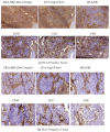
(a) Tissue architecture in HLA-I positive tumors (Phase I). Tumor tissue samples obtained from a patient diagnosed with non-small cell lung cancer (NSCLC) were immunostained with monoclonal antibodies against HLA-I, β2-microglobulin, HLA class II, CD8, CD3, and CD45 molecules. Most of the cancer cells are HLA-I positive and remain in close contact with HLA-I positive immune cells. The tissue structure is “permissive”, allowing TILs to enter the tumor mass, producing a significant CD8+ infiltration, and permitting a direct contact with cancer cells. Tumor parenchyma and stroma cannot be distinguished when immunostained for HLA-I expression. This tissue organization pattern in HLA-I positive tumors differs from that observed in HLA-I negative tumors depicted in Figure 2b; (b) Tissue architecture in HLA-I negative tumors (Phase II). In contrast to Figure 2a, this lung cancer tissue, obtained from another patient, is negative for both HLA-I and -II. T cells and other leukocytes are restricted exclusively to the peritumoral stroma that surrounds the tumor nest in a “non-permissive” tissue structure. There is a clear separation between tumor parenchyma and surrounding stroma. Tumor nodes are composed exclusively of HLA-I negative tumor cells without any infiltrating immune cells (phase of “immunological silence”).
Figure 3
Figure 3
Variations in the percentage of lymphocyte subpopulations and natural killer (NK) cells in different areas of tumor and “normal” adjacent tissues measured by flow cytometry. DNTT—distant non tumor tissue, ATT—adjacent tumor tissue, TT—tumor tissue. The treg cell subset was defined as CD127low CD25bright CD4+. The number of Treg was calculated as a percentage of total number of CD4+ cells, while the CD8+ DR+ and CD8+CD39+ cell levels are shown as a percentage of CD8+ cells. NK cells were determined by the selection of CD45+ CD3− CD20− cells in the FSClow/SSClow gate [31]. Tregs and CD8+CD39+ lymphocytes show increased presence closer to the tumor nest, while the percentage of NK cells is lower in TT as compared to other areas.
Figure 4
Figure 4
Changes in the percentage of CD56bright CD16− NK cells with impaired cytotoxic activity in different areas of the tumor and “normal” adjacent tissues analyzed by flow cytometry. TT is enriched by CD56bright CD16− NK cells, the percentage of which gradually increases from DNTT to TT. DNTT—distant non tumor tissue, ATT—adjacent tumor tissue, TT—tumor tissue. Tumor samples were obtained from primary lung tumors of non-treated patients by excision of a tumor mass fragment. ATT samples were taken from tumor-adjacent lung tissue located at approximately 1 cm from the periphery of the tumor without macroscopic signs of a tumor. DNTT and ATT samples were thoroughly analyzed to guarantee the complete absence of epithelial tumor cells. The NK cell subset was determined by the selection of CD45+ CD3− CD20− cells in the FSClow/SSClow gate and calculated as a percentage of all NK cells [31].

References

    1. Boesen M., Svane I.M., Engel A.M., Rygaard J., Thomsen A.R., Werdelin O. CD8+ T cells are crucial for the ability of congenic normal mice to reject highly immunogenic sarcomas induced in nude mice with 3-methylcholantrene. Clin. Exp. Immunol. 2000;121:210–215. doi: 10.1046/j.1365-2249.2000.01292.x. - DOI - PMC - PubMed
    1. Boon T., Coulie P., Van Den Eynde B.J., Van Der Bruggen P. Human T cell responses against melanoma. Annu. Rev. Immunol. 2006;24:175–208. doi: 10.1146/annurev.immunol.24.021605.090733. - DOI - PubMed
    1. Romero I., Garrido C., Algarra I., Collado A., Garrido F., Garcia-Lora A.M. T lymphocytes restrain spontaneous metastasis in permanent dormancy. Cancer Res. 2014;74:1958–1968. doi: 10.1158/0008-5472.CAN-13-2084. - DOI - PubMed
    1. Kessler B.M., Glas R., Ploegh H.L. MHC class I antigen processing regulated by cytosolic proteolysis-short cuts that alter peptide generation. Mol. Immunol. 2012;39:171–179. doi: 10.1016/S0161-5890(02)00100-1. - DOI - PubMed
    1. Romero P., Coulie P. Adaptive T cell immunity and tumor antigen recognition. In: Rees R., editor. Tumor Immunology and Immunotherapy. Oxford University Press; Oxford, UK: 2014. pp. 1–14.