

Diagnosis and treatment of cystic lung disease
- PMID: 28264540
- PMCID: PMC5339473
- DOI: 10.3904/kjim.2016.242
Diagnosis and treatment of cystic lung disease
Abstract
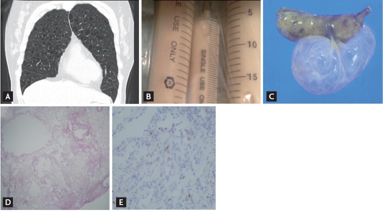
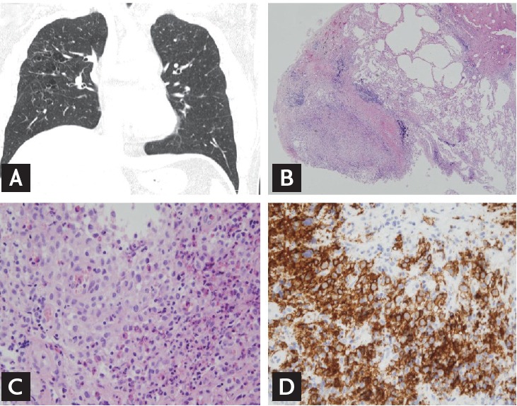
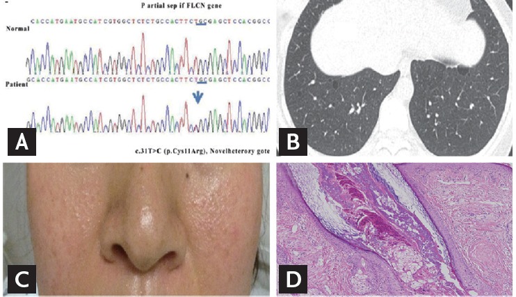
Cystic lung disease (CLD) is a group of lung disorders characterized by the presence of multiple cysts, defined as air-filled lucencies or low-attenuating areas, bordered by a thin wall (usually < 2 mm). The recognition of CLDs has increased with the widespread use of computed tomography. This article addresses the mechanisms of cyst formation and the diagnostic approaches to CLDs. A number of assessment methods that can be used to confirm CLDs are discussed, including high-resolution computed tomography, pathologic approaches, and genetic/ serologic markers, together with treatment modalities, including new therapeutic drugs currently being evaluated. The CLDs covered by this review are lymphangioleiomyomatosis, pulmonary Langerhans cell histiocytosis, Birt-Hogg-Dube syndrome, lymphocytic interstitial pneumonia/follicular bronchiolitis, and amyloidosis.
Keywords: Birt-Hogg-Dube syndrome; Cystic lung disease; Histiocytosis, Langerhans-cell; Lymphangioleiomyomatosis.
Conflict of interest statement
No potential conflict of interest relevant to this article was reported.
Figures
Similar articles
-
Diffuse Cystic Lung Diseases.Respir Care. 2020 Jan;65(1):111-126. doi: 10.4187/respcare.07117. Epub 2019 Oct 15. Respir Care. 2020. PMID: 31615921 Review.
-
Diffuse Cystic Lung Disease: A Clinical Guide to Recognition and Management.Chest. 2025 Feb;167(2):529-547. doi: 10.1016/j.chest.2024.08.008. Epub 2024 Aug 19. Chest. 2025. PMID: 39168181 Review.
-
Diffuse Cystic Lung Diseases: Diagnostic Considerations.Semin Respir Crit Care Med. 2016 Jun;37(3):457-67. doi: 10.1055/s-0036-1580690. Epub 2016 May 27. Semin Respir Crit Care Med. 2016. PMID: 27231867 Review.
-
Lymphangioleiomyomatosis and Other Cystic Lung Diseases.Immunol Allergy Clin North Am. 2023 May;43(2):359-377. doi: 10.1016/j.iac.2023.01.003. Immunol Allergy Clin North Am. 2023. PMID: 37055093 Free PMC article. Review.
-
[Diffuse cystic lung disease].Ther Umsch. 2024 Feb;81(1):16-20. Ther Umsch. 2024. PMID: 38655829 Review. German.
Cited by
-
Bronchoalveolar Lavage as a Diagnostic Tool in an Atypical Pulmonary Langerhans Cell Histiocytosis.Diagnostics (Basel). 2022 Jun 4;12(6):1394. doi: 10.3390/diagnostics12061394. Diagnostics (Basel). 2022. PMID: 35741204 Free PMC article.
-
Management of a rare case of lymphangioleiomyomatosis complicated by recurrent pneumothorax.BMJ Case Rep. 2024 Sep 19;17(9):e260369. doi: 10.1136/bcr-2024-260369. BMJ Case Rep. 2024. PMID: 39304215 Free PMC article.
-
Review of research on pulmonary cysts in sjögren's syndrome.J Cardiothorac Surg. 2025 Jul 18;20(1):306. doi: 10.1186/s13019-025-03540-5. J Cardiothorac Surg. 2025. PMID: 40682117 Free PMC article. Review.
-
Rho-Associated Protein Kinase Activity Is Required for Tissue Homeostasis in the Xenopus laevis Ciliated Epithelium.J Dev Biol. 2024 Jun 11;12(2):17. doi: 10.3390/jdb12020017. J Dev Biol. 2024. PMID: 38921484 Free PMC article.
-
Characterization by Quantitative Serum Proteomics of Immune-Related Prognostic Biomarkers for COVID-19 Symptomatology.Front Immunol. 2021 Sep 8;12:730710. doi: 10.3389/fimmu.2021.730710. eCollection 2021. Front Immunol. 2021. PMID: 34566994 Free PMC article.
References
-
- Hansell DM, Bankier AA, MacMahon H, McLoud TC, Muller NL, Remy J. Fleischner Society: glossary of terms for thoracic imaging. Radiology. 2008;246:697–722. - PubMed
-
- Copley SJ, Wells AU, Hawtin KE, et al. Lung morphology in the elderly: comparative CT study of subjects over 75 years old versus those under 55 years old. Radiology. 2009;251:566–573. - PubMed
Publication types
MeSH terms
Supplementary concepts
LinkOut - more resources
Full Text Sources
Other Literature Sources
Medical