. 2017 Mar 6;18(1):2.
doi: 10.1186/s12858-017-0077-1.

Systematic substitutions at BLIP position 50 result in changes in binding specificity for class A β-lactamases

Affiliations

Systematic substitutions at BLIP position 50 result in changes in binding specificity for class A β-lactamases

Carolyn J Adamski et al. BMC Biochem. .

Abstract

Background: The production of β-lactamases by bacteria is the most common mechanism of resistance to the widely prescribed β-lactam antibiotics. β-lactamase inhibitory protein (BLIP) competitively inhibits class A β-lactamases via two binding loops that occlude the active site. It has been shown that BLIP Tyr50 is a specificity determinant in that substitutions at this position result in large differential changes in the relative affinity of BLIP for class A β-lactamases.

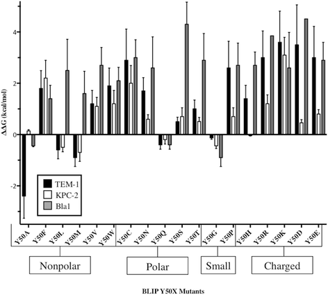
Results: In this study, the effect of systematic substitutions at BLIP position 50 on binding to class A β-lactamases was examined to further explore the role of BLIP Tyr50 in modulating specificity. The results indicate the sequence requirements at position 50 are widely different depending on the target β-lactamase. Stringent sequence requirements were observed at Tyr50 for binding Bacillus anthracis Bla1 while moderate requirements for binding TEM-1 and relaxed requirements for binding KPC-2 β-lactamase were seen. These findings cannot be easily rationalized based on the β-lactamase residues in direct contact with BLIP Tyr50 since they are identical for Bla1 and KPC-2 suggesting that differences in the BLIP-β-lactamase interface outside the local environment of Tyr50 influence the effect of substitutions.

Conclusions: Results from this study and previous studies suggest that substitutions at BLIP Tyr50 may induce changes at the interface outside its local environment and point to the complexity of predicting the impact of substitutions at a protein-protein interaction interface.

Keywords: Beatmusic; Binding specificity; Systematic substitutions; β-lactamase.


Figures
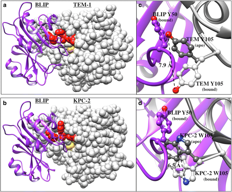
Fig. 1
Fig. 1
Structural representation of the interaction between BLIP and β-lactamases. BLIP is shown as a purple ribbon with Tyr50BLIP shown as stick. TEM-1 a and KPC-2 b β-lactamases are shown as white spheres with the catalytic Ser70 in yellow and positions 107, 129 and 216 (that make contact with Tyr50BLIP) are shown in red. PDB codes: 1JTG and 3E2K. Alignment of apo (gray) and bound (white) TEM-1 c and KPC-2 d structures shown in ribbon with position 105β-lactamase shown as stick. BLIPY50 is shown as a purple stick in the bound form. The measurement provides the distance 105β-lactamase moves upon binding to BLIP. PDB codes 1BTL and 2OV5 (apo) and 1JTG and 3E2K (bound). Images generated with Chimera
Fig. 2
Fig. 2
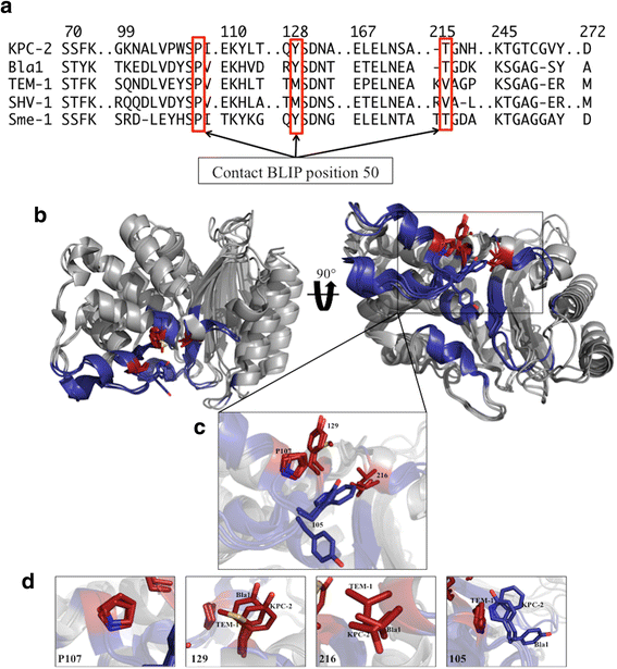
Alignment of class A β-lactamase residues at the BLIP interface. a The alignment is based on the structure of class A β-lactamase residues found at the BLIP interface as defined by the TEM-1/BLIP complex X-ray structure. The positions that contact BLIP position 50 are boxed in red. PDB codes used for the structural alignment are as follows: 2OV5 for KPC-2, 3QHY for Bla1, 1BTL for TEM-1, 1SHV for SHV-1 and 1DY6 for SME-1. Structural alignment performed in Chimera [41]. b β-lactamase structures are shown as grey ribbon and were aligned using MacPyMOL. Interface residues are shown in navy blue with β-lactamase position 105 shown as blue sticks. Residues that make direct contact with Tyr50BLIP (107, 129 and 216) are shown as red sticks. A global structural alignment of TEM-1, Bla1 and KPC-2 β-lactamases is shown in two orientations. c A close-up view of an alignment of the β-lactamase residues that make contact with Tyr50BLIP. β-lactamase position 105 is also shown as stick model as it has been shown to make structural rearrangements upon binding to BLIP [14, 16, 35]. d A close up alignment of β-lactamase positions 107, 129, 216 and 105 are shown with changes in orientation made for ease of viewing the structural alignment. Residues are labeled with their corresponding β-lactamase. PDB codes used for generation of images were as follows: 1BTL for TEM-1, 3QHY for Bla1 and 2OV5 for KPC-2. Images generated in MacPyMOL
Fig. 3
Fig. 3
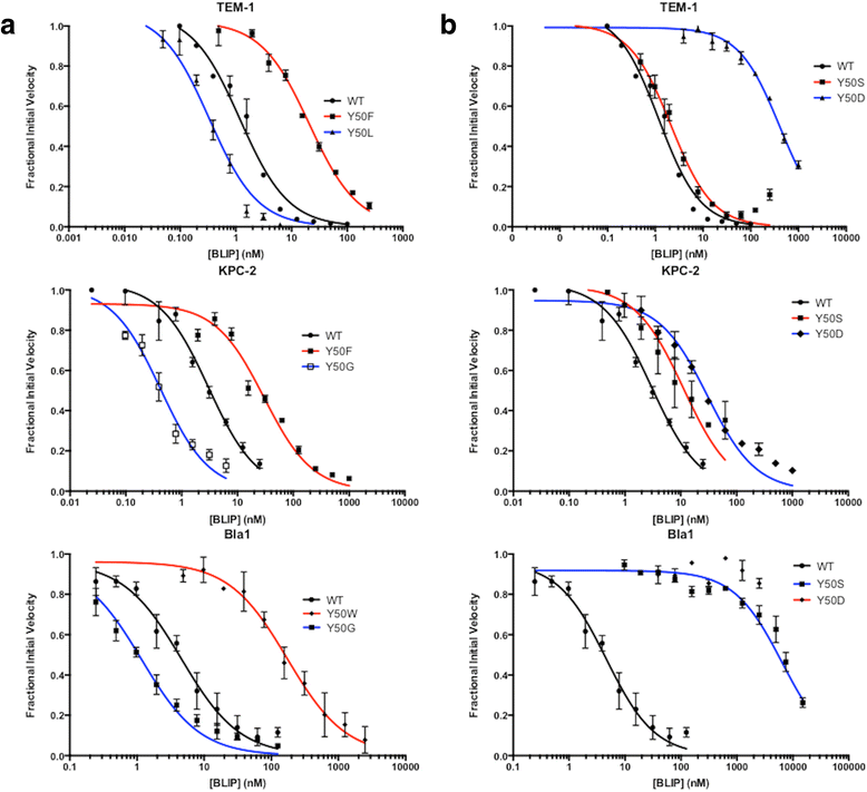
Determination of inhibition constants of BLIP mutants for binding TEM-1, KPC-2 and Bla1. The concentration of BLIP is shown on a log scale on the x-axes and fractional initial velocity is shown on the y-axes. Inhibition curves are shown for wild type BLIP in black. Inhibition curves for BLIP mutants that showed tighter binding than wild type are shown in blue while curves for mutants that showed weaker binding are shown in red
Fig. 4
Fig. 4
Comparison of ΔΔG values of BLIP mutants for binding TEM-1, KPC-2 and Bla1 β-lactamases. BLIP Y50X mutants are shown on the x-axis and the calculated change in free energy is shown on the y-axis. Values for TEM-1 are shown in black, KPC-2 in white and Bla1 in gray
Fig. 5
Fig. 5
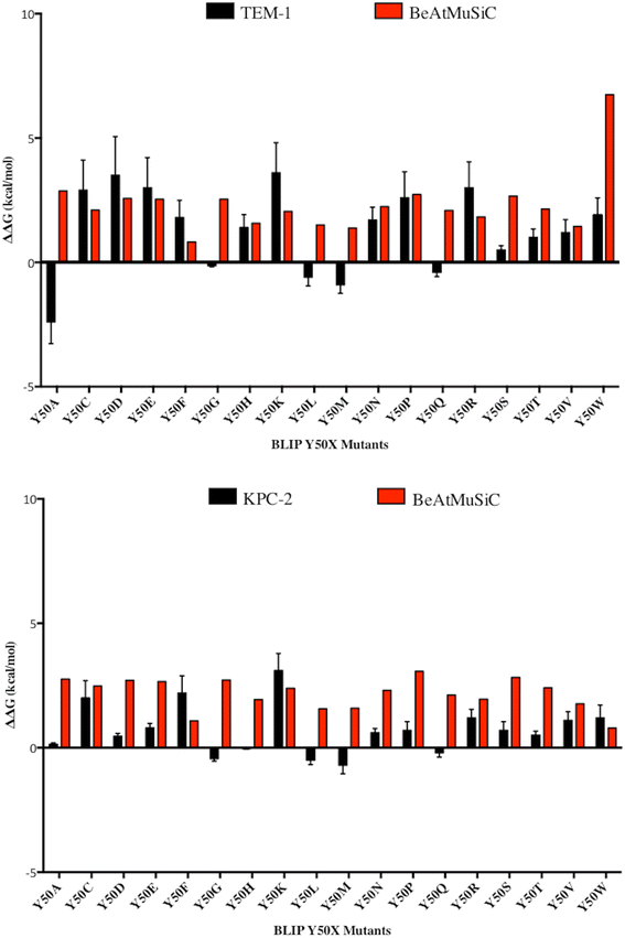
Comparison of experimental and predicted ΔΔG values for BLIP Y50 variants. A comparison of the experimental and predicted ΔΔG values of the BLIP Y50X mutants for binding TEM-1 are shown on the top panel. A comparison of the experimental and predicted ΔΔG values of the BLIP Y50X mutants for binding KPC-2 are shown on the bottom panel. The BeAtMuSiC server was used to generate predicted ΔΔG values. The predicted values are shown in red and the experimental values are shown in black

Similar articles

Cited by

References

    1. Kuzu G, Keskin O, Gursoy A, Nussinov R. Constructing structural networks of signaling pathways on the proteome scale. Curr Opin Struc Biol. sciencedirect; 2012;22:367–377 - PubMed
    1. Jubb H, Higueruelo A, Winter A, Blundell T. Structural biology and drug discovery for protein–protein interactions. Trends Pharmacol Sci. 2012;33:241–248. - PubMed
    1. Shoichet BK, Baase WA, Kuroki R, Matthews BW. A relationship between protein stability and protein function. Proc Nat Acad Sci. 1995;92(2):452–456. doi: 10.1073/pnas.92.2.452. - DOI - PMC - PubMed
    1. Selzer T, Albeck S, Schreiber G. Rational design of faster associating and tighter binding protein complexes. Nat. Struct. Biol. 2000;7:537–41. - PubMed
    1. Kim DE, Chivian D, Baker D. Protein structure prediction and analysis using the Robetta server. Nucleic Acids Res. 2004;32(Web Server issue):31. - PMC - PubMed

Publication types

MeSH terms

LinkOut - more resources