Review
. 2018 Jan 2;10(1):a027813.
doi: 10.1101/cshperspect.a027813.

Role of Polarity Proteins in the Generation and Organization of Apical Surface Protrusions

Affiliations
Review

Role of Polarity Proteins in the Generation and Organization of Apical Surface Protrusions

Gerard Apodaca. Cold Spring Harb Perspect Biol. .

Abstract

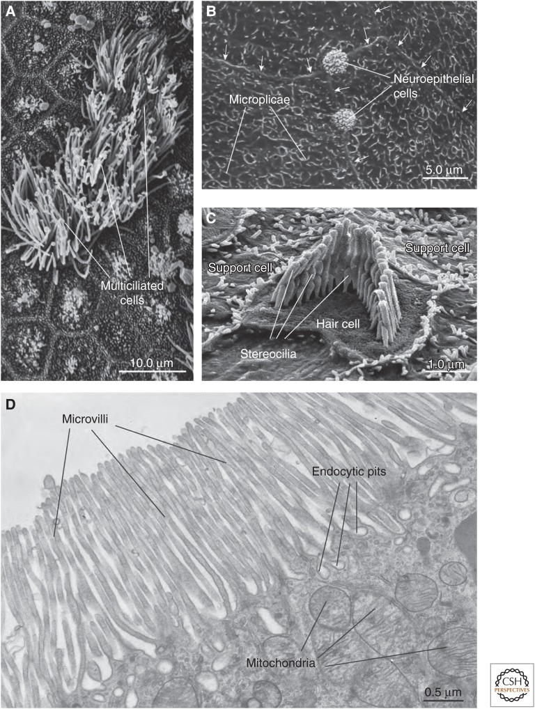
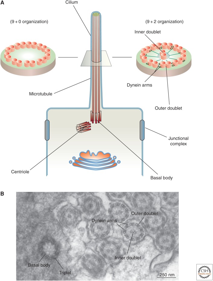
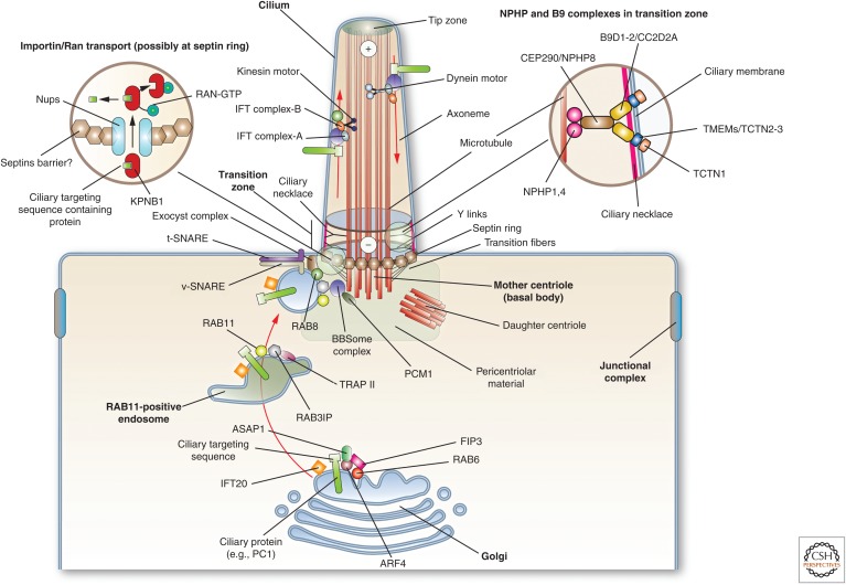
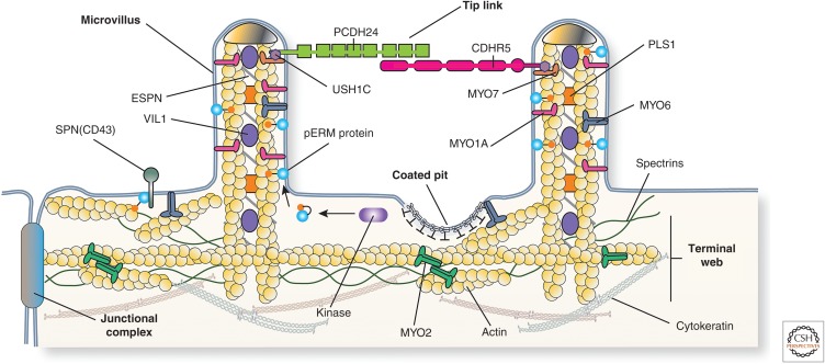
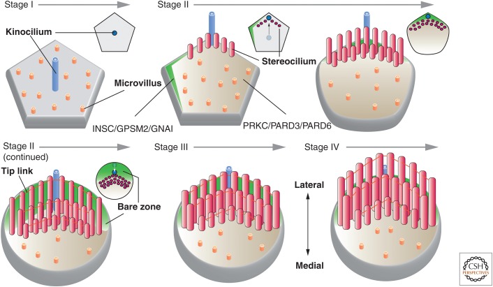
Protruding from the apical surfaces of epithelial cells are specialized structures, including cilia, microplicae, microvilli, and stereocilia. These contribute to epithelial function by cushioning the apical surface, by amplifying its surface area to facilitate nutrient absorption, and by promoting sensory transduction and barrier function. Despite these important roles, and the diseases that result when their formation is perturbed, there remain significant gaps in our understanding of the biogenesis of apical protrusions, or the pathways that promote their organization and orientation once at the apical surface. Here, I review some general aspects of these apical structures, and then discuss our current understanding of their formation and organization with respect to proteins that specify apicobasolateral polarity and planar cell polarity.


Figures

Figure 1.
Figure 1.
Survey of apical surface protrusions found in epithelial cells. (A) Scanning electron micrograph (SEM) of the apical surface of the rat trachea showing multiciliated cells. Adjacent cells are nonciliated or have rudimentary cilia. (B) SEM of the mucosal surface of the rat proximal urethra. Microplicae are observed at the apical surface of umbrella cells found in this region. Arrows mark the junctional ring of adjacent cells. The apical surfaces of neuroepithelial cells, which are covered by an apical tuft of short microvilli, are interspersed between adjacent cells. (C) SEM of the cochlea of the adult mouse showing a hair cell with associated “hair bundle,” which is comprised of stereocilia arranged in a stair-step configuration. The apical surfaces of adjacent support cells are studded with microvilli. (D) Transmission electron micrograph of microvilli at the apical surfaces of the rat proximal tubule epithelial cells. Endocytic pits and mitochondria are marked. (Electron micrograph in panel C was kindly provided by Jonathan Franks, Center for Biological Imaging, University of Pittsburgh; and micrographs in panels A, B, and D were kindly provided by Wily G. Ruiz, Kidney Imaging Core, University of Pittsburgh.) (Figure is from Apodaca and Gallo 2013; adapted, with permission, from the authors.)
Figure 2.
Figure 2.
Structure of cilia. (A) Cilia, which project from the apical surface of epithelial cells, have two types of organization. Motile cilia have a 9+2 organization in which a ring of nine microtubule doublets surrounds an inner microtubule doublet. Inner rays and dynein motor arms are present. Immotile cilia, such as so-called “primary cilia” have an outer ring of nine doublets, but lack the inner doublet, dynein arms, and rays. In both types of cilia, a basal body, derived from the centriole, is present and in cross section is comprised of nine bundles of microtubules arranged as triplets (see panel B). (B) Transmission electron micrograph of the zebrafish larval pronephros, which contains multiciliated cells that project cilia into the lumen of this tubular organ. A cross section through a basal body and through the axoneme highlights the structures described above. (Electron micrograph in panel B was kindly provided by Wily Ruiz, Kidney Imaging Core, University of Pittsburgh.) (Panel A from Apodaca and Gallo 2013; adapted, with permission, from the authors.)
Figure 3.
Figure 3.
Mechanisms of protein transport into cilia. At the Golgi, cilia resident proteins are recognized and packaged for delivery. In the case of PC1, its ciliary targeting sequence is recognized by ARF4, and is then delivered to RAB11-positive endosomes by way of a RAB6-, ARF4-, ASAP1-, and FIP3-dependent mechanism. The only intraflagellar transport (IFT) protein localized to the Golgi, IFT20, may have a role in promoting protein delivery to the cilium. At RAB11-positive endosomes, RAB11 recruits the TRAPII complex and the RAB8 GEF RAB3IP. The latter interacts with the basal body–localized BBSome complex (through the BBS1 subunit), and also recruits and activates RAB8. The BBSome is recruited by PCM1. RAB8 along with the exocyst complex and SNAREs, promotes the docking and fusion of the endocytic vesicle at the base of the cilium. Entry into the cilium is regulated at the transition zone by a septin ring, and the Y-links formed by the NPHP and B9 protein complexes. In addition, import into the cilium may depend on a RAN-GTP/GDP gradient (cytoplasmic RAN-GDP is not shown), which regulates KPNB1 binding to ciliary proteins and their subsequent transport across cilia-associated nucleoporins. Once in the axoneme, the ciliary proteins move along the microtubules by interactions with IFT particles and motor proteins. Anterograde traffic (directed toward the tip of the cilium) is mediated by IFT-B and KIF3 motor complex, whereas retrograde is mediated by IFT-A and the dynein-2 motor complex. The BBSome regulates the IFT complexes assembly at the basal body and its turnaround from anterograde to retrograde transport. (Figure from Apodaca and Gallo 2013; adapted, with permission, from the authors.)
Figure 4.
Figure 4.
Protein constituents of the epithelial cell brush border. The brush border of enterocytes and kidney proximal tubule epithelial cells is comprised of a regular array of microvilli. Each microvillus contains a core of actin, which is associated with MYO1A, ESPN, PLS1, VIL1, and ERM proteins such as EZR. In their active, phosphorylated state, ERM proteins link the actin cytoskeleton to the membrane, in some cases via the membrane protein SPN (alias CD43). The microvillus actin terminates in “rootlets” that associate with actin, spectrins, MYO2, and cytokeratins to form the terminal web. Adjacent microvilli are linked to one another by tip links, which are comprised of the protocadherins PCDH24 and CDHR5. These proteins are anchored to the actin filaments by way of MYO7 and the adaptor protein USH1C. (Figure from Apodaca and Gallo 2013; adapted, with permission, from the authors.)
Figure 5.
Figure 5.
Organization and function of hair cells in the inner ear. (A) Scanning electron micrograph of mucosal surface of adult mouse cochlea after removal of the overlying tectorial membrane. The position of the three rows of outer hair cells (OHC1-3) with associated hair bundle is indicated. The single row of inner hair cells is not shown. Hair cells are surrounded by support cells. The orientation of the lateromedial axis is shown. (B) (Left panel) The basal surface of the hair cell rests on the basilar membrane, while its apical stereocilia, surrounded by K+-rich endolymph fluid, insert into the tectorial membrane. Stereocilia are attached to one another via tip links. (Right panel) Sound waves, propagated through the cochlear fluid, cause an upward deflection of the basilar membrane, generating a shear force between the tectorial membrane and the stereocilia. The bending of the stereocilia promotes the opening of a mechanosensitive, nonselective cation channel. The inward movement of K+ ions through this channel depolarizes the cell, stimulating the opening of voltage-gated Ca2+ channels, which increase cytosolic Ca2+. The subsequent release of neurotransmitters triggers an action potential in the afferent neuronal processes. (Panel B from Apodaca and Gallo 2013; adapted, with permission, from the authors.)
Figure 6.
Figure 6.
Polarity proteins in epithelial cells. (A) Apicobasolateral polarity proteins. The apical-most Crumbs complex includes the transmembrane proteins CRB1-3 and the cytoplasmic proteins INADL/MUPP1 and MPP5. The other apical complex is the Par complex, which is comprised of PRKC, PARD3, and PARD6. PRKC phosphorylates CRB and PARD3, promoting apical identity. In contrast, PRKC-dependent phosphorylation of LLGL stimulates its degradation, and thus represses basolateral identity. Basolateral polarity is promoted by the Scribble complex, comprised of SCRB, DLG, and LLGL. Additional basolateral polarity proteins include YWHAE/Z and MARK2/3, and the Drosophila Yurt (Yrt)/Coracle (Cora) complex, which includes Yrt, Cora, neurexin4 (Nrx4), and Na+/K+-ATPase α-subunit (NaKα). (Panel A from Apodaca and Gallo 2013; adapted, with permission, from the authors.) (B) Planar cell polarity (PCP) proteins in Drosophila wing blade epithelium. An actin-based “hair” extends from the apical surface of each epithelial cell. The localization of the core PCP anterior complex (Flamingo, Van Gogh, Prickle), the core PCP posterior complex (Frizzled, Dishevelled, and Diego), and PCP effector proteins (Fuzzy, Inturned, and Fritz) is shown at the apical surfaces of wing epithelial cells. A portion of the interface between adjacent cells is magnified in the inset, which depicts the interaction between opposing PCP protein subcomplexes and PCP effectors. The gradient of the PCP effector protein Multiple wing hairs is indicated (black indicates higher expression). (C) Localization of core PCP proteins in vertebrate nodal cells. (D) Localization of core PCP proteins in the vertebrate cochlear hair cell and associated support cells.
Figure 7.
Figure 7.
Models for ciliogenesis. Cilia are formed by one of two mechanisms: “intracellular ciliogenesis” or “extracellular ciliogenesis.” The former is depicted in the larger panel and the latter in the smaller panel found in the upper left of the figure. Both should be viewed from left to right. The proteins and associated binding partners depicted in this figure are culled from numerous studies, and may vary depending on organism, cell type, and mechanism of ciliogenesis. (Larger panel) In “intracellular ciliogenesis,” a cilia precursor is formed in the cytoplasm and is subsequently delivered to the apical surface where it undergoes docking. Initially, a mother centriole with attached distal appendages accumulates RAB11A-positive distal appendage vesicles, which subsequently recruit EHD1/3, triggering a fusion event that results in the formation of a “ciliary vesicle.” Concomitantly, the centriole discharges CEP90 and CCP110, marking the conversion of the centriole to the basal body. Either before, or subsequent to ciliary vesicle formation, FUZ recruits DVL, the exocyst subunit EXOC4, and eventually RSG1 to the vesicle surface. In a similar fashion, RAB11 recruits the RAB8 exchange factor RAB3IP. In the next step, protein components from the Golgi (e.g., IFT20, ciliary membrane proteins, and PCP core proteins such as VANGL2), from recycling endosomes (e.g., RAB8), and from the cytoplasm (e.g., IFT and transition zone proteins) arrive at the ciliary vesicle and begin to assemble the axoneme and transition zone. This step is marked by the presence of the ciliary sheath, which will ultimately give rise to the periciliary membrane, and covers the inner ciliary membrane. Expansion of both membranes likely requires continued input from endosomes and Golgi. The ciliary precursor undergoes fusion in a process called “docking,” followed by further building of the axoneme and transition zone. The cilium is associated with both microtubules and actin filaments. (Smaller panel) Extracellular ciliogenesis is observed in cells with multiple cilia. Here, the centriole is delivered to a position just below the apical surface, undergoes maturation, and a ciliary vesicle/sheath is formed. The latter undergoes fusion with the apical plasma membrane, a step that is followed by assembly of the transition zone and axoneme at the cell surface.
Figure 8.
Figure 8.
Biogenesis of the epithelial brush border. In Drosophila, Crumbs may trigger brush-border morphogenesis by interactions with Moesin, the single ERM protein in the fly, as well as the βheavy isoform of spectrin (SPTAN/BN in vertebrates). An additional pathway for brush-border formation may require the activity of the STK11 kinase. In the presence of the pseudokinase STRADA and the cofactor CAB39, STK11 exits the nucleus and promotes the translocation of the kinase STK26 from the Golgi apparatus to the apical pole of the cell. Here, STK26 phosphorylates T567 in EZR, which could act to further promote brush-border formation by stabilizing the actin cytoskeleton and proteins at the apical cell surface. The translocation and activation of STK26 is dependent on PLD-induced production of phosphatidic acid, which promotes association of RAPGEF, a GEF for the small Ras-family GTPase RAP2A, with the cell surface. In turn, RAP2A recruits the TNIK kinase, which promotes translocation of STK26 to the plasma membrane, presumably as a result of TNIK-induced STK26 phosphorylation. Tip links are described in Figure 4. (Figure from Apodaca and Gallo 2013; adapted, with permission, from the authors.)
Figure 9.
Figure 9.
Stages of stereociliogenesis. Stage I: After the hair cell precursor establishes its apicobasolateral axis of polarity, a single kinocilium extends from the center of the cell. At this stage, the hair cell has a pentagonal or hexagonal shape. Stage II: The kinocilium undergoes a centrifugal displacement, positioning it at the periphery of the cell next to the tip of a polarized surface domain marked by INSC/GPSM2/GNAI and opposite of a domain enriched in PRKC/PARD3/PARD6. Stereocilia are seen to emerge in this stage, forming a “V”-shaped, staircase-like pattern as additional rows of cilia form. The vertices of each row point toward the kinocilium. Subsequently, the kinocilium undergoes a second migration, placing it midway between the cell center and cell periphery. The INSC/GPSM2/GNAI domain is now positioned in the lateral portion of the apical surface, establishing a “bare zone,” which derives its name from the lack of microvilli in this region. Stereocilia track the kinocilium and end up positioned at the border of the bare zone and the PRKC/PARD3/PARD6 domain. By the end of stage II, the stereocilia are connected by tip links and the hair cell apical surface has achieved an ovoid-shaped morphology. Stage III: The stereocilia stop growing in height and instead grow wider as additional actin filaments are packed into each stereocilium and cross-linked. Stage IV: The stereocilia continue to lengthen until they reach their final height. The small images at the upper right of each stage show an en face view of the position of the kinocilium and stereocilia at the indicated stage of development.

Similar articles

Cited by

References

    1. Adler PN, Zhu C, Stone D. 2004. Inturned localizes to the proximal side of wing cells under the instruction of upstream planar polarity proteins. Curr Biol 14: 2046–2051. - PubMed
    1. Al Jord A, Lemaitre AI, Delgehyr N, Faucourt M, Spassky N, Meunier A. 2014. Centriole amplification by mother and daughter centrioles differs in multiciliated cells. Nature 516: 104–107. - PubMed
    1. Ameen N, Apodaca G. 2007. Defective CFTR apical endocytosis and enterocyte brush border in myosin VI-deficient mice. Traffic 8: 998–1006. - PubMed
    1. Anderson RA. 1977. Actin filaments in normal and migrating corneal epithelial cells. Invest Ophthalmol Vis Sci 16: 161–166. - PubMed
    1. Andrews PM. 1976. Microplicae: Characteristic ridge-like folds of the plasmalemma. J Cell Biol 68: 420–429. - PMC - PubMed

Publication types

MeSH terms

LinkOut - more resources