. 2017 Apr 15;144(8):1544-1553.
doi: 10.1242/dev.143255. Epub 2017 Mar 6.

IFT56 regulates vertebrate developmental patterning by maintaining IFTB complex integrity and ciliary microtubule architecture

Affiliations

IFT56 regulates vertebrate developmental patterning by maintaining IFTB complex integrity and ciliary microtubule architecture

Daisy Xin et al. Development. .

Abstract

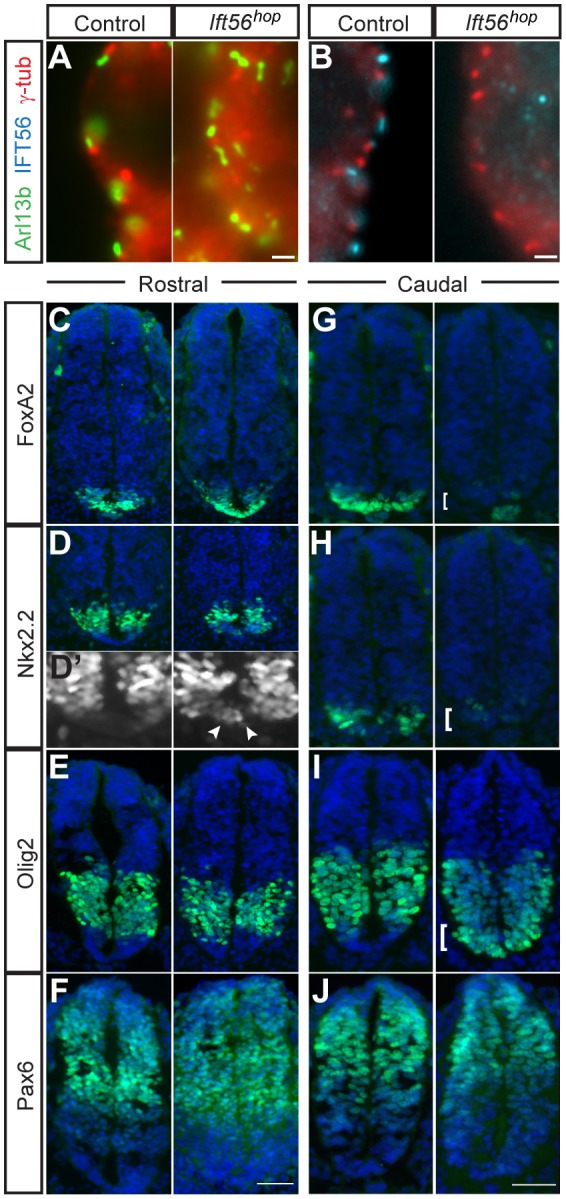
Cilia are key regulators of animal development and depend on intraflagellar transport (IFT) proteins for their formation and function, yet the roles of individual IFT proteins remain unclear. We examined the Ift56hop mouse mutant and reveal novel insight into the function of IFT56, a poorly understood IFTB protein. Ift56hop mice have normal cilia distribution but display defective cilia structure, including abnormal positioning and number of ciliary microtubule doublets. We show that Ift56hop cilia are unable to accumulate Gli proteins efficiently, resulting in developmental patterning defects in Shh signaling-dependent tissues such as the limb and neural tube. Strikingly, core IFTB proteins are unable to accumulate normally within Ift56hop cilia, including IFT88, IFT81 and IFT27, which are crucial for key processes such as tubulin transport and Shh signaling. IFT56 is required specifically for the IFTB complex, as IFTA components and proteins that rely on IFTA function are unaffected in Ift56hop cilia. These studies define a distinct and novel role for IFT56 in IFTB complex integrity that is crucial for cilia structure and function and, ultimately, animal development.

Keywords: Cilia; Hedgehog signaling; IFT56; IFTB; Intraflagellar transport; Microtubule structure; TTC26.


Conflict of interest statement

Competing interests

The authors declare no competing or financial interests.

Figures

Fig. 1.
Fig. 1.
Ift56hop neural tube cilia show reduced sensitivity to Shh signaling. (A,B) Ift56hop neural tubes form a similar number of cilia as controls but lack IFT56 protein. Neural tube sections were stained for Arl13b (A, green) to mark ciliary axonemes or IFT56 (B, cyan) and γ-tubulin to mark basal bodies (A,B, red). (C-J) E10.5 neural tube sections were stained with DAPI (blue) and for markers of neuronal progenitors (green). FoxA2 (C,G) is expressed normally in the rostral (forelimb region) floor plate in Ift56hop neural tubes but dramatically reduced caudally (hindlimb region, bracket). A subset of V3 interneuron progenitors (Nkx2.2+) are misplaced and found at the Ift56hop midline rostrally (D,D′, arrowheads) and almost completely absent caudally (H, bracket). Motor neuron progenitors (Olig2+) are unaffected in rostral Ift56hop sections (E) but are specified throughout the entire ventral caudal neural tube (I, bracket). Pax6 appears unaffected throughout the Ift56hop neural tube (F,J). n=3 biological replicates each from control and mutant, with three sections per replicate analyzed. Scale bars: 2 μm in A,B; 100 μm in C-J.
Fig. 2.
Fig. 2.
Ift56hop mutants develop preaxial polydactyly due to impaired Gli3R function. (A,B) E18.5 Ift56hop forelimbs (A) develop a duplication of the anterior digit (two phalanges: P1, P2) and hindlimbs (B) either form an extra anterior digit (not shown) or a digit with more posterior morphology (three phalanges: P1, P2, P3) preaxially. Alizarin Red marks bone and Alcian Blue marks cartilage. n=5 biological replicates each from control and mutant for skeletal analyses. (C) Ift56hop limb mesenchyme forms cilia but lacks IFT56 protein. Cilia were stained for acetylated α-tubulin (red) to mark ciliary axonemes and IFT56 (cyan). (D) At E11.5, Ift56hop limb buds express Shh in a posterior domain similar to controls. (E) Gli1, a direct positive target of Shh, is expressed in a relatively normal posterior domain in Ift56hop. By contrast, Grem1, which is normally inhibited by Gli3R in anterior limbs, shows an ectopic anterior domain of expression in Ift56hop limb buds (F, bracket). n=3 biological replicates each from control and mutant, with three sections per replicate analyzed. Scale bars: 0.5 mm in A; 1 mm in B; 2 μm in C; 0.1 mm in D-F.
Fig. 3.
Fig. 3.
Ciliary localization of Gli proteins is disrupted in Ift56hop cells. (A,B) Ciliary levels of Smo are unchanged in Ift56hop MEF cilia in response to activation of the Shh pathway via SAG treatment. Control n=12 and mutant n=10 cilia counted in representative MEF line. (C,D) In Ift56hop cilia, mean Gli2 tip accumulation is significantly decreased and over half of the mutant cilia completely lack Gli2. Control n=17 and mutant n=20 cilia counted in representative MEF line. (E,F) Similar to Gli2, mean Gli3 accumulation is significantly decreased in Ift56hop cilia and ∼80% of mutant cilia lack Gli3 at the distal tip. Control n=36 and mutant n=21 cilia counted in representative MEF line. (G,H) Limb mesenchyme cilia were assayed for Gli2 (green) and for Arl13b (red) to mark ciliary axonemes. Representative images are shown of the posterior limb bud of caudal (hindlimb) regions. (G′,H′) Gli2-only channel in black and white. (I) Ift56hop mutants displayed fewer cilia with high levels of Gli2 and more cilia with low and absent Gli2. Control n=92 and mutant n=89 cilia counted. (B,D,F) ns, not significant; ***P<0.001, ****P<0.0001 by two-tailed Student's t-test with unequal variances. All data are calculated as mean±s.d. n=4 biological replicates each from control and mutant for MEFs and tissue, and three sections per replicate were analyzed in tissue. Scale bars: 3 μm in A,C,E; 2 μm in G-H′.
Fig. 4.
Fig. 4.
The integrity of the IFTB complex breaks down in Ift56hop cilia. (A,C,E) MEFs were immunostained for specific IFT proteins (green), acetylated α-tubulin to mark axonemes (red) and γ-tubulin to mark basal bodies (blue). (A,B) Although axonemal levels of IFT88 were comparable to controls, a pool of IFT88 accumulated at the base of Ift56hop MEF cilia (arrowheads). Control n=9 and mutant n=13 cilia counted in representative MEF line. (C,D) Axonemal levels of IFT81 were significantly decreased and a pool of IFT81 was found at the base of Ift56hop cilia (arrowheads). Control n=15 and mutant n=15 cilia counted in representative MEF line. (E,F) IFT27 did not pool at the base of Ift56hop cilia but showed ∼50% reduction in ciliary axonemes. Control n=21 and mutant n=22 cilia counted in representative MEF line. (G-L) Limb mesenchyme cilia were immunostained for specific IFT proteins (green), Arl13b to mark axonemes (red) and γ-tubulin to mark basal bodies (blue). In vivo analyses support the MEF results, showing a significant reduction of IFT81 (G-I) and IFT27 (J-L) in Ift56hop limb mesenchyme cilia. n=4 biological replicates each from control and mutant; three sections per replicate were analyzed. (G′,H′,J′,K′) The IFT81-only or IFT27-only channels are shown in black and white. (I,L) Both IFT81 and IFT27 within Ift56hop mutant limb cilia are significantly decreased compared with control cilia. n=35 cilia from each control and mutant counted for each IFT. ****P<0.0001, ***P<0.001, *P<0.05 by two-tailed Student's t-test with unequal (B,D,F) or equal (I,L) variances. All data are calculated as mean±s.d. Scale bars: 3 μm in A,C,E; 2 μm in G-H′,J-K′.
Fig. 5.
Fig. 5.
Ciliary axonemal microtubule organization and number are altered in Ift56hop mutants. (A,B) TEM on E11.5 neural tube cilia revealed normal basal body docking and ciliary outgrowth in Ift56hop mutants. (C,D) Transverse sections through the basal body showed a normal structure of 9+0 triplet microtubules in mutants. (E-H) Axonemal transverse sections revealed reduced numbers of microtubule doublets in Ift56hop cilia (e.g. 8+0 and 7+0) and a disruption of the normal circular arrangement of doublets. All imaging and analyses were performed in the posterior neural tube along the ventral wall. n=2 sections analyzed for each sample, with three biological replicates of each control and mutant. Scale bars: 500 nm.
Fig. 6.
Fig. 6.
IFTA components and IFTA-dependent proteins localize normally in Ift56hop cilia. (A-D) MEFs were immunostained for specific IFT proteins (green), acetylated α-tubulin to mark axonemes (red) and γ-tubulin to mark basal bodies. IFT140 (A,B) and IFT122 (C,D) proteins localize normally and are found at similar levels in Ift56hop and control cilia. Control n=14 and mutant n=16 (A,B) or control n=9 and mutant n=13 (C,D) cilia were counted in representative MEF lines. n=4 biological replicates each from control and mutant, and two technical replicates were analyzed. All data are represented as mean±s.d. (E-G) AC3, Arl13b and PC2 are crucial for cilia function and do not require IFT56 for their localization. MEFs were immunostained for acetylated α-tubulin (red), specific ciliary proteins (green) and with DAPI (blue). Insets show higher magnification of ciliary protein expression in black and white. Scale bars: 3 μm in A,C; 2 μm in E-G,1 μm in insets.

Similar articles

Cited by

References

    1. Badano J. L., Mitsuma N., Beales P. L. and Katsanis N. (2006). The ciliopathies: an emerging class of human genetic disorders. Annu. Rev. Genomics Hum. Genet. 7, 125-148. 10.1146/annurev.genom.7.080505.115610 - DOI - PubMed
    1. Berbari N. F., Kin N. W., Sharma N., Michaud E. J., Kesterson R. A. and Yoder B. K. (2011). Mutations in Traf3ip1 reveal defects in ciliogenesis, embryonic development, and altered cell size regulation. Dev. Biol. 360, 66-76. 10.1016/j.ydbio.2011.09.001 - DOI - PMC - PubMed
    1. Bhogaraju S., Cajanek L., Fort C., Blisnick T., Weber K., Taschner M., Mizuno N., Lamla S., Bastin P., Nigg E. A. et al. (2013). Molecular basis of tubulin transport within the cilium by IFT74 and IFT81. Science 341, 1009-1012. 10.1126/science.1240985 - DOI - PMC - PubMed
    1. Blacque O. E., Perens E. A., Boroevich K. A., Inglis P. N., Li C., Warner A., Khattra J., Holt R. A., Ou G., Mah A. K. et al. (2005). Functional genomics of the cilium, a sensory organelle. Curr. Biol. 15, 935-941. 10.1016/j.cub.2005.04.059 - DOI - PubMed
    1. Blacque O. E., Cevik S. and Kaplan O. I. (2008). Intraflagellar transport: from molecular characterisation to mechanism. Front. Biosci. 13, 2633-2652. 10.2741/2871 - DOI - PubMed

Publication types

MeSH terms