. 2017 Apr 3;214(4):1153-1167.
doi: 10.1084/jem.20160880. Epub 2017 Mar 6.

Inflammatory monocytes require type I interferon receptor signaling to activate NK cells via IL-18 during a mucosal viral infection

Affiliations

Inflammatory monocytes require type I interferon receptor signaling to activate NK cells via IL-18 during a mucosal viral infection

Amanda J Lee et al. J Exp Med. .

Abstract

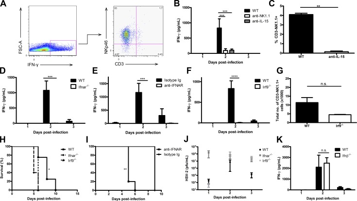
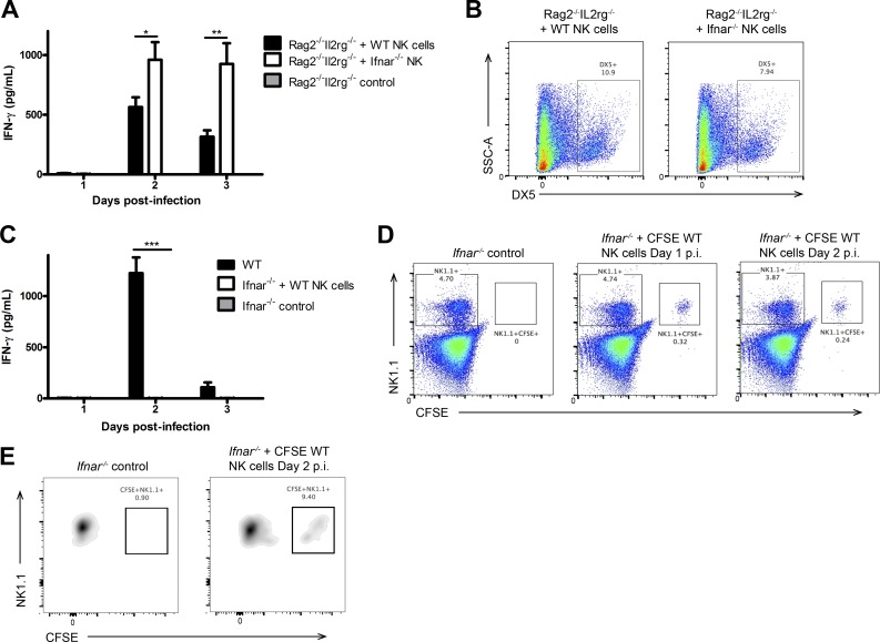
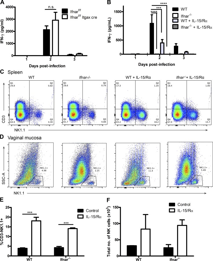
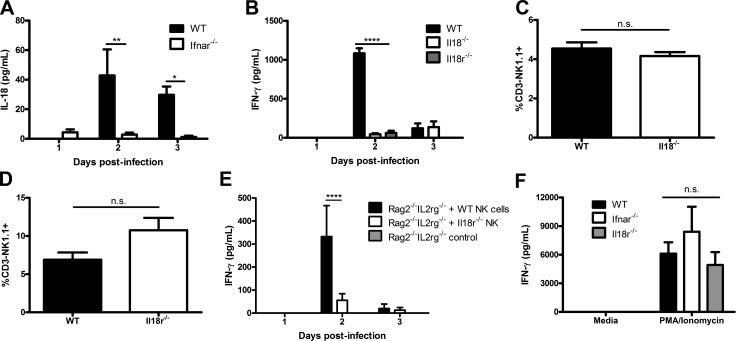
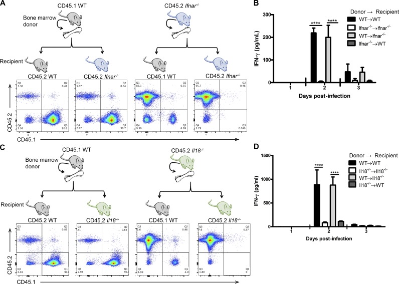
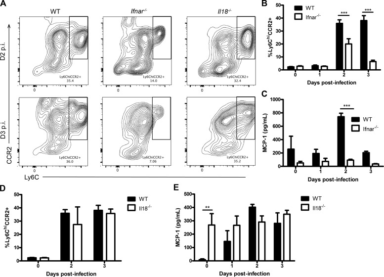
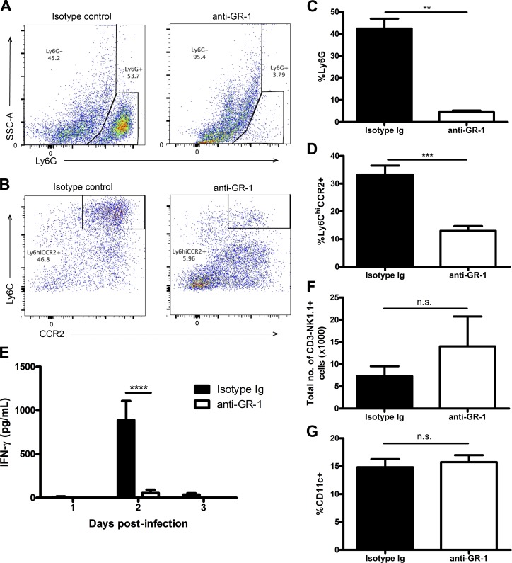
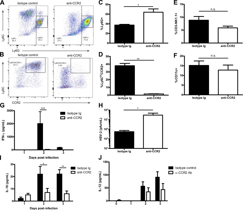
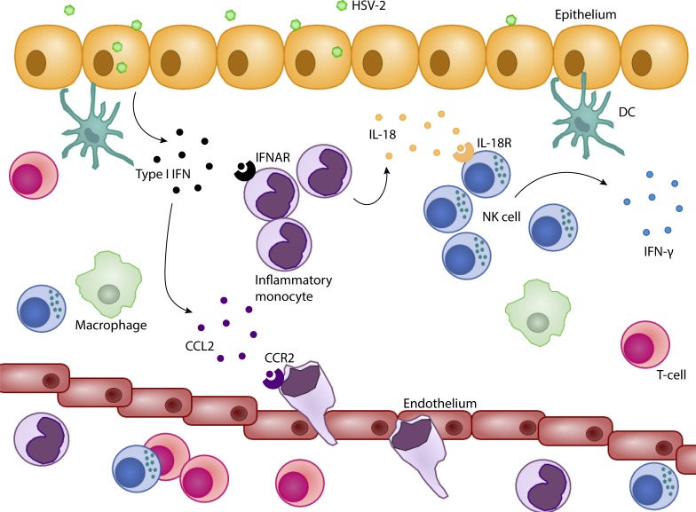
The requirement of type I interferon (IFN) for natural killer (NK) cell activation in response to viral infection is known, but the underlying mechanism remains unclear. Here, we demonstrate that type I IFN signaling in inflammatory monocytes, but not in dendritic cells (DCs) or NK cells, is essential for NK cell function in response to a mucosal herpes simplex virus type 2 (HSV-2) infection. Mice deficient in type I IFN signaling, Ifnar-/- and Irf9-/- mice, had significantly lower levels of inflammatory monocytes, were deficient in IL-18 production, and lacked NK cell-derived IFN-γ. Depletion of inflammatory monocytes, but not DCs or other myeloid cells, resulted in lower levels of IL-18 and a complete abrogation of NK cell function in HSV-2 infection. Moreover, this resulted in higher susceptibility to HSV-2 infection. Although Il18-/- mice had normal levels of inflammatory monocytes, their NK cells were unresponsive to HSV-2 challenge. This study highlights the importance of type I IFN signaling in inflammatory monocytes and the induction of the early innate antiviral response.


Figures

Figure 1.
Figure 1.
Type I IFN receptor and its respective signaling through IRF9 is required for NK cell IFN-γ production during HSV-2 infection. (A) WT B6 mice were infected with 104 pfu HSV-2 intravaginally (ivag). On day 2 p.i., vaginal tracts were processed and examined for NKp46, CD3, and IFN-γ expression. (B) WT B6 mice were depleted of NK cells using anti-NK1.1 antibody or IL-15 using anti–IL-15 antibody and then infected with 104 pfu HSV-2 ivag. Day 1–3 p.i. vaginal lavages were examined for IFN-γ levels (n = 5). (C) Peripheral blood was examined for NK cells in mice given anti–IL-15 antibody (n = 5). (D) WT and Ifnar−/− mice were infected with 104 pfu HSV-2 ivag, and vaginal lavages were examined for IFN-γ production on days 1–3 p.i. (n = 3; repeated twice with similar results). (E) WT B6 mice were administered anti-IFNAR antibody or the respective isotype-matched control Ig on days −1 through 2 i.p. and then infected with 104 pfu HSV-2 ivag. Their vaginal lavages were examined for IFN-γ content (n = 4; repeated once with similar results). (F) WT and Irf9−/− mice were infected with 104 pfu HSV-2 ivag, and vaginal lavages were examined for IFN-γ levels on days 1–3 p.i. (n = 3; repeated once with similar results). (G) WT and Irf9−/− mice were infected with 104 pfu HSV-2 ivag. At day 3 p.i., the vaginal mucosa was examined for CD3-NK1.1+ NK cells (n = 3; repeated once with similar results). (H) WT, Ifnar−/−, and Irf9−/− mice were infected with 104 pfu HSV-2 ivag and followed for survival (n = 5; repeated once with similar results). (I) WT mice were administered anti-IFNAR antibody or the respective isotype control Ig on days −1 through 2 and infected with 104 pfu HSV-2 ivag. Mice were followed for survival (n = 4). (J) After infection with HSV-2, vaginal lavages were collected from WT, Ifnar−/−, and Irf9−/− mice and assessed for HSV-2 level via plaque assay (n = 5). (K) WT and Ifnb−/− mice were infected with 104 pfu HSV-2 ivag. Day 1-3 vaginal washes were collected and examined for IFN-γ amount (n = 4). Data in B, D–F, and K are displayed as mean ± SEM and were analyzed using two-way ANOVA: n.s., not significant; ***, P < 0.001; ****, P < 0.0001. Data in C and G are displayed as mean ± SEM and were analyzed using an unpaired Student’s t test and a Mann–Whitney test (for nonparametric data), respectively: n.s., not significant; **, P < 0.01. Data in J are displayed as mean ± SEM and were analyzed using one-way ANOVA. Data in H and I were analyzed using a log-rank test: *, P < 0.05; **, P < 0.01.
Figure 2.
Figure 2.
IFNAR is not required directly on NK cells to activate their IFN-γ production. (A) NK cells were isolated from WT or Ifnar−/− spleens and adoptively transferred into Rag2−/−Il2rg−/− mice i.v. 24 h after transfer, mice were infected with 104 pfu HSV-2 ivag, and on days 1–3, vaginal lavages were examined for IFN-γ levels (n = 3; repeated twice with similar results). (B) Spleens were collected on day 3 p.i. and analyzed for DX5 expression. (C) NK cells were isolated from WT spleens, CFSE-labeled, and then adoptively transferred into Ifnar−/− mice. 24 h after transfer, Ifnar−/− mice given WT NK cells, and WT controls were infected with 104 pfu HSV-2 ivag. Day 1–3 p.i. vaginal lavages were examined for IFN-γ content (n = 3; repeated once with similar results). (D) Spleens were examined for CFSE+NK1.1+ adoptively transferred cells on days 1 and 2 p.i. (representative of two independent experiments). (E) Vaginal tissue from Ifnar−/− mice with or without adoptive transfer of CFSE-labeled NK cells was examined on day 2 p.i. for CFSE+NK1.1+ cells (representative of two independent experiments). Vaginal cells were first gated on the CD45+CD3NK1.1+ population and then examined for CFSE expression. Data in A and C are displayed as mean ± SEM and were analyzed using two-way ANOVA: *, P < 0.05; **, P < 0.01; ***, P < 0.001.
Figure 3.
Figure 3.
CD11c+ cells are not required to respond to type I IFN or trans-present IL-15 during the activation of NK cell IFN-γ production during infection. (A) Ifnarf/f and Ifnarf/f Itgax-cre mice were infected with 104 pfu HSV-2 ivag and examined for IFN-γ in the vaginal lavages collected days 1–3 p.i. (n = 5; repeated once with similar results). (B) WT and Ifnar−/− mice were given 0.5 µg IL-15 in complex with 1 µg IL-15Rα i.p. and subsequently infected with 5 × 10⁴ pfu HSV-2 ivag on the same day. Vaginal washes were collected on days 1–3 p.i. and examined for IFN-γ levels (n = 4; repeated once with similar results). (C and D) Spleen (C; n = 3) and vaginal (D; n = 2) tissue were collected on day 3 p.i. and examined for CD45+CD3NK1.1+ NK cells as shown in the representative flow plots. (E and F) Flow data are quantitatively shown for spleen (E) and vaginal mucosa (F). Data in A, B, E, and F are displayed as mean ± SEM and were analyzed using two-way ANOVA: n.s., not significant; ***, P < 0.001; ****, P < 0.0001.
Figure 4.
Figure 4.
IL-18 and IL-18R are both required for NK cell IFN-γ production during HSV-2 infection. (A) WT and Ifnar−/− mice were infected with 104 pfu HSV-2 ivag, and day 1–3 vaginal lavages were examined for IL-18. Data were normalized to Il18−/− data (n = 5; repeated once with similar results). (B) WT, Il18−/−, and Il18r1−/− B6 mice were infected with 104 pfu HSV-2 ivag, and on day 1–3 p.i. vaginal lavages were examined for IFN-γ content (n = 5; repeated once with similar results). (C and D) Vaginal tissue was isolated on day 3 p.i. and examined for CD45+CD3NK1.1+ cells in Il18−/− (C; n = 3) and Il18r1−/− (D; n = 3) mice. (E) WT and Il18r1−/− NK cells were isolated from the spleen and adoptively transferred into Rag2−/−Il2rg−/− mice i.v. 24 h p.i. The mice were infected with 104 pfu HSV-2 ivag. On days 1–3 p.i., vaginal lavages were collected and examined for IFN-γ (n = 6). (F) NK cells were isolated from WT, Ifnar−/−, and Il18r−/− spleens and stimulated with 50 ng/ml PMA and 500 ng/ml ionomycin for 24 h ex vivo. Supernatants were collected and assayed for IFN-γ (n = 3). Data in A, B, and E are displayed as mean ± SEM and were analyzed using two-way ANOVA: *, P < 0.05; **, P < 0.01; ****, P < 0.0001. Data in C and D are displayed as mean ± SEM and were analyzed using an unpaired Student’s t test: n.s., not significant. Data in F are displayed as mean ± SEM and were analyzed using one-way ANOVA: n.s., not significant.
Figure 5.
Figure 5.
The hematopoietic cell compartment is required to respond to type I IFN and produce IL-18 to activate IFN-γ production during HSV-2 infection. (A) WT and Ifnar−/− mice were lethally irradiated and reconstituted with either WT or Ifnar−/− bone marrow. Mice were allowed to reconstitute for 6–8 wk, and peripheral blood was assessed for reconstitution by examining the frequency of cells expressing CD45.1 or CD45.2. (B) Mice were then infected with 5 × 10⁴ pfu HSV-2 ivag. On days 1–3 p.i., vaginal lavages were examined for IFN-γ content (n = 3; repeated once with similar results). (C) WT and Il18−/− mice were lethally irradiated and reconstituted with either WT or Il18−/− bone marrow. 6–8 wk after reconstitution, mice were examined for CD45.1 and CD45.2 expression. (D) Mice were then infected with 104 pfu HSV-2 ivag. On days 1–3 p.i., vaginal lavages were examined for IFN-γ (n = 7). Data in B and D are displayed as mean ± SEM and were analyzed using two-way ANOVA: ****, P < 0.0001.
Figure 6.
Figure 6.
Decreased vaginal inflammatory monocyte infiltration in Ifnar−/− mice during HSV-2 infection. (A) WT, Ifnar−/−, and Il18−/− mice were infected with 104 pfu HSV-2, and vaginal tissue was collected at baseline and on days 0–3 p.i. and examined for inflammatory monocytes, defined as CD45+CD11cCD11b+Ly6GLy6ChiCCR2+ cells. Day 2 and 3 p.i. data are shown in representative flow plots. (B) Flow cytometry data were quantified and graphically represented (n = 3; repeated once with similar results). (C) Vaginal lavages were collected on days 0–3 p.i. and examined for MCP-1 (n = 3; repeated once with similar results). (D) WT and Il18−/− mice were infected with HSV-2 ivag, and vaginal tissue was collected at baseline and on days 2 and 3 p.i. and examined for inflammatory monocytes (n = 3). (E) WT and Il18−/− mice were infected with 104 pfu HSV-2 ivag, and on days 0–3 p.i., vaginal lavages were examined for MCP-1 (n = 4). Data in B–E are displayed as mean ± SEM and were analyzed using two-way ANOVA: **, P < 0.01; ***, P < 0.001.
Figure 7.
Figure 7.
Inflammatory monocytes are required for activating NK cell IFN-γ production during HSV-2 infection. (A and B) WT B6 mice were given an anti–GR-1 antibody or its respective isotype-matched control Ig and infected with 104 pfu HSV-2 ivag. Vaginal tissue was examined for neutrophil (CD45+CD11cCD11b+Ly6G+) and inflammatory monocyte (CD45+CD11cCD11b+Ly6GLy6ChiCCR2+) depletion on day 3 p.i. Representative flow plots of vaginal neutrophil and inflammatory monocyte populations are shown (respectively in A and B). (C and D) Vaginal neutrophil (C) and inflammatory monocyte (D) populations are displayed graphically (n = 5; repeated once with similar results). (E) On days 1–3 p.i., vaginal washes were examined for IFN-γ (n = 5; repeated twice with similar results). (F and G) Vaginal tissue collected on day 3 p.i. was also examined for total vaginal NK cell number (F; CD45+CD3NK1.1+; n = 5) and proportion of CD11c+ cells (G; CD45+CD11c+; n = 5; repeated once with similar results). Data in C, D, F, and G are displayed as mean ± SEM and were analyzed using an unpaired Student’s t test in D and G and a Mann–Whitney test (for nonparametric data) in C and F: n.s., not significant; **, P < 0.01; ***, P < 0.001. Data in E are displayed as mean ± SEM and were analyzed using two-way ANOVA: ****, P < 0.0001.
Figure 8.
Figure 8.
Inflammatory monocytes are required for activation of NK cell IFN-γ production during infection. (A–D) WT B6 mice were given an anti-CCR2 antibody or the respective isotype-matched control Ig to deplete inflammatory monocytes and then infected with 104 pfu HSV-2 ivag. Vaginal tissue was collected on day 3 p.i. and examined for neutrophil and inflammatory monocyte populations. Representative flow plots are respectively shown in A and B and graphically in C and D (n = 5). (E and F) On day 3 p.i., vaginal cells were also examined for NK cells (E; n = 5) and CD11c+ cells (F; n = 5). (G) On days 0–3 p.i., vaginal lavages were examined for IFN-γ levels (n = 4; repeated once with similar results). (H) On day 2 p.i., vaginal washes were examined for HSV-2 viral titers using a plaque assay method (n = 4; repeated once with similar results). (I and J) Day 0–3 p.i. vaginal lavages were also examined for IL-18 levels (I; n = 4; repeated once with similar results) and IL-12 levels (J; n = 5). Data in C–F and H are displayed as mean ± SEM and were analyzed using an unpaired Student’s t test in F and a Mann-Whitney test (for nonparametric data) in C–E and H: n.s., not significant; *, P < 0.05; **, P < 0.01. Data in G, I, and J are displayed as mean ± SEM and were analyzed using two-way ANOVA: *, P < 0.05; **, P < 0.01; ***, P < 0.001.
Figure 9.
Figure 9.
Type I IFN activates NK cell IFN-γ production through the stimulation of IL-18 production in inflammatory monocytes during vaginal HSV-2 infection. Rapidly after HSV-2 infection, type I IFN is produced in the vaginal mucosa. Type I IFN induces CCL2 production from a cell type or types and recruits inflammatory monocytes to the site of infection. Type I IFN binds IFNAR on inflammatory monocytes and signals through IRF9 to induce their release of IL-18. IL-18 then ligates to IL-18R on NK cells to induce their production of IFN-γ.

Similar articles

Cited by

References

    1. Ashkar A.A., and Rosenthal K.L.. 2003. Interleukin-15 and natural killer and NKT cells play a critical role in innate protection against genital herpes simplex virus type 2 infection. J. Virol. 77:10168–10171. 10.1128/JVI.77.18.10168-10171.2003 - DOI - PMC - PubMed
    1. Baranek T., Manh T.P., Alexandre Y., Maqbool M.A., Cabeza J.Z., Tomasello E., Crozat K., Bessou G., Zucchini N., Robbins S.H., et al. . 2012. Differential responses of immune cells to type I interferon contribute to host resistance to viral infection. Cell Host Microbe. 12:571–584 (published erratum appears in Cell Host Microbe 2013. 13:372) . 10.1016/j.chom.2012.09.002 - DOI - PubMed
    1. Barr D.P., Belz G.T., Reading P.C., Wojtasiak M., Whitney P.G., Heath W.R., Carbone F.R., and Brooks A.G.. 2007. A role for plasmacytoid dendritic cells in the rapid IL-18-dependent activation of NK cells following HSV-1 infection. Eur. J. Immunol. 37:1334–1342. 10.1002/eji.200636362 - DOI - PMC - PubMed
    1. Bogunovic D., Byun M., Durfee L.A., Abhyankar A., Sanal O., Mansouri D., Salem S., Radovanovic I., Grant A.V., Adimi P., et al. . 2012. Mycobacterial disease and impaired IFN-γ immunity in humans with inherited ISG15 deficiency. Science. 337:1684–1688. 10.1126/science.1224026 - DOI - PMC - PubMed
    1. Brandstadter J.D., Huang X., and Yang Y.. 2014. NK cell-extrinsic IL-18 signaling is required for efficient NK-cell activation by vaccinia virus. Eur. J. Immunol. 44:2659–2666. 10.1002/eji.201344134 - DOI - PMC - PubMed

Publication types

MeSH terms