. 2017 Apr 21;85(5):e00991-16.
doi: 10.1128/IAI.00991-16. Print 2017 May.

Promotion and Rescue of Intracellular Brucella neotomae Replication during Coinfection with Legionella pneumophila

Affiliations

Promotion and Rescue of Intracellular Brucella neotomae Replication during Coinfection with Legionella pneumophila

Yoon-Suk Kang et al. Infect Immun. .

Abstract

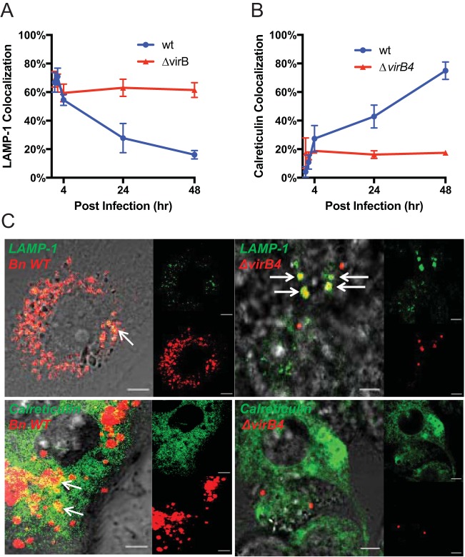
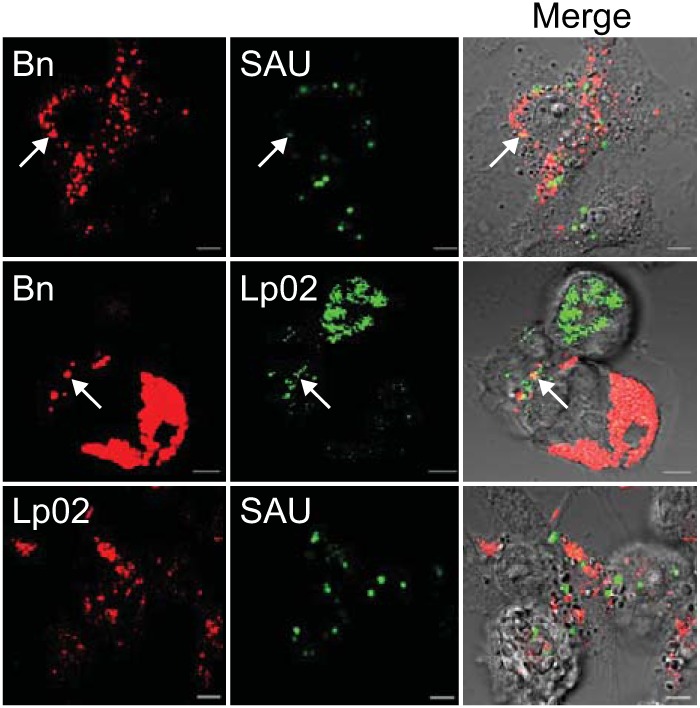
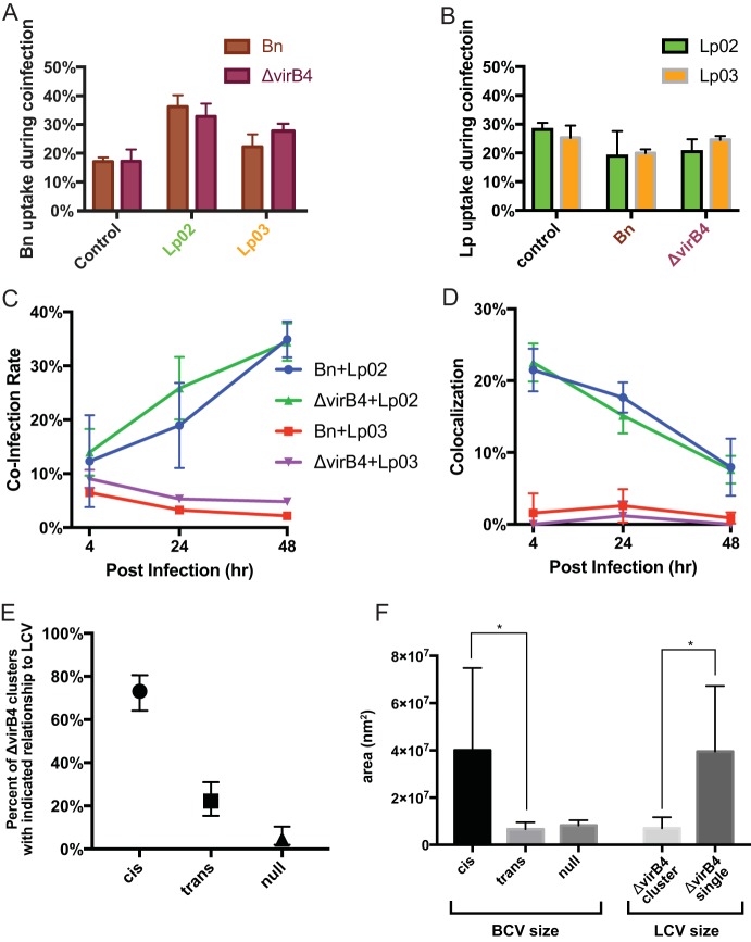
We established a new Brucella neotomaein vitro model system for study of type IV secretion system-dependent (T4SS) pathogenesis in the Brucella genus. Importantly, B. neotomae is a rodent pathogen, and unlike B. abortus, B. melitensis, and B. suis, B. neotomae has not been observed to infect humans. It therefore can be handled more facilely using biosafety level 2 practices. More particularly, using a series of novel fluorescent protein and lux operon reporter systems to differentially label pathogens and track intracellular replication, we confirmed T4SS-dependent intracellular growth of B. neotomae in macrophage cell lines. Furthermore, B. neotomae exhibited early endosomal (LAMP-1) and late endoplasmic reticulum (calreticulin)-associated phagosome maturation. These findings recapitulate prior observations for human-pathogenic Brucella spp. In addition, during coinfection experiments with Legionella pneumophila, we found that defective intracellular replication of a B. neotomae T4SS virB4 mutant was rescued and baseline levels of intracellular replication of wild-type B. neotomae were significantly stimulated by coinfection with wild-type but not T4SS mutant L. pneumophila Using confocal microscopy, it was determined that intracellular colocalization of B. neotomae and L. pneumophila was required for rescue and that colocalization came at a cost to L. pneumophila fitness. These findings were not completely expected based on known temporal and qualitative differences in the intracellular life cycles of these two pathogens. Taken together, we have developed a new system for studying in vitroBrucella pathogenesis and found a remarkable T4SS-dependent interplay between Brucella and Legionella during macrophage coinfection.

Keywords: Brucella; Legionella pneumophila; coinfection; fluorescent image analysis; intracellular pathogens; pathogenesis; phagosomes; reporter genes; type IV secretion system.


Figures

FIG 1
FIG 1
Intracellular growth of wild-type and ΔvirB4 mutant B. neotomae. (A) CFU from lysed macrophages per microplate well at indicated times postinfection. (B) Intracellular growth of lux operon-labeled B. neotomae wild-type (wt) and ΔvirB4 mutant. Cells of the mouse J774A.1 or human THP-1 macrophage cell lines adherent to 96-well plates were infected with luminescent Brucella cells at an MOI of 1, treated with gentamicin to kill extracellular bacteria, and incubated for the indicated time points. (C) Intracellular replication of proD/tdTomato-expressing B. neotomae wild-type, ΔvirB4 mutant, and ΔvirB4/virB4 (virB4c) complemented strains in J774A.1 cells. The data shown are the mean ± standard deviation (SD) from at least three replicates for experiments performed in parallel and are representative of two independent experiments.
FIG 2
FIG 2
Phagosome trafficking. (A) Percentage of phagosomes containing the proD/tdTomato B. neotomae (Bn) wild type (wt) or ΔvirB4 mutant cololocalizing with LAMP-1::mTurquoise2-labeled compartments during infection of J744A.1 cells. (B) Percentage of phagosomes containing proD/tdTomato B. neotomae wild-type or ΔvirB4 mutant cololocalizing with mTurquoise2::calreticulin-labeled compartments during infection of J744A.1 cells. Total scored events for each data point were used for Fisher's exact test contingency analysis as described in the text. (C) Representative confocal laser scanning microscope images of cells of the proD/tdTomato B. neotomae wild type or ΔvirB4 mutant colocalizing with either LAMP-1::mTurquoise2 or mTurquoise2::calreticulin at 24 h p.i. For each panel, the lower right inset shows B. neotomae signal pseudocolored red, the upper right inset shows eukaryotic fusion protein signal pseudocolored green, and the left panel shows an enlarged, merged image of the two signals superimposed on a differential interference contrast (DIC) image. Arrows indicate examples of colocalization events associated with a yellow merged overlap signal. Scale bars, 5 μm.
FIG 3
FIG 3
Intracellular colocalization of bacterial pathogens. Shown are confocal laser scanning microscope images of J774A.1 cells coinfected for 24 h with proD::tdTomato-expressing Brucella neotomae (Bn) and eGFP-expressing Staphylococcus aureus (SAU), proD/tdTomato-expressing B. neotomae and proD/mClover-expressing L. pneumophila Lp02, or eGFP-expressing S. aureus and proD/mNeptune2-expressing Lp02. Pseudocolored confocal images from green (mClover) and red (tdTomato) or far-red (mNeptune2) channels superimposed on differential interference contrast (DIC) images demonstrated distinct fluorescent signals originating from the different coinfecting intracellular pathogens. Colocalization is indicated by yellow coloration in the merged images (arrows highlight examples). The images shown are representative of at least 20 cells imaged from two independent experiments. Scale bars, 5 μm.
FIG 4
FIG 4
Use of luminescence and fluorescence reporters to independently assess intracellular growth of coinfecting pathogens. Intracellular growth was simultaneously assessed by luminescence (A) and fluorescence (B) during macrophage infection at an MOI of 1 in a 96-well microplate format for coinfections with luminescent B. neotomae (Bn) (wild-type [wt] or ΔvirB4 mutant strains) and fluorescent Lp02, Lp03, or S. aureus. Shown are the mean and SD of results from 6 replicate wells per data point; results are representative of two independent experiments. (C) Effect of Lp02 MOI on its ability to stimulate intracellular replication of the B. neotomae wild type and ΔvirB4 mutant. J774A.1 macrophages were infected with luminescent B. neotomae at an MOI of 1 and Lp02 at MOI of 1, 2, 5, or 10 for the indicated times. Shown are the mean and SD of results from 6 replicate wells per data point; results are representative of two independent experiments. (D) Intracellular growth in macrophages of the B. neotomae ΔvirB4 strain coinfected with Lp02, Lp03, or S. aureus was assessed by CFU determination. Shown are the mean and SD of results from 6 replicate wells per data point.
FIG 5
FIG 5
Colocalization of B. neotomae and L. pneumophila during macrophage coinfection. Coinfection of J774A.1 macrophages with the B. neotomae wild type (Bn wt) or ΔvirB4 mutant expressing proD/tdTomato and L. pneumophila Lp02 or Lp03 expressing proD/mClover. At indicated time points, cells were fixed and examined by confocal microscopy. Shown are merged pseudocolored images obtained using the red (tdTomato) and green (mClover) settings. Size bars, 5 μm. Areas enclosed in circles are magnified 3× in insets to demonstrate either presence (yellow merged signal) or absence of pathogen species overlap.
FIG 6
FIG 6
Quantitative coinfection analysis. (A) The percentage of J774A.1 cells infected by the B. neotomae wild type (Bn wt) or ΔvirB4 mutant at 4 h p.i. in the presence or absence of coinfection with L. pneumophila Lp02 or Lp03 or alternatively (B) the percentage of J774A.1 cells infected with Lp02 or Lp03 at 4 h p.i. in the presence or absence of the B. neotomae wild type or ΔvirB4 mutant. The data shown are means and SD of data obtained from two independent experiments with at least 50 J774A.1 cells examined in each replicate. Scored events from both trials were combined for Fisher's exact test contingency analysis described in the text. (C) J774A.1 cells were infected with indicated B. neotomae and L. pneumophila strains at an MOI of 1. The percentage of J774A.1 cells coinfected with both indicated B. neotomae and L. pneumophila strains is shown. Data points are mean percentage of coinfection ± SD of data obtained from two independent experiments with a minimum of 50 host cells scored in each. Scored events from both trials were combined for Fisher's exact test contingency analysis described in the text. (D) Colocalization percentages of indicated strains of B. neotomae and L. pneumophila were compared at different times postinfection. The colocalization percentage was calculated by examining coinfected macrophages using confocal microscopy, counting the number of phagosomes with colocalized pathogens (yellow merged signal) as the numerator and counting all phagosomes with B. neotomae and/or L. pneumophila as the denominator. The data shown are means ± SD from two independent experiments with an average of >150 cells scored in total per data point. Total scored events for each data point were used for Fisher's exact test contingency analysis described in the text. (E) Relationship of B. neotomae ΔvirB4 bacterial clusters to Lp02 in coinfected J774A.1 cells 24 h postinfection. Clusters (defined as a B. neotomae ΔvirB4-containing vacuole or BCV containing ≥4 B. neotomae bacteria or ≥3 μm in diameter) that overlapped with Lp02-containing vacuoles (LCV) were considered “cis,” those that did not were considered “trans,” and rare clusters present in a macrophage without apparent LCV were considered “null.” The results shown are cumulative percentage data from 108 randomly selected clusters scored over two independent coinfection experiments with 95% confidence intervals shown calculated using the Wilson-Brown method (57). (F) Mean surface area ± SD of B. neotomae ΔvirB4 clusters found in cis or trans to Lp02 or in cells without apparent LCV (null) 24 h p.i. or mean surface area ± SD of LCV in host cells 24 h p.i. containing any number of ΔvirB4 clusters or in host cells only containing BCV with <4 ΔvirB4 bacteria, designated “single.” P < 0.0001 for bracketed comparisons (*) of BCV and LCV size, respectively.

Similar articles

Cited by

References

    1. Dalrymple-Champneys W. 1960. Brucella infection and undulant fever in man. Oxford University Press, London, United Kingdom.
    1. Watarai M, Makino S, Shirahata T. 2002. An essential virulence protein of Brucella abortus, VirB4, requires an intact nucleoside-triphosphate-binding domain. Microbiology 148:1439–1446. doi:10.1099/00221287-148-5-1439. - DOI - PubMed
    1. Delrue RM, Martinez-Lorenzo M, Lestrate P, Danese I, Bielarz V, Mertens P, De Bolle X, Tibor A, Gorvel JP, Letesson JJ. 2001. Identification of Brucella spp. genes involved in intracellular trafficking. Cell Microbiol 3:487–497. doi:10.1046/j.1462-5822.2001.00131.x. - DOI - PubMed
    1. Celli J, de Chastellier C, Franchini DM, Pizarro-Cerda J, Moreno E, Gorvel JP. 2003. Brucella evades macrophage killing via VirB-dependent sustained interactions with the endoplasmic reticulum. J Exp Med 198:545–556. doi:10.1084/jem.20030088. - DOI - PMC - PubMed
    1. Starr T, Child R, Wehrly TD, Hansen B, Hwang S, Lopez-Otin C, Virgin HW, Celli J. 2012. Selective subversion of autophagy complexes facilitates completion of the Brucella intracellular cycle. Cell Host Microbe 11:33–45. doi:10.1016/j.chom.2011.12.002. - DOI - PMC - PubMed

Publication types