. 2017 Apr 15;130(8):1475-1485.
doi: 10.1242/jcs.198937. Epub 2017 Mar 6.

Missing-in-metastasis protein downregulates CXCR4 by promoting ubiquitylation and interaction with small Rab GTPases

Affiliations

Missing-in-metastasis protein downregulates CXCR4 by promoting ubiquitylation and interaction with small Rab GTPases

Lushen Li et al. J Cell Sci. .

Abstract

Surface expression of chemokine receptor CXCR4 is downregulated by missing-in-metastasis protein (MIM; also known as MTSS1), a member of the inverse BAR (I-BAR)-domain protein family that recognizes and generates membranes with negative curvature. Yet, the mechanism for the regulation is unknown. Here, we show that MIM forms a complex with CXCR4 by binding to E3 ubiquitin ligase AIP4 (also known as ITCH) in response to stromal cell-derived factor 1 (SDF-1; also known as CXCL12). Overexpression of MIM promoted CXCR4 ubiquitylation, inhibited cellular response to SDF-1, caused accumulation and aggregation of multivesicular bodies (MVBs) in the cytoplasm, and promoted CXCR4 sorting into MVBs in a manner depending on binding to AIP4. In response to SDF-1, MIM also bound transiently to the small GTPase Rab5 at 5 min and to Rab7 at 30 min. Binding to Rab7 requires an N-terminal coiled-coil motif, deletion of which abolished MIM-mediated MVB formation and CXCR4 internalization. Our results unveil a previously unknown property of MIM that establishes the linkage of protein ubiquitylation with Rab-guided trafficking of CXCR4 in endocytic vesicles.

Keywords: AIP4; CXCR4; MIM; MVBs; Rab7; Ubiquitylation.


Conflict of interest statement

Competing interests

The authors declare no competing or financial interests.

Figures

Fig. 1.
Fig. 1.
MIM attenuates the chemotactic response to SDF-1 and promotes CXCR4 internalization. (A) HeLa cells expressing MIM–GFP or GFP were plated in the upper chamber of Transwell plates of which the lower chamber was filled with medium containing SDF-1 at concentrations as indicated. As a control, one plate was filled with medium supplemented with 15 nM SDF-1 plus 2 μM AMD3100 (SDF-1+AMD). After 16 h of incubation, migrated cells were stained with Crystal Violet, inspected, photographed and counted under a 20× objective lens. Scale bars: 500 µm. (B) Quantification of cell migration was based on three independent experiments as described in A. (C,D) Cells expressing GFP or MIM–GFP were treated with 100 nM SDF-1 for 15 min and subjected to flow cytometric analysis after staining with PE-conjugated monoclonal antibody directed against CXCR4. As a negative control, cells were also stained with PE–IgG antibody (isotype ctrl). Ctrl, no SDF-1. (E) The percentage of CXCR4 remaining on the cell surface after SDF-1 stimulation was calculated based on normalization to that of cells without SDF-1 treatment. (F–I) HeLa cells expressing MIM–GFP or GFP were treated with 100 nM SDF-1 for the times (min) indicated and then analyzed by western blotting for the presence of phosphorylated Erk1/2 (F, P-Erk1/2), phosphorylated p38 (G, P-p38), GTP-Rac1 (H) and GTP-Cdc42 (I). Images shown are representative of three independent experiments. All the data shown represent the mean±s.e.m. (n=3). **P<0.01 and ***P<0.001 (Student's t-test).
Fig. 2.
Fig. 2.
MIM promotes CXCR4 degradation through AIP4-mediated ubiquitylation. (A) Cells derived from the bone marrow of either WT or MIM KO mice were pretreated with 500 μg/ml cycloheximide, followed by exposure to 100 nM SDF-1 for the times as indicated. The protein level of CXCR4 within treated cells was estimated by western blotting. (B) Cells co-expressing Myc–CXCR4, and MIM–GFP or GFP were treated with cycloheximide and SDF-1 as above. The amount of Myc–CXCR4 in treated cells was estimated by western blotting. (C,D) Cells expressing MIM–GFP or GFP were co-transfected with HA–ubiquitin and Myc–CXCR4 plasmids. The transient transfectants were lysed, and cell lysates were analyzed for Myc–CXCR4 ubiquitylation by IP with antibody against Myc antibody followed by western blotting for HA (C) and vice versa (D). (E) Cells expressing MIM–GFP or GFP were transfected with Flag–AIP4 and Myc–CXCR4 plasmids. The transient transfectants were stimulated with 100 nM SDF-1 for 30 min, lysed and subsequently subjected to IP with antibody against Myc followed by western blotting for AIP4. (F) Bone marrow cells from MIM KO or WT mice were treated with SDF-1 and subjected to IP with antibody against CXCR4, followed by western blotting for AIP4. Images shown are representative of three independent experiments. All the data shown represent the mean±s.e.m. (n=3). **P<0.01 and ***P<0.001 (Student's t-test). IB, immunoblot; Ub, ubiquitin.
Fig. 3.
Fig. 3.
MIM forms a complex with CXCR4 and AIP4. (A,B) MIM–GFP and GFP cells were transfected with Myc–CXCR4 plasmid and stimulated with 100 nM SDF-1 for 30 min. The lysates of treated cells were analyzed by IP with antibody against Myc followed by western blotting for GFP (A) or vice versa (B). (C) MIM–GFP and GFP cells were transfected with Myc–CXCR4 and Flag–AIP4 plasmids. The interaction between MIM–GFP and Flag–AIP4 was analyzed by IP using antibodies as indicated. (D) MIM–GFP or GFP cells were transfected with Flag–AIP4 and stimulated with 100 nM SDF-1 for 30 min. The treated cells were co-stained with antibodies against GFP (green) and Flag (red) and inspected by confocal microscopy. The proportion of Flag–AIP4 that colocalized with GFP or MIM–GFP was estimated by calculating MOC using ImageJ software, as described in the Materials and Methods. (E) Cells co-expressing MIM–GFP and Myc–CXCR4, or GFP and Myc–CXCR4 were stimulated with SDF-1, stained for GFP (green) and Myc (red), and the proportion of Myc–CXCR4 that colocalized with GFP or GFP-MIM quantified as described in D. All boxed areas within cells represent the zoomed area shown in the insets. Images shown are representative of three independent experiments. All the data shown represent the mean±s.e.m. (n=3). **P<0.01 and ***P<0.001 (Student's t-test). IB, immunoblot.
Fig. 4.
Fig. 4.
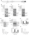
Analysis of the effects of MIM variants on the response to SDF-1. (A) Schematic presentation of mouse MIM protein. CCD, coiled-coil domain; I-BAR, inverse BAR domain; SRD, serine-rich domain; PRD, proline-rich domain; and WH2, WASP homology 2 domain. (B-D) Cells expressing different combinations of MIM, AIP4 and CXCR4 proteins were treated with SDF-1 for 30 min, and then subjected to IP using antibodies as indicated for detection of the interaction between MIM variants and AIP4 (B and C) or Myc–CXCR4 (D). (E) Cells co-expressing MIM–GFP and Myc–CXCR4 were treated with either siRNA against AIP4 (SiRNA-AIP4) or control siRNA (Ctrl-SiRNA) and were further treated with 100 nM SDF-1 for 30 min. The interaction between MIM–GFP and Myc–CXCR4 in the treated cells was analyzed by co-immunoprecipitation. As a positive control, cells co-expressing MIM–GFP, Myc–CXCR4 and Flag–AIP4 were also analyzed in parallel. The data represent mean±s.e.m. (n=3). ***P<0.001 (Student's t-test). (F) Cells co-expressing HA–ubiquitin, Myc–CXCR4, and GFP or different MIM–GFP variants were stimulated with 100 nM SDF-1 for 30 min. Ubiquitylated Myc–CXCR4 proteins were analyzed by co-immunoprecipitation as indicated. (G) Cells expressing different MIM–GFP variants were analyzed for their chemotactic response to 100 nM SDF-1 as described in the legend of Fig. 1A. (H) Cells co-expressing Myc–CXCR4 with GFP or MIM–GFP variants were treated with 100 nM SDF-1 for 15 min. The surface amount of Myc–CXCR4 was estimated using flow cytometry. All the data represent mean±s.e.m. (n=3). **P<0.01 and *** P<0.001; ns, not significant (Student's t-test). IB, immunoblot.
Fig. 5.
Fig. 5.
MIM promotes MVB formation and sorting of CXCR4 into MVBs. (A) Cells co-expressing Flag–AIP4 with MIM–GFP or GFP were stimulated with 100 nM SDF-1 for 30 min, co-stained for GFP (green) and Flag (red) using antibodies. (B,C) Cells co-expressing Myc–CXCR4 with MIM–GFP or GFP were treated with and without SDF-1 for 30 min, stained for GFP (green) and CD63 (blue) with antibodies (B), or for Myc (red) and CD63 (green) with antibodies (C). All the stained cells were analyzed by confocal microscopy. Quantification of the proportion of red staining that colocalized with green staining was estimated based on the MOC. All the data represent mean±s.e.m. of three independent experiments in which 50 cells were examined in each experiment. ***P<0.001; NS, not significant (Student's t-test). All boxed areas within cells represent the zoomed area shown in the insets. Ctrl, no SDF-1.
Fig. 6.
Fig. 6.
MIM binds to Rab5 and Rab7 in response to SDF-1. (A) Cells expressing MIM–GFP were stimulated with 100 nM SDF-1 for 5, 30 and 90 min. The stimulated cells were lysed, and proteins were precipitated with antibody against MIM. The precipitates were than blotted for Rab5 and Rab7 using antibodies. (B) Cells expressing MIM–GFP were treated with 100 nM SDF-1 for 30 min. The lysates of treated cells were incubated with antibody against Rab7, and the precipitates were blotted for MIM using an antibody. (C) The interaction between endogenous MIM and Rab7 was analyzed by co-immunoprecipitation in Raw264 cells under conditions with and without treatment with 100 nM SDF-1 for 30 min. (D,E) Cells expressing different MIM variants were stimulated with 100 nM SDF-1 for 30 min. Interaction between MIM variants and Rab7 in the treated cells was analyzed by co-immunoprecipitation with antibodies as indicated. (F) Cells expressing GFP, or MIM–GFP or MIM-I-BAR–GFP were treated with SDF-1 for 15 min. CXCR4 internalization was estimated by flow cytometry. The data represent mean±s.e.m. (n=3). IB, immunoblot.

References

    1. Agarwal E., Robb C. M., Smith L. M., Brattain M. G., Wang J., Black J. D. Chowdhury S. (2017). Role of Akt2 in regulation of metastasis suppressor 1 expression and colorectal cancer metastasis. Oncogene. [Epub ahead of print] doi: 10.1038/onc.2016.46010.1038/onc.2016.460 - DOI - PMC - PubMed
    1. Bedford M. T., Chan D. C. and Leder P. (1997). FBP WW domains and the Abl SH3 domain bind to a specific class of proline-rich ligands. EMBO J. 16, 2376-2383. 10.1093/emboj/16.9.2376 - DOI - PMC - PubMed
    1. Bhandari D., Robia S. L. and Marchese A. (2009). The E3 ubiquitin ligase atrophin interacting protein 4 binds directly to the chemokine receptor CXCR4 via a novel WW domain-mediated interaction. Mol. Biol. Cell 20, 1324-1339. 10.1091/mbc.E08-03-0308 - DOI - PMC - PubMed
    1. Cao M., Zhan T., Ji M. and Zhan X. (2012). Dimerization is necessary for MIM-mediated membrane deformation and endocytosis. Biochem. J. 446, 469-475. 10.1042/BJ20120329 - DOI - PMC - PubMed
    1. Chen H. I. and Sudol M. (1995). The WW domain of Yes-associated protein binds a proline-rich ligand that differs from the consensus established for Src homology 3-binding modules. Proc. Natl. Acad. Sci. USA 92, 7819-7823. 10.1073/pnas.92.17.7819 - DOI - PMC - PubMed

Publication types

MeSH terms