. 2017 Apr 28;292(17):7223-7232.
doi: 10.1074/jbc.M116.743765. Epub 2017 Mar 6.

The hydrogen peroxide hypersensitivity of OxyR2 in Vibrio vulnificus depends on conformational constraints

Affiliations

The hydrogen peroxide hypersensitivity of OxyR2 in Vibrio vulnificus depends on conformational constraints

Inseong Jo et al. J Biol Chem. .

Abstract

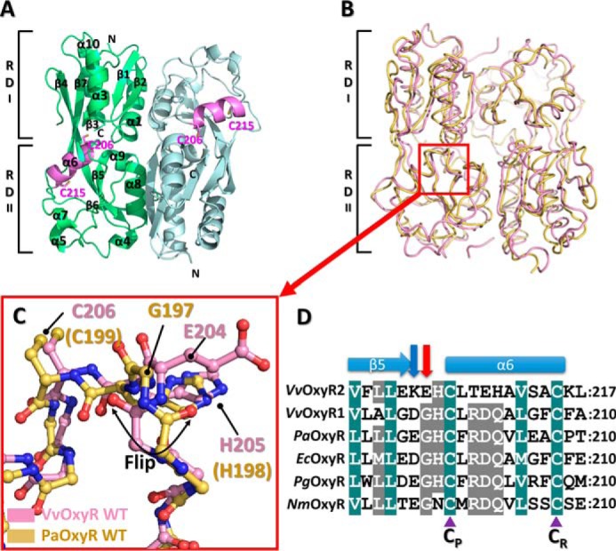
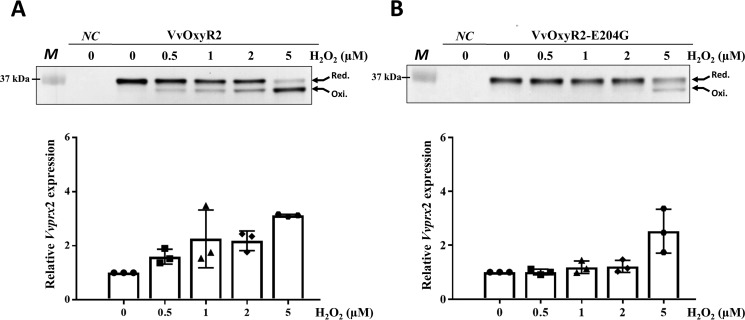
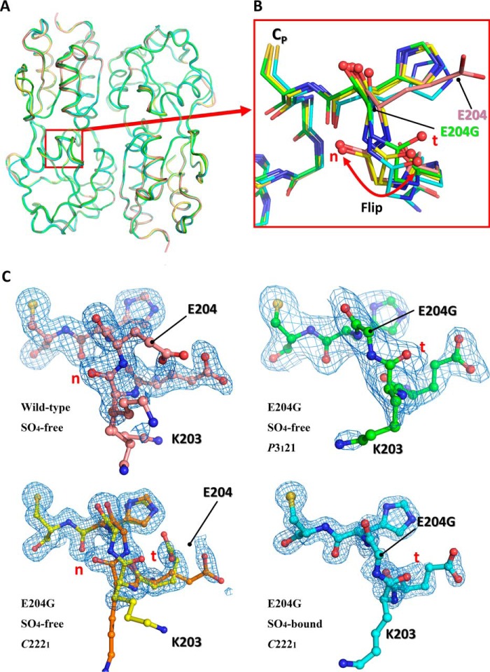
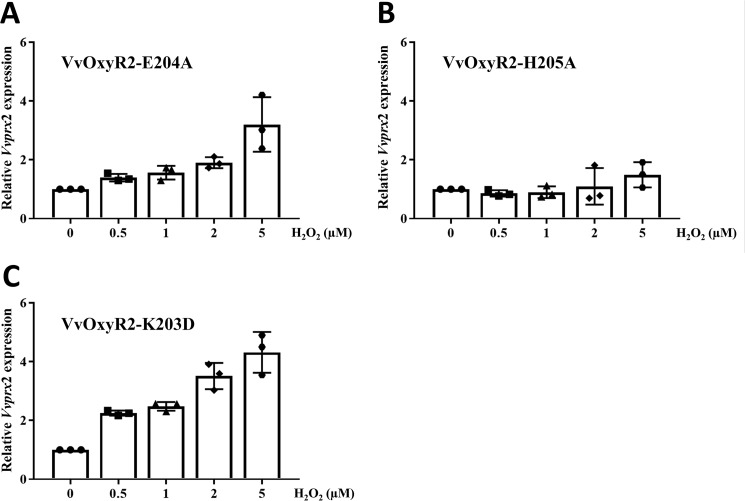
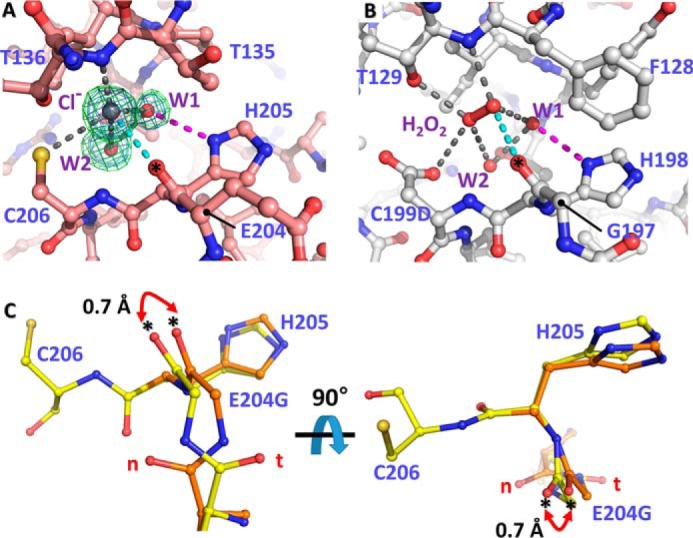
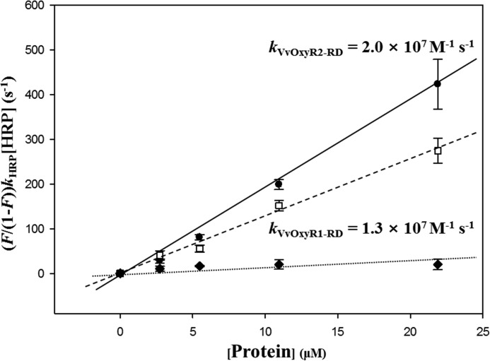
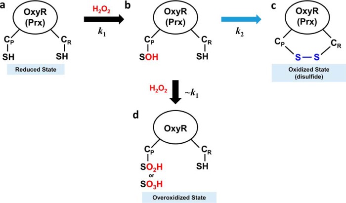
Most Gram-negative bacteria respond to excessive levels of H2O2 using the peroxide-sensing transcriptional regulator OxyR, which can induce the expression of antioxidant genes to restore normality. Vibrio vulnificus has two distinct OxyRs (OxyR1 and OxyR2), which are sensitive to different levels of H2O2 and induce expression of two different peroxidases, Prx1 and Prx2. Although OxyR1 has both high sequence similarity and H2O2 sensitivity comparable with that of other OxyR proteins, OxyR2 exhibits limited sequence similarity and is more sensitive to H2O2 To investigate the basis for this difference, we determined crystal structures and carried out biochemical analyses of OxyR2. The determined structure of OxyR2 revealed a flipped conformation of the peptide bond before Glu-204, a position occupied by glycine in other OxyR proteins. Activity assays showed that the sensitivity to H2O2 was reduced to the level of other OxyR proteins by the E204G mutation. We solved the structure of the OxyR2-E204G mutant with the same packing environment. The structure of the mutant revealed a dual conformation of the peptide bond before Gly-204, indicating the structural flexibility of the region. This structural duality extended to the backbone atoms of Gly-204 and the imidazole ring of His-205, which interact with H2O2 and invariant water molecules near the peroxidatic cysteine, respectively. Structural comparison suggests that Glu-204 in OxyR2 provides rigidity to the region that is important in H2O2 sensing, compared with the E204G structure or other OxyR proteins. Our findings provide a structural basis for the higher sensitivity of OxyR2 to H2O2 and also suggest a molecular mechanism for bacterial regulation of expression of antioxidant genes at divergent concentrations of cellular H2O2.

Keywords: OxyR; Vibrio vulnificus; antioxidant; bacterial transcription; crystal structure; crystallography; hydrogen peroxide; reactive oxygen species (ROS); sensitivity.


Conflict of interest statement

The authors declare that they have no conflicts of interest with the contents of this article

Figures

Figure 1.
Figure 1.
Structure of the wild-type VvOxyR2-RD. A, dimeric assembly of wild-type VvOxyR2-RD under reduced conditions. Protomers are highlighted in green or light blue, except in the inter-cysteine α-helical regions (pink). The conserved cysteines (Cys-206 and Cys-215) are represented as sticks. Secondary structural elements and the subdomains RD-I and RD-II are labeled. B, comparison of VvOxyR2-RD dimers (pink) with P. aeruginosa OxyR-RD in the reduced state (yellow; PDB code 4Y0M): The red box indicates the region near the peroxidatic cysteine (Cys-206) and the Glu-204-containing loop. C, close-up view of the region indicated by the red box in B. The regions near the peroxidatic cysteine of VvOxyR2-RD (pink) and PaOxyR-RD (yellow) are shown in a ball-and-stick representation. The flipped peptide bond is indicated with a double-headed arrow. D, sequence alignment of VvOxyR2 with other OxyRs, focusing on the region containing the conserved cysteine residues (V. vulnificus (Vv), P. aeruginosa (Pa), and N. meningitidis (Nm)). The secondary structures are displayed above the sequence. Glu-204 or its equivalent residue and Lys-203 or its equivalent residue are indicated by a red or a blue arrow, respectively. Two conserved cysteine residues (peroxidatic cysteine (CP) and resolving cysteine (CR)) are indicated by purple triangles. The strictly conserved amino acid residues are indicated by cobalt blue boxes, and the moderately conserved amino acid residues are indicated by gray boxes.
Figure 2.
Figure 2.
Effects of the Glu-204 mutation and transition to glycine on H2O2 sensing at low levels in VvOxyR2. The VvoxyR2 mutants containing plasmids expressing VvOxyR2 (A) or VvOxyR2 (E204G), in which Glu204 was mutated to glycine (B), were grown anaerobically to A600 = 0.3 and exposed to various concentrations of H2O2, as indicated, for 30 s (top panels) or 3 min (bottom panels) before harvesting. The cells (top panels) were then precipitated with TCA and alkylated with fresh AMS buffer for 1 h at 37 °C. Alkylated VvOxyR2 proteins were resolved by non-reducing SDS-PAGE and immunoblotted using anti-VvOxyR2 antibody. The two redox states for each VvOxyR2 (reduced and oxidized) are shown with arrows. The negative control (NC) is the VvoxyR2 mutant expressing an empty vector. M, protein size marker. Total RNAs (bottom panels) were isolated, and the relative Vvprx2 transcript levels were estimated by quantitative real-time PCR analyses. The Vvprx2 mRNA level under anaerobic conditions (H2O2 of 0) was set to 1. Error bars from three independent experiments represent S.D.
Figure 3.
Figure 3.
Structural comparison of the wild-type VvOxyR-RD and the VvOxyR2-RD (E204G) variant. A, structural superposition of the wild-type VvOxyR2-RD (pink) and the three VvOxyR2-RD (E204G) variants that are named SO4-free P3121 (green), SO4-free high-resolution (yellow and orange), and SO4-bound (cyan). Regions containing the peroxidatic cysteine and the Glu-204-containing loop are indicated by a red box. B, a close-up view of the red boxed region in A; all residues are drawn as ball-and-stick representations. A flipped peptide bond is indicated with a double-headed arrow. t and n letters in red indicate the typical conformation observed in the most OxyR proteins and the noncanonical conformation only observed in the wild-type VvOxyR2, respectively. C, 2FoFc electron density maps (blue mesh) around the Glu-204-containing of the wild-type (top left, pink), SO4-free high-resolution E204G (bottom left, yellow and orange), SO4-free P3121 E204G (top right, green), and SO4-bound E204G variant (bottom right, cyan) are contoured at 1.0 σ. t and n letters in red indicate the typical conformation observed in the most OxyR proteins and the noncanonical conformation only observed in the wild-type VvOxyR2, respectively.
Figure 4.
Figure 4.
Effects of the Glu-204 and His-205 mutations on H2O2 sensing at low levels in VvOxyR2. The VvoxyR2 mutants containing plasmids expressing VvOxyR2 (E204A) (A), VvOxyR2 (H205A) (B), and VvOxyR2 (K203D) (C) were grown anaerobically to an A600 of 0.3 and exposed to various concentrations of H2O2, as indicated, for 3 min. Total RNA isolation and quantitative real-time PCR analyses were performed as described previously. The Vvprx2 mRNA level under anaerobic conditions (H2O2 = 0) was set as 1. All data are presented as mean ± S.D. (error bars); n = 3.
Figure 5.
Figure 5.
Structural variation of VvOxyR2 His-205 at the H2O2 and water binding sites near the peroxidatic cysteine residue. The putative chloride ion in VvOxyR2 (A) and the H2O2 molecule in P. aeruginosa OxyR (B; PDB code 4X6G) are bound near the peroxidatic cysteine (C206) or the mutated aspartic acid (C199D) residue. The polar interactions involved in binding the chloride ion (gray ball) or H2O2 (red ball and stick) are indicated by dotted lines. The bound water molecules (W1 and W2) are indicated by red balls. The 2FoFc (blue mesh) and FoFc (green mesh) omit maps of chloride ion and water molecules are contoured at 1.0 σ and 3.0 σ, respectively. C, two orthogonal views of the SO4-free high-resolution structure of VvOxyR2 E204G variant. The alternative residues are presented by yellow or orange ball-and-stick representations. The difference points in dual conformation are indicated by red dihedral arrows. t and n letters in red indicate the typical conformation observed in the most OxyR proteins and the noncanonical conformation only observed in the wild-type VvOxyR2, respectively. *, carbonyl atom between Glu-204 (or Gly-204) and His-205.
Figure 6.
Figure 6.
Determination of the second-order rate constants of VvOxyR2-RD and VvOxyR1-RD. Reaction mixtures containing HRP and various concentrations of VvOxyR2-RD (●) or VvOxyR1-RD (□) (0–21.9 μm) were exposed to H2O2. BSA (♦) was used as a negative control. The ratio of HRP oxidation was determined by measuring the absorbance at 403 nm. kVvOxyR1-RD and kVvOxyR2-RD were determined from the slope of a plot of the equation, (F/(1-F))kHRP[HRP] versus [VvOxyR1-RD or VvOxyR2-RD]. The fitting lines were calculated by the least square methods, and the error bars reflect the S.D. of three independent experiments.
Figure 7.
Figure 7.
Schematic representation of an oxidation mechanism for OxyR and Prx protein. a, the OxyR or Prx in the reduced state has two free thiols at the CP and CR. b, H2O2 rapidly reacts with CP, resulting in CP-SOH at the second-order reaction rate constant of k1. c, CP-SOH then forms a disulfide bond with CR, resulting in the oxidized state with a disulfide bond at the reaction rate constant of k2. d, in an alternative to c, CP-SOH is further oxidized by additional H2O2, resulting in the overoxidized state with CP-SO2H or CP-SO3H. Due to the structural similarity with OxyR in the reduced state, the second-order reaction rate constant would be close to k1 (∼k1).

Similar articles

Cited by

References

    1. Storz G., and Imlay J. A. (1999) Oxidative stress. Curr. Opin. Microbiol. 2, 188–194 - PubMed
    1. Stadtman E. R. (2001) Protein oxidation in aging and age-related diseases. Ann. N.Y. Acad. Sci. 928, 22–38 - PubMed
    1. Imlay J. A., Chin S. M., and Linn S. (1988) Toxic DNA damage by hydrogen peroxide through the Fenton reaction in vivo and in vitro. Science 240, 640–642 - PubMed
    1. Storz G., Tartaglia L. A., Farr S. B., and Ames B. N. (1990) Bacterial defenses against oxidative stress. Trends Genet. 6, 363–368 - PubMed
    1. Schell M. A. (1993) Molecular biology of the LysR family of transcriptional regulators. Annu. Rev. Microbiol. 47, 597–626 - PubMed

Publication types

MeSH terms

LinkOut - more resources