. 2017 Apr 28;292(17):6938-6951.
doi: 10.1074/jbc.M117.778431. Epub 2017 Mar 6.

Structure/activity relationship of thapsigargin inhibition on the purified Golgi/secretory pathway Ca2+/Mn2+-transport ATPase (SPCA1a)

Affiliations

Structure/activity relationship of thapsigargin inhibition on the purified Golgi/secretory pathway Ca2+/Mn2+-transport ATPase (SPCA1a)

Jialin Chen et al. J Biol Chem. .

Abstract

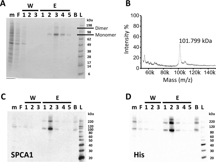
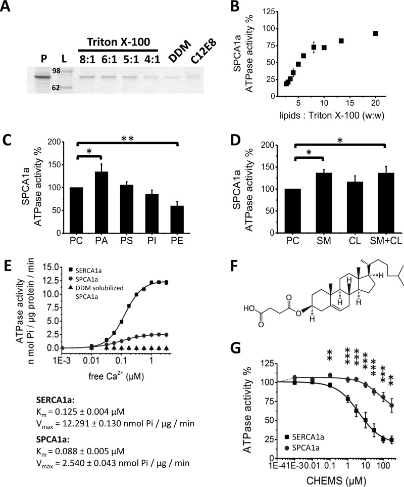
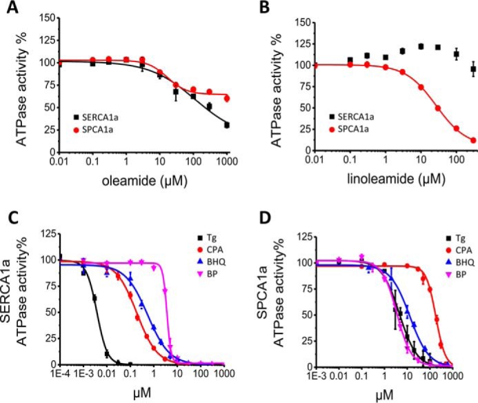
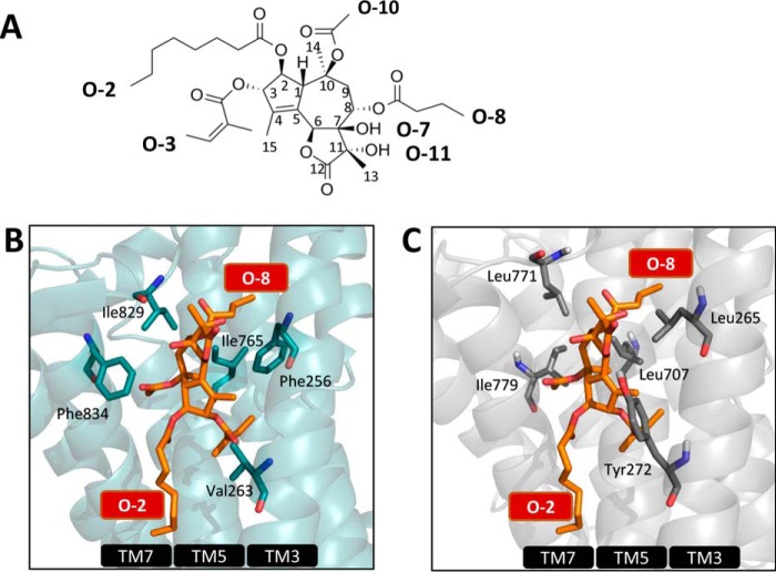
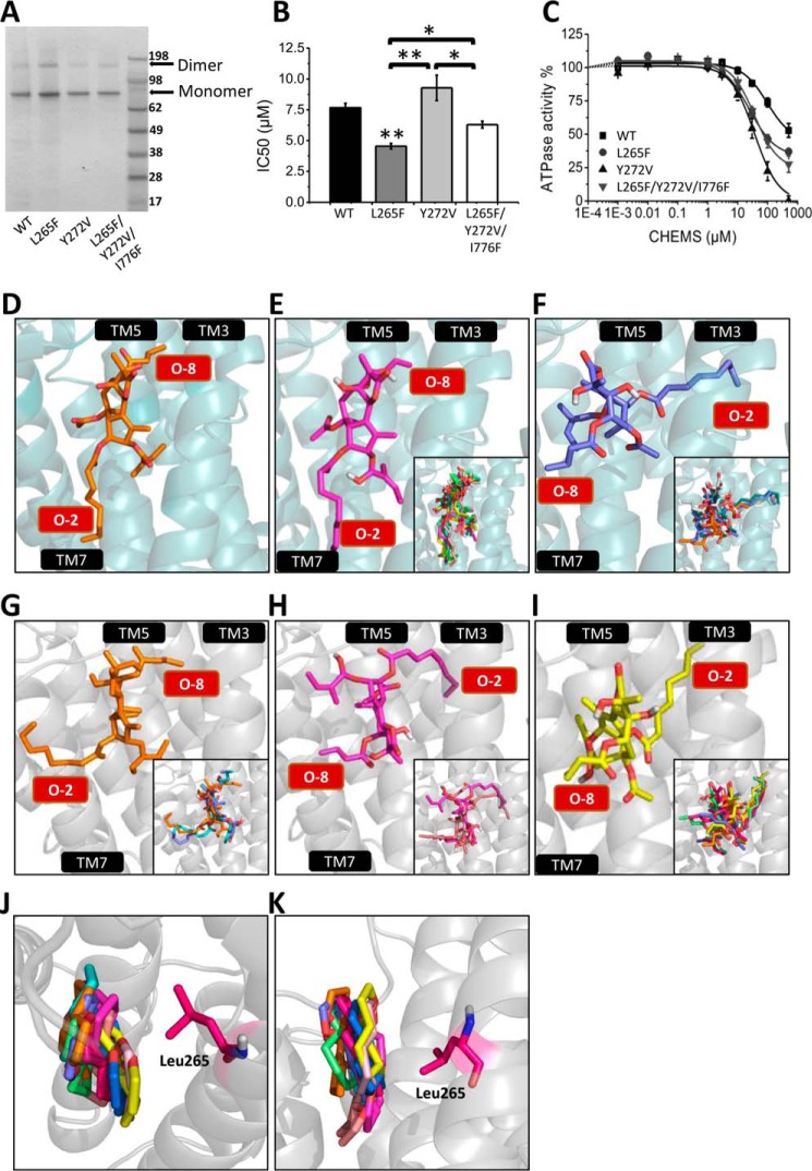
The Golgi/secretory pathway Ca2+/Mn2+-transport ATPase (SPCA1a) is implicated in breast cancer and Hailey-Hailey disease. Here, we purified recombinant human SPCA1a from Saccharomyces cerevisiae and measured Ca2+-dependent ATPase activity following reconstitution in proteoliposomes. The purified SPCA1a displays a higher apparent Ca2+ affinity and a lower maximal turnover rate than the purified sarco(endo)plasmic reticulum Ca2+-ATPase (SERCA1a). The lipids cholesteryl hemisuccinate, linoleamide/oleamide, and phosphatidylethanolamine inhibit and phosphatidic acid and sphingomyelin enhance SPCA1a activity. Moreover, SPCA1a is blocked by micromolar concentrations of the commonly used SERCA1a inhibitors thapsigargin (Tg), cyclopiazonic acid, and 2,5-di-tert-butylhydroquinone. Because tissue-specific targeting of SERCA2b by Tg analogues is considered for prostate cancer therapy, the inhibition of SPCA1a by Tg might represent an off-target risk. We assessed the structure-activity relationship (SAR) of Tg for SPCA1a by in silico modeling, site-directed mutagenesis, and measuring the potency of a series of Tg analogues. These indicate that Tg and the analogues are bound via the Tg scaffold but with lower affinity to the same homologous cavity as on the membrane surface of SERCA1a. The lower Tg affinity may depend on a more flexible binding cavity in SPCA1a, with low contributions of the Tg O-3, O-8, and O-10 chains to the binding energy. Conversely, the protein interaction of the Tg O-2 side chain with SPCA1a appears comparable with that of SERCA1a. These differences define a SAR of Tg for SPCA1a distinct from that of SERCA1a, indicating that Tg analogues with a higher specificity for SPCA1a can probably be developed.

Keywords: Golgi; calcium transport; cholesterol; cyclopiazonic acid; membrane transporter reconstitution; molecular modeling; prostate cancer; protein purification; thapsigargin.


Conflict of interest statement

The authors declare that they have no conflicts of interest with the contents of this article

Figures

Figure 1.
Figure 1.
Purification of the human SPCA1a isoform by affinity chromatography. Gel stained for an SDS-PAGE (A), mass spectrometry analysis (B), and immunoblotting (C and D) of hSPCA1a fractions collected during affinity purification. The yeast membrane fractions (m), the flow-through after binding to Ni-NTA beads (F), the flow-through of each washing step (W1–3), each elution fraction (E1–5), and the beads (B) after elution were collected. Equal volumes of each fraction were separated via SDS-PAGE. Immunoblotting was performed with antibodies against SPCA1a (C) or the C-terminal His tag (D). Images are representative for a minimum of n = 3 experiments. L, ladder.
Figure 2.
Figure 2.
Optimization of SPCA1a reconstitution into proteoliposomes. A, SDS-polyacrylamide gel stained with SPCA1a proteoliposomes generated with the detergents Triton X-100, DDM, or C12E8. Same volume of samples was loaded on the gel after reconstitution. The lipid/Triton X-100 ratios (w/w) are indicated above the lanes. B, comparison of the SPCA1a ATPase activity following reconstitution with a different (w/w) ratio of lipids/Triton X-100. C and D, 20% weight percent of the indicated lipid(s) were supplemented to PC for SPCA1a reconstitution, and the maximal Ca2+-dependent ATPase activity was determined at 1 μm free Ca2+ concentration (n ≥ 3). The ATPase activities were normalized to the activity when only PC was used. Comparison of the specific SPCA1a ATPase activities in the presence of indicated phospholipids (C), CL, SM, or the combination of CL and SM (1:1 molar ratio) (D). E, comparison of the specific ATPase activities between SERCA1a and SPCA1a in proteoliposomes, and SPCA1a in lipid-free, DDM-solubilized state (n = 3). The DDM-solubilized SPCA1a was purified without supplement of PC in the buffers. F, structure of CHEMS. G, dose-responses of CHEMS on reconstituted SERCA1a and SPCA1a ATPase activities. P, purified SPCA1a; L, ladder; PA, phosphatidic acid; PE, phosphatidylethanolamine; PS, phosphatidylserine; PI, phosphatidylinositol; CL, cholesterol; SM, sphingomyelin; CHEMS, cholesteryl hemisuccinate. *, p < 0.05; **, p < 0.01; ***, p < 0.001.
Figure 3.
Figure 3.
Dose-response effects of SPCA1a and SERCA1a inhibitors. Dose-response curves of oleamide (A) and linoleamide (B) on SERCA1a and SPCA1a. Dose-response curves for the inhibition of SERCA1a (C) and SPCA1a (D) by Tg, CPA, BHQ, and BP. n = 3 for all experiments.
Figure 4.
Figure 4.
Establishing the structure-activity relationship of Tg in SPCA1a. A, structure of Tg with the numbering of the carbon atoms and side chains indicated. B, Tg pocket in SERCA1a in the E2 conformation with bound Tg (1IWO (41)) with important residues shown as blue sticks. The same view of the SPCA1a homology model is shown in C, and the residues that are different between SPCA1a and SERCA1a are labeled.
Figure 5.
Figure 5.
Modeling Tg into the binding pocket of SPCA1a. SDS-polyacrylamide gel staining (A), IC50 values of Tg (B), and CHEMS dose-response curves (C) of the reconstituted SPCA1a WT and L265F, Y272V, and L265F/Y272V/I776F mutants. D, crystal structure of SERCA1a-Tg complex (1IWO (41)). E and F, Tg docking simulation performed on the SERCA1a E2 crystal structure without Tg (3W5C (46)) (side-chain torsion rotations were assigned to Glu-255, Phe-256, Gln-259, Leu-260, Val-263, Ile-765, Val-769, Ile-829, Phe-834, and Met-838), rendering two conformational populations: model 1 (E) and model 2 (F). G–I, Tg docking simulation was performed on the SPCA1a homology model (side-chain torsion rotations were assigned to Leu-264, Leu-265, Leu-269, Tyr-272, Leu-707, Ile-711, Ile-770, Leu-771, Ile-776, and Ile-779), rendering three conformational populations (G–I), which are corresponding to two major models according to the orientation of the Tg O-2 and O-8 side chains: model 1 (G) and model 2 (H and I). The O-2 and O-8 chains are indicated in red text boxes. Top (J) and side (K) view of the superimposed 10 Tg scaffolds from the docking result of G–I. The residue Leu-265 is highlighted. TM, transmembrane helix. One-way ANOVA was performed followed by a post hoc Fisher test. *, p < 0.05; **, p < 0.01.

References

    1. Berridge M. J., Bootman M. D., and Roderick H. L. (2003) Calcium signalling: dynamics, homeostasis and remodelling. Nat. Rev. Mol. Cell Biol. 4, 517–529 - PubMed
    1. Monteith G. R., McAndrew D., Faddy H. M., and Roberts-Thomson S. J. (2007) Calcium and cancer: targeting Ca2+ transport. Nat. Rev. Cancer 7, 519–530 - PubMed
    1. Palmgren M. G., and Nissen P. (2011) P-type ATPases. Annu. Rev. Biophys. 40, 243–266 - PubMed
    1. Vangheluwe P., Sepúlveda M. R., Missiaen L., Raeymaekers L., Wuytack F., and Vanoevelen J. (2009) Intracellular Ca2+- and Mn2+-transport ATPases. Chem. Rev. 109, 4733–4759 - PubMed
    1. Behne M. J., Tu C. L., Aronchik I., Epstein E., Bench G., Bikle D. D., Pozzan T., and Mauro T. M. (2003) Human keratinocyte ATP2C1 localizes to the Golgi and controls Golgi Ca2+ stores. J. Invest. Dermatol. 121, 688–694 - PubMed

MeSH terms

LinkOut - more resources