. 2017 Apr;48(4):907-914.
doi: 10.1161/STROKEAHA.116.016174. Epub 2017 Mar 6.

Absent Filling of Ipsilateral Superficial Middle Cerebral Vein Is Associated With Poor Outcome After Reperfusion Therapy

Affiliations

Absent Filling of Ipsilateral Superficial Middle Cerebral Vein Is Associated With Poor Outcome After Reperfusion Therapy

Sheng Zhang et al. Stroke. 2017 Apr.

Abstract

Background and purpose: Our aim was to study the effect of drainage of cortical veins, including the superficial middle cerebral vein (SMCV), vein of Trolard, and vein of Labbé on neurological outcomes after reperfusion therapy.

Methods: Consecutive ischemic stroke patients who underwent pretreatment computed tomographic perfusion and 24-hour computed tomographic perfusion or magnetic resonance perfusion after intravenous thrombolysis were included. We defined "absent filling of ipsilateral cortical vein" (eg, SMCV-) as no contrast filling of the vein across the whole venous phase on 4-dimensional computed tomographic angiography in the ischemic hemisphere.

Results: Of 228 patients, SMCV-, vein of Trolard- and vein of Labbé- were observed in 50 (21.9%), 27 (11.8%), and 32 (14.0%) patients, respectively. Only SMCV- independently predicted poor outcome (3-month modified Rankin Scale score of >2; odds ratio, 2.710; P=0.040). No difference was found in reperfusion rate after treatment between patients with and without SMCV- (P>0.05). In patients achieving major reperfusion (≥80%), there was no difference in 24-hour infarct volume, or rate of poor outcome between patients with and without SMCV- (P>0.05). However, in those without major reperfusion, patients with SMCV- had larger 24-hour infarct volume (P=0.011), higher rate of poor outcome (P=0.012), and death (P=0.032) compared with those with SMCV filling. SMCV- was significantly associated with brain edema at 24 hours (P=0.037), which, in turn, was associated with poor 3-month outcome (P=0.002).

Conclusions: Lack of SMCV filling contributed to poor outcome after thrombolysis, especially when reperfusion was not achieved. The main deleterious effect of poor venous filling appears related to the development of brain edema.

Keywords: brain imaging; edema; outcome; reperfusion; vein.


Figures

Figure 1
Figure 1
Identification of cortical veins. SMCV, superficial middle cerebral vein; VOT, vein of Trolard; VOL, vein of Labbé. A: According to locations of the 3 cortical veins (“Y” structure) shown on the venous peak-phase 3D CTV, the appearance of contrast flow in the targeted cortical vein was defined as the presence of the targeted cortical vein (marked as SMCV+/VOT+/VOL+). B: Visual assessment for cortical veins, eg. ipsilateral superficial middle cerebral vein (SMCV) on 4D CTA. The presence (red arrow) and the absence (red broken circle) of cortical veins were displayed. Based on venous output function (VOF) curve, the contrast in the SMCV of non-ischemic hemisphere appeared in the early venous phase, enhanced at the maximum of the peak venous phase, and disappeared at the late venous phase (yellow arrow). In comparison, no contrast in the ischemic hemispheric SMCV was shown in any of these three time points. C: Example of the presence of SMCV, VOT and VOL in axial, coronal, and sagittal planes of both hemispheres across the whole venous phase (SMCV+/VOT+/VOL+). D: Example of absent filling of SMCV, VOT and VOL in the ischemic hemisphere across the whole venous phase (SMCV-/VOT-/VOL-), in axial, coronal, and sagittal planes.
Figure 2
Figure 2
Matching hypoperfusion area on Tmax map with the drainage territory of the selected cortical vein on 4D CTA. A: The representative drainage territories of superficial middle cerebral vein (SMCV, green), vein of Labbé (VOL, red), and superior sagittal sinus (blue) which receives the drainage of vein of Trolard (VOT) and was then used for matching with VOT, due to the location variation of VOT. B, C and D show the ipsilateral hypoperfusion (colored voxels) on Tmax map located within the drainage territories of SMCV (B), VOT (C) and VOL (D), respectively. The suspected location of the ipsilateral targeted cortical vein (yellow box) can be simultaneously viewed on Tmax map.
Figure 3
Figure 3
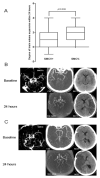
The difference in brain edema expansion within 24 hours between patients with SMCV- and SMCV+ among those without major reperfusion. A: Brain edema expansion was significantly higher in SMCV-, in comparison of SMCV+. B: A 48-year-old female with baseline CT-ASPECT of 7 (NIHSS:7) and acute left proximal middle cerebral artery occlusion (MCAO), presented with SMCV+. Even without recanalization after IVT, brain edema was mildly enlarged (score from 1 to 2). She achieved good outcome (3-month mRS:1). C: A 49-year-old male with baseline CT-ASPECT of 6 (NIHSS:9) and acute right MCAO, presented with ipsilateral SMCV-. Without recanalization, he had enlargement of brain edema (score from 1 to 5). He died within one week after stroke onset.
Figure 4
Figure 4
The hypothesized pathogenesis of venous outflow dysfunction. Due to large arterial occlusion seen on 4D CTA (a) and hypoperfusion seen on Tmax map (b), venous flow is subsequently reduced, leading to microthrombi formation and occlusion in venules, which is visualized as the absent filling of draining cortical vein (c). Due to occlusion in venules, the subsequent resist of cerebrospinal fluid absorption will result in the elevation of venous pressure, which might increase fluid leakage from the disrupted tight junctions of endothelial cells (EC) into the perivascular space, leading to the development of brain edema, as shown on non-contrast CT (d).

References

    1. Zhang JH, Badaut J, Tang JP, Obenaus A, Hartman R, Pearce WJ. The vascular neural network-a new paradigm in stroke pathophysiology. Nature Reviews Neurology. 2012;8:711–716. - PMC - PubMed
    1. Ishikawa M, Kusaka G, Yamaguchi N, Sekizuka E, Nakadate H, Minamitani H, et al. Platelet and leukocyte adhesion in the microvasculature at the cerebral surface immediately after subarachnoid hemorrhage. Neurosurgery. 2009;64:546–553. discussion 553-544. - PubMed
    1. Sasaki M, Honmou O, Radtke C, Kocsis JD. Development of a middle cerebral artery occlusion model in the nonhuman primate and a safety study of i.V. Infusion of human mesenchymal stem cells. PLoS One. 2011;6:e26577. - PMC - PubMed
    1. Parthasarathy R, Kate M, Rempel JL, Liebeskind DS, Jeerakathil T, Butcher KS, et al. Prognostic evaluation based on cortical vein score difference in stroke. Stroke. 2013;44:2748–2754. - PMC - PubMed
    1. van den Wijngaard IR, Wermer MJ, Boiten J, Algra A, Holswilder G, Meijer FJ, et al. Cortical venous filling on dynamic computed tomographic angiography: A novel predictor of clinical outcome in patients with acute middle cerebral artery stroke. Stroke. 2016;47:762–767. - PubMed