. 2017 Mar 21;114(12):E2310-E2318.
doi: 10.1073/pnas.1700280114. Epub 2017 Mar 6.

Deep-sea vent phage DNA polymerase specifically initiates DNA synthesis in the absence of primers

Affiliations

Deep-sea vent phage DNA polymerase specifically initiates DNA synthesis in the absence of primers

Bin Zhu et al. Proc Natl Acad Sci U S A. .

Abstract

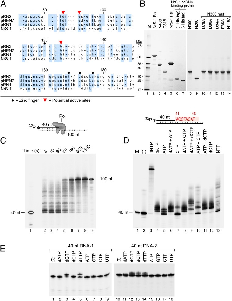
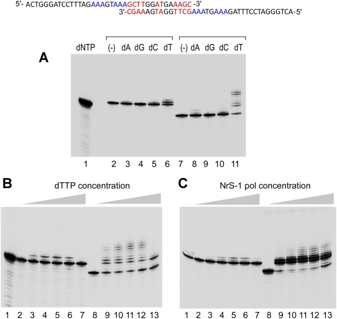
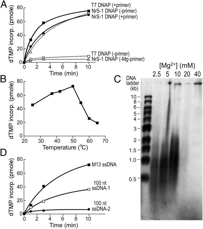
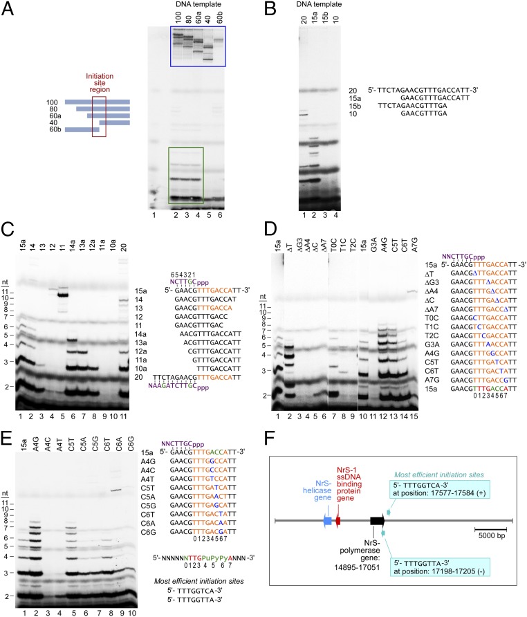
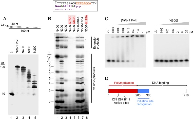
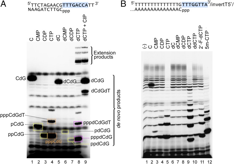
A DNA polymerase is encoded by the deep-sea vent phage NrS-1. NrS-1 has a unique genome organization containing genes that are predicted to encode a helicase and a single-stranded DNA (ssDNA)-binding protein. The gene for an unknown protein shares weak homology with the bifunctional primase-polymerases (prim-pols) from archaeal plasmids but is missing the zinc-binding domain typically found in primases. We show that this gene product has efficient DNA polymerase activity and is processive in DNA synthesis in the presence of the NrS-1 helicase and ssDNA-binding protein. Remarkably, this NrS-1 DNA polymerase initiates DNA synthesis from a specific template DNA sequence in the absence of any primer. The de novo DNA polymerase activity resides in the N-terminal domain of the protein, whereas the C-terminal domain enhances DNA binding.

Keywords: NrS-1; helicase; primase; prim–pol; ssDNA-binding protein.


Conflict of interest statement

The authors declare no conflict of interest.

Figures

Fig. 1.
Fig. 1.
An unusual DNA polymerase from deep-sea vent phage NrS-1. (A) NrS-1 gene protein 28 shares weak homology to archaeal prim–pols. Alignment between NrS-1 gene protein 28 and three prim–pols encoded by archaeal plasmids (10) was made using CLC Sequence Viewer 6, and the regions containing homology are shown. Identical residues among archaeal prim–pols and those among prim–pols and NrS-1 gene protein 28 are highlighted by blue background. (B) SDS/PAGE gels showing purified proteins analyzed in this work. Lane 1, protein size marker; lane 2, NrS-1 polymerase; lane 3, N-terminal 400 residues of NrS-1 polymerase (N400); lane 4, C-terminal 318 residues of NrS-1 polymerase; lane 5, NrS-1 helicase; lane 6, NrS-1 ssDNA-binding protein; lane 7, NrS-1 ssDNA-binding protein (His-tag removed); lane 8, N-terminal 300 residues of NrS-1 polymerase (N300); lane 9, N-terminal 200 residues of NrS-1 polymerase (N200); lane 10, N300-D78A; lane 11, N300-D80A; lane 12, N300-D84A; lane 13, N300-E85A; lane 14, N300-H115A. All proteins carry an N-terminal His-tag except that in lane 7. (C) Extension of a primer by NrS-1 polymerase. A primed-template (40/100 nt) substrate in which the primer was 5′-32P–labeled and was incubated at 100 nM with 200 nM NrS-1 polymerase and 0.5 mM four dNTPs at 50 °C. Aliquots were removed at increasing time and analyzed on a 10% (wt/vol) TBE-urea gel (lanes 2–8). Lanes 1 and 9 contain 5′-32P–labeled 40 nt (primer) and the 100 nt complete complement to the template strand, respectively. (D) Nonspecific incorporation by NrS-1 polymerase. A primed-template (40/100 nt) substrate in which the primer was 5′-32P–labeled was incubated at 100 nM with 200 nM NrS-1 polymerase and 0.5 mM of various (d)NTPs at 50 °C for 30 min and then analyzed on a 10% (wt/vol) TBE-urea gel. Lane 1 is the 5′-32P–labeled 40 nt marker. Shown in red is the 5′ part of the DNA sequence to be synthesized downstream of the primer. (E) 100 nM each of the two 5′-32P–labeled 40-nt DNA strand (40 nt DNA-1 sequence 5′-TTTAGGTACCGGTGCCTAGCAGAAGGCCTAATTCTGCAAA-3′; 40 nt DNA-2 sequence 5′-TTTGCAGAATTAGGCCTTCTGCTAGGCACCGGTACCTAAA-3′) was incubated with 200 nM NrS-1 polymerase and 0.5 mM various (d)NTPs at 50 °C for 30 min and reaction products analyzed on a 10% (wt/vol) TBE-urea gel.
Fig. S1.
Fig. S1.
DNA terminal extension by NrS-1 DNA polymerase. (A) Each of the two DNA strands [5′-ACTGGGATCCTTTGCACACTCAAGTATCCAACTTTCAGAACC-3′ (lanes 2–6) and 5′-ACTGGGATCCTTTAGAAAGTAAAGCTTGGATGAAAGC-3′ (lanes 7–11)] was labeled at its 5′ end with 32P. We mixed 100 nM of each template with 200 nM NrS-1 polymerase in the presence of 0.5 mM of the following dNTPs: none (lanes 2 and 7), dATP (lanes 3 and 8), dGTP (lanes 4 and 9), dCTP (lanes 5 and 10), and dTTP (lanes 6 and 11). Reaction mixtures were incubated at 50 °C for 30 min, and then the products were analyzed on a 10% (wt/vol) polyacrylamide TBE-urea gel. Lane 1 contains a 5′-32P–labeled 40-nt DNA, same as that used in Fig. 1E, Right. A potential intermolecular secondary structure formed by the 37-nt DNA (template for lanes 7–11) was shown, in which the complementary sequences are indicated in red and the templates for dTMP incorporation are indicated in blue. (B) Terminal extension of two DNA strands. Each of two DNA strands [5′-TTTGCAGAATTAGGCCTTCTGCTAGGCACCGGTACCTAAA-3′ (lanes 2–7) and 5′-ACTGGGATCCTTTAGAAAGTAAAGCTTGGATGAAAGC-3′ (lanes 8–13)] was labeled at its 5′ end with 32P. We mixed 100 nM of each template with 200 nM of NrS-1 DNA polymerase in the presence of the following amounts of dTTP: 0 mM (lanes 2 and 8), 0.1 mM (lanes 3 and 9), 0.4 mM (lanes 4 and 10), 1.1 mM (lanes 5 and 11), 3.3 mM (lanes 6 and 12), and 10 mM (lanes 7 and 13). Lane 1 is the same as lane 1 in A. Reaction mixtures were incubated at 50 °C for 30 min, and then the products were analyzed on a 10% (wt/vol) polyacrylamide TBE-urea gel. (C) Terminal extension of two DNA strands. Each of two DNA strands [5′-TTTGCAGAATTAGGCCTTCTGCTAGGCACCGGTACCTAAA-3′ (lanes 2–7) and 5′-ACTGGGATCCTTTAGAAAGTAAAGCTTGGATGAAAGC-3′ (lanes 8–13)] was labeled at its 5′ end with 32P. We mixed 100 nM of each template with 1 mM dTTP and the following amounts of NrS-1 DNA polymerase: 0 nM (lanes 2 and 8), 12 nM (lanes 3 and 9), 37 nM (lanes 4 and 10), 111 nM (lanes 5 and 11), 333 nM (lanes 6 and 12), and 1,000 nM (lanes 7 and 13). Reaction mixtures were incubated at 50 °C for 30 min, and then the products were analyzed on a 10% (wt/vol) polyacrylamide TBE-urea gel. Lane 1 is the same as lane 1 in A.
Fig. 2.
Fig. 2.
NrS-1 DNA polymerase synthesizes DNA de novo. (A) 10 nM M13 ssDNA or primed-M13 ssDNA (annealed with M13 primer M2, 5′-CCCAG TCACG ACGTT-3′) was incubated with 100 nM T7 DNA polymerase (solid square, with primer; open square, without primer) or NrS-1 DNA polymerase (solid circle, with primer; open circle, without primer) in the presence of 250 μM each of dATP, dGTP, and dCTP and 10 μM 3H-dTTP; [Mg2+] was 5 mM for all assays except that no Mg2+ was added for the assay shown by an open triangle, and the incorporation of dTMP into DNA at various time points at 37 °C (for T7 DNA polymerase) or 50 °C (for NrS-1 DNA polymerase) was measured by DE81 (Whatman) filter-binding assay. (B) Effect of temperature on de novo DNA synthesis by NrS-1 DNA polymerase. Reaction conditions were as described for A using 10 nM M13 ssDNA template and 100 nM NrS-1 DNA polymerase. Reactions were carried out for 10 min at the indicated temperatures, and then the incorporation of dTMP into DNA at various time points was measured by DE81 (Whatman) filter-binding assay. (C) De novo DNA synthesis products catalyzed by NrS-1 DNA polymerase on M13 ssDNA were analyzed on 0.8% alkaline agarose gel. Reaction conditions were as described for A, except that nucleotide mixture was 250 μM each of dATP, dCTP, and dTTP and 25 μM α-32P-dGTP, and the indicated amounts of MgCl2 were used. Reactions were carried out for 10 min at 50 °C. (D) De novo DNA synthesis by NrS-1 DNA polymerase on the templates M13 ssDNA (solid square), 100 nt template-1 (open square), and -2 (solid circle) (ssDNA-1 sequence 5′-TAGACTGAATAGTTAAATAGGCAGATATAAAATGGTCAAACGTTCTAGAACTATGTAGGTTTTGCAGAATTAGGCCTTCTGCTAGGCACCGGTACCTAAA-3′; ssDNA-2 sequence 5′-TTTAGGTACCGGTGCCTAGCAGAAGGCCTAATTCTGCAAAACCTACATAGTTCTAGAACGTTTGACCATTTTATATCTGCCTATTTAACTATTCAGTCTA-3′). Reaction conditions were as described for A, except for the DNA templates used.
Fig. 3.
Fig. 3.
NrS-1 DNA polymerase initiates DNA synthesis at specific template sequences. (A) Gel analysis of products synthesized by NrS-1 DNA polymerase on ssDNA-1 (described in Fig. 2D) and templates that are truncated forms of ssDNA-1. We incubated 10 μM 100 nt ssDNA-1 (or its 80, 60, or 40 nt 3′ fragments or its 60 nt 5′ fragment) with 200 nM NrS-1 polymerase in the presence of 250 μM each of dATP, dCTP, and dTTP and 25 μM α-32P-dGTP at 50 °C for 30 min. The products were separated on a 25% (wt/vol) TBE-urea gel and analyzed by phosphoimager. Extended DNA templates are indicated by the blue box, and the abortive 2–5-nt products are indicated by the green box. The region that contains the potential initiation site is indicated by the red box. (B) We incubated 10 μM of either 20-nt, 15-nt, or 10-nt DNA fragments (derived from the sequence within the red box of A) with 200 nM NrS-1 polymerase in the presence of 250 μM each dATP dCTP, and dTTP and 25 μM α-32P-dGTP at 50 °C for 30 min. And the products were separated on a 25% (wt/vol) TBE-urea gel and analyzed by phosphoimager. (C) DNA templates derived from template 15a in B were tested for whether they could support de novo synthesis by NrS-1 polymerase. Assay conditions were the same as that described for B. The sequence in orange is the deduced recognition sequence for initiation of de novo synthesis by NrS-1 polymerase. The sequence of the synthesized product is shown in purple (with the radioactively labeled nucleotide shown in green). Nucleotides in the products are numbered from 5′ to 3′. (D) Same assay as that described in C, except that various DNA templates were used that are derived from template 15a, with one nucleotide deletion or replacement (shown in blue) within the recognition region (shown in orange), were tested for their efficiency to support de novo synthesis. Based on these results, the strictly required sequence for template recognition for de novo DNA synthesis is shown at the bottom in red, and those sequences that do not show strict specificity are shown in green. The 8 nt of the recognition sequence are numbered from 5′ to 3′ as 0–7. (E) Same assay as that described in C, except that various DNA templates were used that are derived from template 15a, with one nucleotide replacement (shown in blue) within the recognition region (shown in orange), were tested for their efficiency to support de novo synthesis. Based on these results, the strictly required sequence for template recognition site for de novo DNA synthesis is shown at the bottom in red, whereas those sequences that are important but do not show strict specificity are shown in green. (F) Position of the NrS-1 DNA polymerase gene and the two most efficient initiation sites in NrS-1 genome.
Fig. 4.
Fig. 4.
Active site and functional subdomains of NrS-1 DNA polymerase. (A) Conditions were as in Fig. 1C, except that each reaction was carried out using either 200 nM full-length NrS-1 DNA polymerase (lane 2) or 500 nM of either the N-terminal fragment, N400 (lane 3), N300 (lane 4), or N200 (lane 5). The ability of each of these enzymes to extend 100 nM of a 40/100 nt primed-template was determined. The 40-mer primer is labeled at its 5′ end with 32P. Lane 1 shows the reaction mixture in the absence of enzyme. (B) 10 μM of the 20-nt DNA template containing the NrS-1 polymerase recognition sequence (shown in orange) was incubated with 500 nM of either NrS-1 polymerase fragment N400 (lane 1), N300 (lane 2), N200 (lane 3), or N300 mutants (D78A, lane 4; D80A, lane 5; D84A, lane 6; E85A, lane 7; H115A, lane 8) in the presence of 250 μM each of dATP, dCTP, and dTTP and 25 μM α-32P-dGTP at 50 °C for 30 min. The products were separated on a 25% (wt/vol) TBE-urea gel and analyzed by phosphoimager. Products larger than 20 nt are the result of extension of the templates (Extension Products), whereas those smaller than 13 nt represent abortive and runoff products synthesized de novo (“de novo products”). The runoff product sequence is shown in purple (with the two radioactively labeled G’s indicated in green). Numbering of the nucleotides in the product is 5′ to 3′. Mutations that abolish NrS-1 polymerase activities are shown in red. (C) In a DNA mobility shift assay, 5 nM of 5′-32P–labeled 15 nt DNA (15a) was incubated with the indicated amounts of either full-length NrS-1 polymerase or N300 fragment in the absence of dNTPs for 20 min, followed by loading on a 10% (wt/vol) TBE native acrylamide gel to detect the mobility shift. After electrophoresis, the gel was analyzed using a phosphoimager. (D) A schematic showing the active site and proposed functional subdomains of NrS-1 DNA polymerase.
Fig. 5.
Fig. 5.
NrS-1 DNA polymerase initiates DNA synthesis exclusively with dNTP. (A) The sequence of the 20-nt DNA template used in this experiment is shown at the top, with the NrS-1 polymerase recognition sequence in blue background and the product sequence shown below the template. We mixed 10 μM of this template with 500 nM NrS-1 DNA polymerase N300 fragment, 250 μM dATP, 250 μM dTTP, and 50 μM α-32P-dGTP. The reaction was initiated by the addition of 250 μM of either C (lane 1), CMP (lane 2), CDP (lane 3), CTP (lane 4), dC (lane 5), dCMP (lane 6), dCDP (lane 7), or dCTP (lane 8). Reaction mixtures were incubated at 50 °C for 30 min. The products then were separated on a 25% (wt/vol) TBE-urea polyacrylamide gel. The gel was then analyzed using a phosphoimager. Lane 9 contains the same reaction mixture as that in lane 8, except that it was treated with 1 U/μL calf-intestinal alkaline phosphatase at 37 °C for 15 min before loading on the gel. The identities of the major product bands on the gel are annotated. (B) The sequence of the 23-nt DNA template used in this experiment is shown at the top, with the NrS-1 polymerase recognition sequence in blue background and the product sequence shown below the template. The template was designed for the specificity of NrS-1 polymerase to produce a product initiated with dCTP and followed by a run of dAMPs (5′-CAAAAAAAAAAAAAA-3′). Reactions were prepared and analyzed as in A above, with the mixtures containing 50 μM α-32P-dATP and 50 μM of either C (lane 2), CMP (lane 3), CDP (lane 4), CTP (lane 5), dC (lane 6), dCMP (lane 7), dCDP (lane 8), dCTP (lane 9), ddCTP (lane 10), 2’-F-dCTP (lane 11), or 5m-CTP (lane 12). Lane 1 contains the control reaction mixture carried out in the absence of any dCTP analogs.
Fig. 6.
Fig. 6.
Coordination between NrS-1 replication proteins. (A) We incubated 10 nM of M13 ssDNA with 250 µM of dATP, dCTP, and dTTP and 25 μM α-32P-dGTP in the absence (lanes 2–6) or presence (lanes 7–11) of 30 μM NrS-1 ssDNA-binding protein (His-tag removed, as shown in Fig. 1B, lane 7). Reactions were carried out with increasing concentrations of NrS-1 DNA polymerase as follows: 12 nM (lanes 2 and 7), 37 nM (lanes 3 and 8), 111 nM (lanes 4 and 9), 333 nM (lanes 5 and 10), and 1,000 nM (lanes 6 and 11). Reactions were carried out at 50 °C for 30 min. The products were separated on a 0.8% alkaline agarose gel, and then the gel was analyzed by a phosphoimager. Lane 1 contains the 5′-32P–labeled DNA size marker. (B) DNA synthesis on a minicircle template to measure leading strand DNA synthesis. The primer template consists of a 110 nt 5′-32P–labeled DNA template annealed to a 70-nt circular template, shown schematically on top of the gel (23). We incubated 50 nM of this template with 200 nM NrS-1 DNA polymerase (lanes 2–5, 7, and 8) and 250 μM four dNTPs in the absence (lanes 2, 3, and 7) or presence (lanes 4, 5, and 8) of 1 μM NrS-1 helicase (shown in Fig. 1B, lane 5). Reaction mixtures were incubated at 50 °C for 30 min. The products were analyzed on a 10% (wt/vol) polyacrylamide TBE-urea gel, and then the gel was analyzed by a phosphoimager. The reaction mixtures in lanes 3 and 5 also contain 1 mM ATP. The reaction mixtures in lanes 9 and 10 contained 500 nM NrS-1 polymerase N300 fragment in place of the full-length NrS-1 polymerase. Lanes 1 and 6 contain the 5′-32P–labeled 110 nt DNA as a marker for no extension.

References

    1. Johansson E, Dixon N. Replicative DNA polymerases. Cold Spring Harb Perspect Biol. 2013;5(6):a012799. - PMC - PubMed
    1. Frick DN, Richardson CC. DNA primases. Annu Rev Biochem. 2001;70:39–80. - PubMed
    1. Kuchta RD, Stengel G. Mechanism and evolution of DNA primases. Biochim Biophys Acta. 2010;1804(5):1180–1189. - PMC - PubMed
    1. Cheetham GM, Steitz TA. Insights into transcription: Structure and function of single-subunit DNA-dependent RNA polymerases. Curr Opin Struct Biol. 2000;10(1):117–123. - PubMed
    1. Liu L, et al. The archaeal DNA primase: Biochemical characterization of the p41-p46 complex from Pyrococcus furiosus. J Biol Chem. 2001;276(48):45484–45490. - PubMed

Publication types

MeSH terms