. 2017 Mar 21;114(12):3109-3114.
doi: 10.1073/pnas.1611717114. Epub 2017 Mar 6.

Clinical concentrations of chemically diverse general anesthetics minimally affect lipid bilayer properties

Affiliations

Clinical concentrations of chemically diverse general anesthetics minimally affect lipid bilayer properties

Karl F Herold et al. Proc Natl Acad Sci U S A. .

Abstract

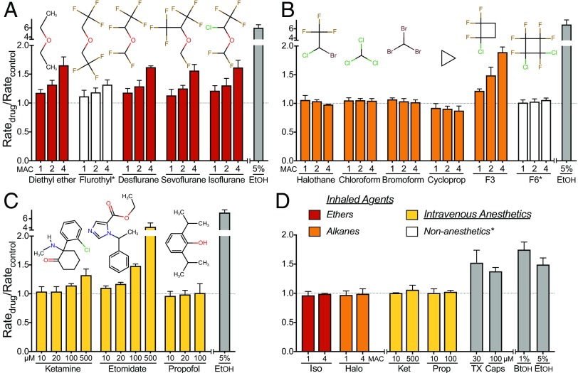
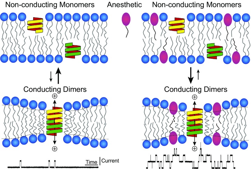
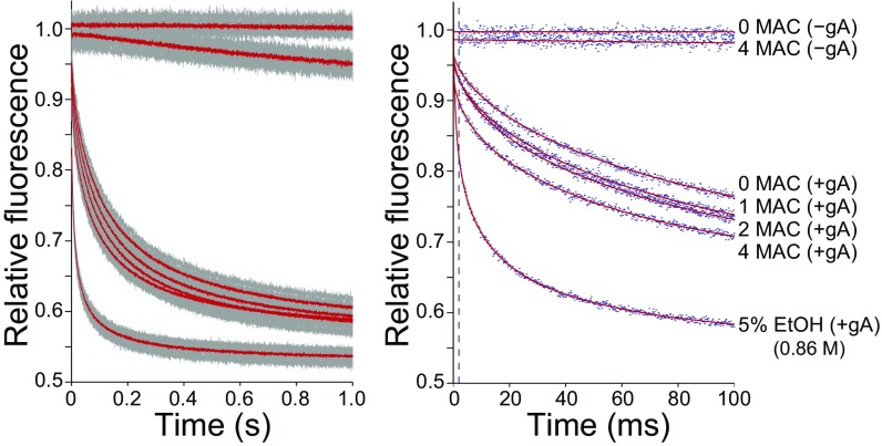
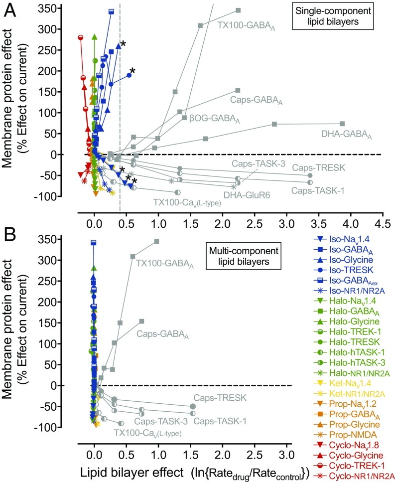
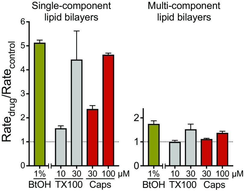
General anesthetics have revolutionized medicine by facilitating invasive procedures, and have thus become essential drugs. However, detailed understanding of their molecular mechanisms remains elusive. A mechanism proposed over a century ago involving unspecified interactions with the lipid bilayer known as the unitary lipid-based hypothesis of anesthetic action, has been challenged by evidence for direct anesthetic interactions with a range of proteins, including transmembrane ion channels. Anesthetic concentrations in the membrane are high (10-100 mM), however, and there is no experimental evidence ruling out a role for the lipid bilayer in their ion channel effects. A recent hypothesis proposes that anesthetic-induced changes in ion channel function result from changes in bilayer lateral pressure that arise from partitioning of anesthetics into the bilayer. We examined the effects of a broad range of chemically diverse general anesthetics and related nonanesthetics on lipid bilayer properties using an established fluorescence assay that senses drug-induced changes in lipid bilayer properties. None of the compounds tested altered bilayer properties sufficiently to produce meaningful changes in ion channel function at clinically relevant concentrations. Even supra-anesthetic concentrations caused minimal bilayer effects, although much higher (toxic) concentrations of certain anesthetic agents did alter lipid bilayer properties. We conclude that general anesthetics have minimal effects on bilayer properties at clinically relevant concentrations, indicating that anesthetic effects on ion channel function are not bilayer-mediated but rather involve direct protein interactions.

Keywords: amphiphiles; anesthetic mechanisms; bilayer modification; gramicidin channel; isoflurane.


Conflict of interest statement

The authors declare no conflict of interest.

Figures

Fig. 1.
Fig. 1.
Effects of general anesthetics on lipid bilayer properties. Normalized fluorescence quench rates of inhaled ether (A) and alkane (B) compounds at concentrations of ∼1, 2, and 4 MAC (minimum alveolar concentration, defined as the concentration that prevents movement in response to a painful stimulus in 50% of subjects, comparable to EC50), and of i.v. anesthetics (C) at 10–500 µM, using single-component lipid bilayer vesicles. White columns represent compounds that do not cause immobility (nonanesthetics*) that were tested at concentrations predicted to produce anesthesia based on their lipid solubility. A normalized quench rate (Ratedrug/Ratecontrol) of 1.0 indicates no significant effect on bulk lipid bilayer properties. Ethanol [EtOH] (gray columns), a known bilayer-modifier at 5% (∼0.86 M), was included as a positive control. Data are expressed as mean ± SD, n = 3–5. [10 and 20 µM values for propofol are from (102).]. (D) Effects of anesthetics on lipid bilayer properties in multicomponent bilayer vesicles. Identical experiments (as in A–C) were performed using LUVs composed of an equimolar mixture of 1,2-dioleoyl-sn-glycero-3-phosphocholine (DC18:1PC), cholesterol and brain sphingomyelin. Normalized fluorescence quench rates of select anesthetics (colored columns) representing each group at both low and high concentrations (1 or 4 MAC for isoflurane [Iso] and halothane [Halo]; 10 or 500 µM for ketamine [Ket]; 10 or 100 µM for propofol [Prop]). Conventional bilayer-modifying molecules (gray columns), such as 30 μM Triton X-100 [TX], 100 μM capsaicin [Caps] and alcohols (1% 1-butanol [BtOH] or 5% ethanol [EtOH]), were included as positive controls, which altered lipid bilayer properties even at low concentrations. Data are expressed as mean ± SD, n = 3–5.
Fig. S1.
Fig. S1.
Hydrophobic coupling between gramicidin and a lipid bilayer. When two gramicidin subunits (yellow and green structures) from opposing bilayer leaflets dimerize, they form a cation selective channel. Amphiphilic molecules such as anesthetics (red symbol) affect the conformational changes of membrane bound proteins by altering lipid bilayer properties and gramicidin serves as a probe to track these changes. An increase in gramicidin activity indicates that the monomer ↔ dimer equilibrium has shifted toward the conducting gramicidin dimers (Lower Right).
Fig. S2.
Fig. S2.
Lipid bilayer-modifying effects of the inhaled anesthetic diethyl ether tested using the gramicidin-based fluorescence assay. (Left) Time course of fluorescence decay of 8-aminonaphthalene-1,3,6-trisulfonic acid (ANTS) over 1 s in the absence (−gA) or presence (+gA) of gramicidin and in the absence or presence of diethyl ether (1, 2 and fourfold multiples of the EC50 for anesthesia defined as minimum alveolar concentration, MAC). Gray dots and solid red lines indicate individual data points and the average of all data, respectively. (Right) The first 100 ms of the normalized fluorescence decay from the same experiment as in the left panel. Blue dots are results from one assay for each condition. Because of the unavoidable dispersity in vesicle sizes, the fluorescence quench time course cannot be described by a single exponential decay, but rather by a stretched exponential decay; the stretched exponential fits to the data (2–100 ms) are denoted by red lines; the dashed vertical line at 2 ms marks the time point at which quench rate was determined. Ethanol (EtOH), a strong bilayer modifier at 5% (∼0.86 M) or (in some experiments) butanol at 1% (∼0.11 M) (27) were included as positive controls.
Fig. 2.
Fig. 2.
Effects of general anesthetics and other amphiphiles on ion channel function compared with their lipid bilayer modifying properties. (A) Plot of anesthetic- and amphiphile-induced changes in specific ion channel function (as a percentage of potentiation or inhibition of ionic current) against changes in lipid bilayer properties measured (or extrapolated) from the fluorescence quench rate in single component DC22:1PC LUVs. The relation between bilayer-modifying effect and alteration of ion-channel function by five representative anesthetics (isoflurane [Iso], halothane [Halo], ketamine [Ket], propofol [Prop], cyclopropane [Cyclo], colored symbols) and other amphiphiles (Triton X-100 [TX100], β-octyl-glucoside [βOG], capsaicin [Caps], docosahexaenoic acid [DHA], gray symbols) is based on results from this study and from published studies on ion channels (, , , , , –122). The horizontal dashed line denotes no change in ion-channel current, and the vertical dashed line shows the threshold for a significant effect on bulk lipid-bilayer properties. All five anesthetics have strong ion channel effects at concentrations at which they have minimal or no bilayer-modifying effects. Gray symbols represent amphiphiles known to strongly alter lipid bilayer properties. A few data points for isoflurane (Iso-Nav1.4, Iso-Glycine and Iso-TRESK, denoted with asterisk) do reach or cross the vertical border, but these bilayer-modifying effects only occur at very high, supratherapeutic concentrations (>4 MAC). (B) Corresponding plot using the changes in fluorescence quench rates for multicomponent lipid bilayer LUVs for isoflurane, halothane, ketamine and propofol (abbreviation and color code as in A), as well as the known bilayer-modifying amphiphiles Triton X-100 and capsaicin (abbreviation as in A; gray symbols). In these multicomponent lipid bilayer experiments, the vertical alignment of the data (corresponding to anesthetics, colored symbols) is much more pronounced compared with the data for single component LUVs (A), confirming that clinical concentrations of anesthetics do not have any lipid bilayer altering effects.
Fig. S3.
Fig. S3.
Effects of “conventional” bilayer-modifying molecules using single- and multicomponent lipid bilayer vesicles. Comparison of normalized fluorescence quench rates of 1-butanol [BtOH] at 1% (∼0.11 M) or Triton X-100 [TX100] and capsaicin [Caps] at low and high concentrations in LUVs formed with either single-component (DC22:1PC) or multicomponent (equimolar mixture of DC18:1PC; cholesterol and sphingomyelin) lipid bilayers.

References

    1. Ueda I, Kamaya H. Kinetic and thermodynamic aspects of the mechanism of general anesthesia in a model system of firefly luminescence in vitro. Anesthesiology. 1973;38(5):425–436. - PubMed
    1. Franks NP, Lieb WR. Where do general anaesthetics act? Nature. 1978;274(5669):339–342. - PubMed
    1. Richards CD, et al. Degenerate perturbations of protein structure as the mechanism of anaesthetic action. Nature. 1978;276(5690):775–779. - PubMed
    1. Franks NP, Lieb WR. Mapping of general anaesthetic target sites provides a molecular basis for cutoff effects. Nature. 1985;316(6026):349–351. - PubMed
    1. Hemmings HC, Jr, et al. Emerging molecular mechanisms of general anesthetic action. Trends Pharmacol Sci. 2005;26(10):503–510. - PubMed

Publication types

MeSH terms