. 2017 Mar 21;114(12):3240-3245.
doi: 10.1073/pnas.1619484114. Epub 2017 Mar 6.

Myosin filament activation in the heart is tuned to the mechanical task

Affiliations

Myosin filament activation in the heart is tuned to the mechanical task

Massimo Reconditi et al. Proc Natl Acad Sci U S A. .

Abstract

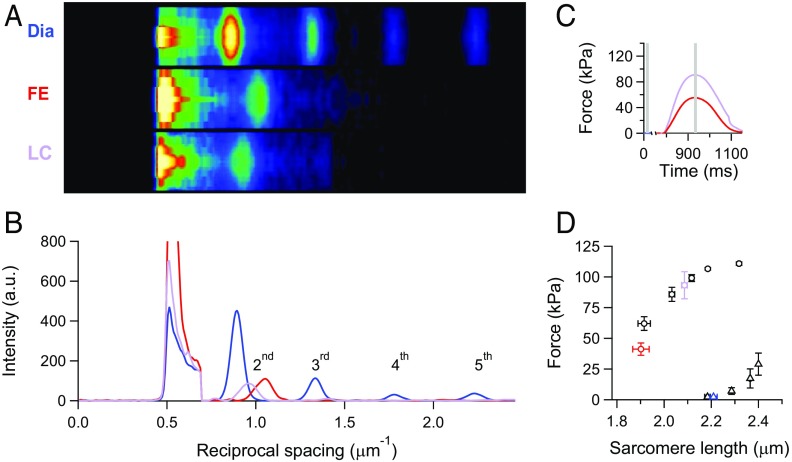
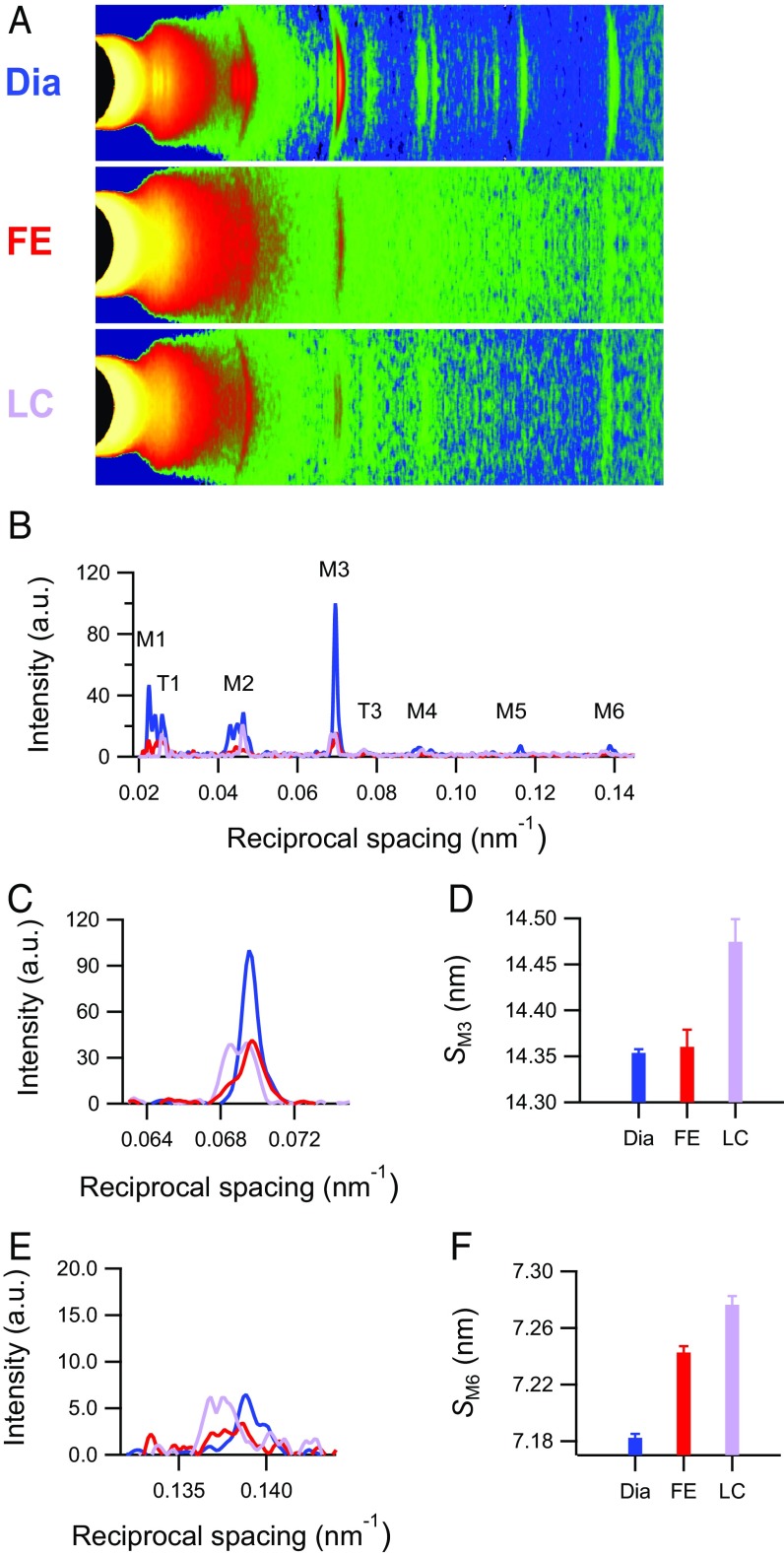
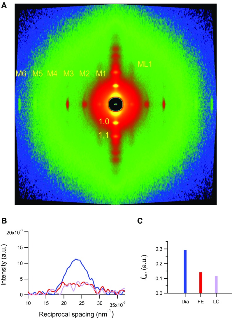
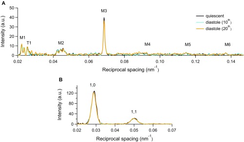
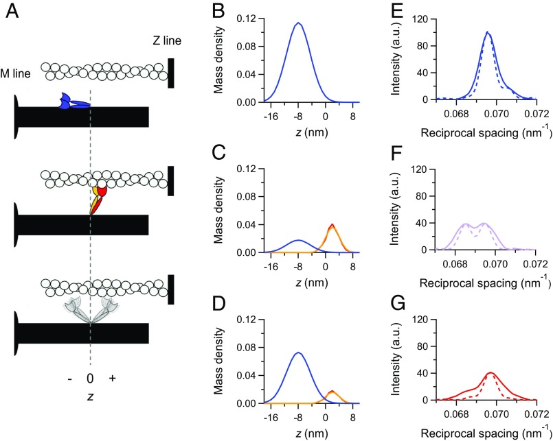
The mammalian heart pumps blood through the vessels, maintaining the dynamic equilibrium in a circulatory system driven by two pumps in series. This vital function is based on the fine-tuning of cardiac performance by the Frank-Starling mechanism that relates the pressure exerted by the contracting ventricle (end systolic pressure) to its volume (end systolic volume). At the level of the sarcomere, the structural unit of the cardiac myocytes, the Frank-Starling mechanism consists of the increase in active force with the increase of sarcomere length (length-dependent activation). We combine sarcomere mechanics and micrometer-nanometer-scale X-ray diffraction from synchrotron light in intact ventricular trabeculae from the rat to measure the axial movement of the myosin motors during the diastole-systole cycle under sarcomere length control. We find that the number of myosin motors leaving the off, ATP hydrolysis-unavailable state characteristic of the diastole is adjusted to the sarcomere length-dependent systolic force. This mechanosensing-based regulation of the thick filament makes the energetic cost of the systole rapidly tuned to the mechanical task, revealing a prime aspect of the Frank-Starling mechanism. The regulation is putatively impaired by cardiomyopathy-causing mutations that affect the intramolecular and intermolecular interactions controlling the off state of the motors.

Keywords: Frank–Starling mechanism; cardiac muscle; heart regulation; myosin filament mechanosensing; small-angle X-ray diffraction.


Conflict of interest statement

The authors declare no conflict of interest.

Figures

Fig. 1.
Fig. 1.
SL measured in a twitching trabecula with ultra-small-angle X-ray diffraction. (A) Meridional slices of 2D diffraction patterns during diastole (Dia) and at the force peak of a systole in either FE or sarcomere LC conditions. Camera length of 30 m; total exposure time of 10 ms for FE and LC and 20 ms for Dia. (B) Meridional intensity profiles from A. Background subtraction starts at ∼0.7 μm−1. Blue, diastole; red, FE twitch; violet, LC twitch. (C) Force of the FE (red) and LC (violet) twitches. Stimulus starts at 775 ms. Gray bars indicate X-ray time windows. (D) Force–SL relations in diastole (triangles) and systole (circles). Black symbols are from ref. ; blue (Dia), red (FE), and violet (LC) symbols are from four trabeculae used in this work. Graph reprinted with permission from ref. .
Fig. S1.
Fig. S1.
Instantaneous force–SL plots for twitches in FE (red) and LC with the feedforward signal either sufficient (black) or insufficient (violet) to prevent shortening. SL changes are recorded on a horizontally mounted trabecula by the striation follower (46). The feedforward signal is an exponential calibrated on the sarcomere shortening recorded in the FE twitch. Circles are the respective Tp values according to the same color code as the traces. The black dashed line is the force–SL relation interpolated on data from Fig. 1D (13). The shaded areas are two ideal loops followed by contractions that start at SL ∼ 2.2 µm in isometric condition and become isotonic when the force attains the level corresponding to the Tp value identified by the color. In terms of the mechanical parameters of the left ventricle beat, the dashed line is the relation between end systolic pressure and end systolic volume, and the loops are two pressure–volume loops starting from the same preload (i.e., end diastolic volume) and with two different afterloads (i.e., aortic pressures). The correspondence between the stress–strain relation (i.e., tension–SL relation) of a myocyte of the ventricular wall and the pressure–volume relation of the whole ventricle has been extensively discussed in ref. .
Fig. 2.
Fig. 2.
X-ray reflections marking the changes in the structure of the myosin filament in a twitching trabecula. (A) Meridional slices and (B) meridional intensity profiles of patterns collected with 1.6-m camera length from four vertically mounted trabeculae. M1–M6 are myosin-based reflections. T1 and T3 indicate troponin-based reflections indexing on a 38-nm axial periodicity. Total exposure times are 150 ms for diastole (Dia), 60 ms for FE, and 90 ms for LC. (C and E) Intensity profiles and (D and F) spacing around the region of M3 and M6 reflections for three conditions as in Fig. 1 (blue, Dia; red, FE; violet, LC).
Fig. S2.
Fig. S2.
Small angle X-ray diffraction from a horizontally mounted trabecula. (A) 2D pattern collected at 1.6 m from the trabecula in diastole (Dia) showing up to the sixth order of the meridional myosin-based reflections, the first-order myosin layer line (ML1), and the equatorial reflections (1,0 and 1,1). Total exposure time was 70 ms. (B) ML1 intensity along the meridional direction in Dia (blue) and the FE (red) and LC (violet) systole. (C) ML1 integrated intensity (same color code as in B).
Fig. S3.
Fig. S3.
Comparison of the X-ray meridional reflections from the rat trabecula and the intact fiber from frog skeletal muscle. Superimposed meridional intensity profiles from a trabecula in diastole (blue; 27 °C) and a resting muscle fiber from Rana esculenta (orange; 4 °C).
Fig. S4.
Fig. S4.
Comparison of X-ray data in the quiescent trabecula and during diastole. Superimposed intensity profiles along the (A) meridional and (B) equatorial axes from the trabecula in the quiescent state (black) and the diastole of the 10th (cyan) and 20th (orange) cycles during 0.5-Hz pacing.
Fig. S5.
Fig. S5.
Results of the simulation with model 1. (A–C) Mass density projection of the myosin motors in the different states for (A) the diastole, (B) the LC twitch, and (C) the FE twitch: blue, motors in the off state; red, actin-attached motors; orange, partner motors. (D–F) M3 intensity profiles calculated from the mass projections in A–C in the corresponding rows (dashed lines) superimposed on the observed intensities (continuous lines; same color code as in Fig. 2C). Details are in the text.
Fig. 3.
Fig. 3.
Load-dependent regulation of the state of myosin motors. (A) Schematic of the motor configurations: blue, off state; red, actin-attached motor of a dimer; orange, its detached partner; gray, detached dimer in the on state. The vertical dashed line intercepts the origin of the axial displacement of the center of mass of the motor (z), which corresponds to the position of the head–rod junction; z is negative for displacements toward the center of the thick filament. (B–D) Axial mass density of the myosin motors in the different states identified by the color code as in A and calculated by the simulation for (B) the diastole, (C) the LC twitch, and (D) the FE twitch. (E–G) Superimposed M3 intensity profiles: observed (continuous lines) and calculated from the mass projections in B–D (dashed lines) (same color code as in Fig. 2C). (C, D, F, and G) The simulated data for the LC and FE twitches are obtained with model 2.

References

    1. Huxley HE, Brown W. The low-angle x-ray diagram of vertebrate striated muscle and its behaviour during contraction and rigor. J Mol Biol. 1967;30(2):383–434. - PubMed
    1. Zoghbi ME, Woodhead JL, Moss RL, Craig R. Three-dimensional structure of vertebrate cardiac muscle myosin filaments. Proc Natl Acad Sci USA. 2008;105(7):2386–2390. - PMC - PubMed
    1. Luther PK, et al. Direct visualization of myosin-binding protein C bridging myosin and actin filaments in intact muscle. Proc Natl Acad Sci USA. 2011;108(28):11423–11428. - PMC - PubMed
    1. Reconditi M, et al. Motion of myosin head domains during activation and force development in skeletal muscle. Proc Natl Acad Sci USA. 2011;108(17):7236–7240. - PMC - PubMed
    1. Stewart MA, Franks-Skiba K, Chen S, Cooke R. Myosin ATP turnover rate is a mechanism involved in thermogenesis in resting skeletal muscle fibers. Proc Natl Acad Sci USA. 2010;107(1):430–435. - PMC - PubMed

Publication types

LinkOut - more resources