Case Reports
. 2017 Feb 20:8:50.
doi: 10.3389/fneur.2017.00050. eCollection 2017.

Remodeling Brain Activity by Repetitive Cervicothoracic Transspinal Stimulation after Human Spinal Cord Injury

Affiliations
Case Reports

Remodeling Brain Activity by Repetitive Cervicothoracic Transspinal Stimulation after Human Spinal Cord Injury

Lynda M Murray et al. Front Neurol. .

Abstract

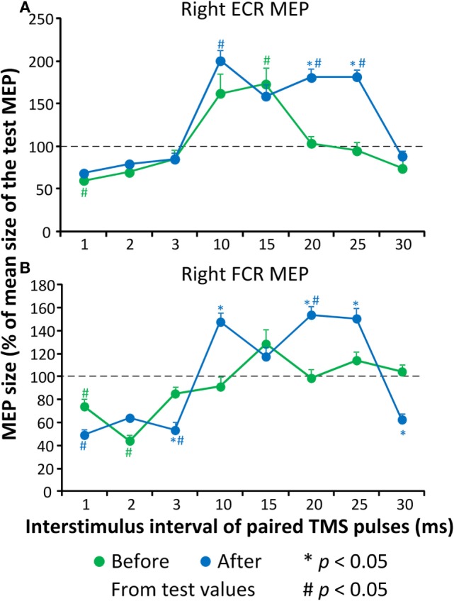
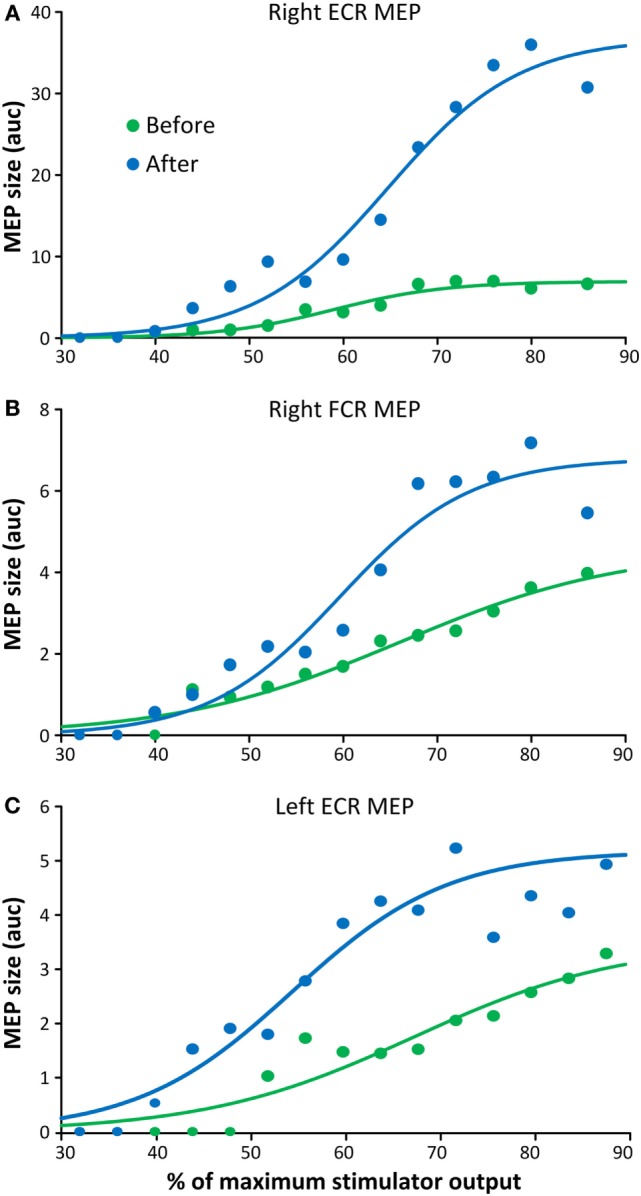
Interventions that can produce targeted brain plasticity after human spinal cord injury (SCI) are needed for restoration of impaired movement in these patients. In this study, we tested the effects of repetitive cervicothoracic transspinal stimulation in one person with cervical motor incomplete SCI on cortical and corticospinal excitability, which were assessed via transcranial magnetic stimulation with paired and single pulses, respectively. We found that repetitive cervicothoracic transspinal stimulation potentiated intracortical facilitation in flexor and extensor wrist muscles, recovered intracortical inhibition in the more impaired wrist flexor muscle, increased corticospinal excitability bilaterally, and improved voluntary muscle strength. These effects may have been mediated by improvements in cortical integration of ascending sensory inputs and strengthening of corticospinal connections. Our novel therapeutic intervention opens new avenues for targeted brain neuromodulation protocols in individuals with cervical motor incomplete SCI.

Keywords: cortical plasticity; corticospinal plasticity; primary motor cortex; repetitive transspinal stimulation; spinal cord injury.


Figures

Figure 1
Figure 1
Protocol of transspinal stimulation for neurorecovery. (A) Repetitive cervicothoracic transspinal stimulation protocol. (B) Illustration of single-pulse transspinal stimulation delivered during the intervention, along with the intensities of daily transspinal stimulation normalized to the baseline transspinal-evoked potential motor threshold. (C) Illustration of single and paired transcranial magnetic stimulation (TMS) pulses for recording motor-evoked potentials (MEPs). Single-pulse TMS at different stimulation intensities was delivered for assembling the MEP recruitment input–output curves. Paired-pulse TMS was used to condition MEPs at different interstimulus intervals (ISIs) of 1, 2, 3, 10, 15, 20, 25, and 30 ms.
Figure 2
Figure 2
Conditioned motor evoked potentials (MEPs) of the right arm. MEPs tested in the resting extensor carpi radialis (ECR) muscle upon single- and paired-pulse transcranial magnetic stimulation (TMS) before (A) and after (B) repetitive cervicothoracic transspinal stimulation. Traces show the averages of 24 test MEPs (green traces) and 12 conditioned MEPs (blue traces) for short- and medium-latency interstimulus intervals.
Figure 3
Figure 3
Cortical excitability measures before and after repetitive cervicothoracic transspinal stimulation. Overall amplitude of extensor carpi radialis (ECR) (A) and flexor carpi radialis (FCR) (B). Motor-evoked potentials (MEPs) from the right arm upon paired-pulse transcranial magnetic stimulation (TMS). Conditioned MEPs are presented as a percentage of the mean size of the homonymous test MEP. Error bars indicate SE. *p < 0.05 for before–after comparisons, #p < 0.05 from homonymous test MEP values.
Figure 4
Figure 4
Corticospinal excitability measures before and after repetitive cervicothoracic transspinal stimulation. Motor-evoked potentials (MEPs) recorded from the right extensor carpi radialis (ECR) (A), right flexor carpi radialis (FCR) (B), and left ECR (C) muscles before and after repetitive transspinal stimulation are depicted as the area under the curve (auc) and are plotted against the percentage of the maximum stimulator output. Before and after repetitive transspinal stimulation, MEP recruitment input–output curves were assembled with single-pulse transcranial magnetic stimulation (TMS) at exactly the same stimulation intensities. A sigmoid fit to the data is also shown. Note the significant increases in MEP sizes after repetitive transspinal stimulation regardless of the stimulation intensity.

Similar articles

Cited by

References

    1. Pascual-Leone A, Valls-Sole J, Wassermann EM, Hallett M. Responses to rapid-rate transcranial magnetic stimulation of the human motor cortex. Brain (1994) 117(4):847–58. - PubMed
    1. Chen R, Classen J, Gerloff C, Celnik P, Wassermann EM, Hallett M, et al. Depression of motor cortex excitability by low-frequency transcranial magnetic stimulation. Neurology (1997) 48(5):1398–403. - PubMed
    1. Rosenzweig ES, Courtine G, Jindrich DL, Brock JH, Ferguson AR, Strand SC, et al. Extensive spontaneous plasticity of corticospinal projections after primate spinal cord injury. Nat Neurosci (2010) 13(12):1505–10.10.1038/nn.2691 - DOI - PMC - PubMed
    1. Courtine G, Song B, Roy RR, Zhong H, Herrmann JE, Ao Y, et al. Recovery of supraspinal control of stepping via indirect propriospinal relay connections after spinal cord injury. Nat Med (2008) 14(1):69–74.10.1038/nm1682 - DOI - PMC - PubMed
    1. Ellaway PH, Catley M, Davey NJ, Kuppuswamy A, Strutton P, Frankel HL, et al. Review of physiological motor outcome measures in spinal cord injury using transcranial magnetic stimulation and spinal reflexes. J Rehabil Res Dev (2007) 44(1):69–76.10.1682/JRRD.2005.08.0140 - DOI - PubMed

Publication types

LinkOut - more resources