Review
. 2017 Feb 20:8:224.
doi: 10.3389/fmicb.2017.00224. eCollection 2017.

Role of Streptococcus pneumoniae Proteins in Evasion of Complement-Mediated Immunity

Affiliations
Review

Role of Streptococcus pneumoniae Proteins in Evasion of Complement-Mediated Immunity

Greiciely O Andre et al. Front Microbiol. .

Abstract

The complement system plays a central role in immune defense against Streptococcus pneumoniae. In order to evade complement attack, pneumococci have evolved a number of mechanisms that limit complement mediated opsonization and subsequent phagocytosis. This review focuses on the strategies employed by pneumococci to circumvent complement mediated immunity, both in vitro and in vivo. At last, since many of the proteins involved in interactions with complement components are vaccine candidates in different stages of validation, we explore the use of these antigens alone or in combination, as potential vaccine approaches that aim at elimination or drastic reduction in the ability of this bacterium to evade complement.

Keywords: Streptococcus pneumoniae; complement system; pneumococcal moonlighting proteins; protein-based vaccines; virulence factors pneumococcal surface proteins.


Figures
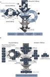
FIGURE 1
FIGURE 1
(A) Complement System activation pathways. The classical pathway is activated by binding of C1q molecules to IgM or IgG. C1s cleaves C4 into C4a and C4b, further degrading C2 when bound with C4b, forming C2b (free molecule) and C2a, which remains bound to C4b forming C4bC2a (C3 convertase of the classical pathway). The lectin pathway is activated in a similar fashion, by binding of MBL molecule to mannose or other sugars on microbial surfaces. It can also be initiated by ficolins or collectins. Upon target binding, MASP-1 autoactivates first, and then it activates MASP-2. MASP-2 then cleaves C4 and both enzymes cleave C2, generating the classical C3 convertase (C4b2a). A third serine protease, MASP-3, is able to activate pro-Factor D (pro-FD) in the resting human blood, favoring alternative complement pathway activation. The Alternative pathway is activated continuously by spontaneous hydrolysis of C3 into C3 (H2O); FB binds to this molecule and is split up by FD, yielding Ba and Bb that remains bound to form C3 (H2O)Bb (the initial alternative pathway C3 convertase). This convertase is able to cleave C3 in the blood, generating C3a and C3b, which attaches to the activating surface and recruits FB. Binding of C3b with the cleavage product of FB, Bb, generates the second C3convertase of the alternative pathway, C3bBb. The amplification loop is initiated when plasmatic C3 is cleaved by C3 convertase from all three pathways yielding more C3b, which may lead to more convertase or promote microbial opsonization. The C5 convertase is formed by association of one C3b molecule to the C3 convertase (in all pathways). This aggregate cleaves C5 molecules into C5a (anaphylatoxin) and C5b, which is the platform for C6 binding. Following the cascade, C7, C8, and C9 (16–18 units) molecules bind to the activated surface to form the membrane attack complex. (B) Pneumococcal virulence factors act on different points of the complement cascades. Each pneumococcal antigen is depicted in relation to the complement components they interact with. PLY, GDAPH and PepO promote activation and depletion of complement components. PspA inhibits activation of both classical (CP) and alternative pathways (AP) C3 convertases; LytA inhibits classical pathway activation. PspC, PepO, Eno, and LytA undermine CP C3 convertase formation. PspC, Phts, LytA, and Tuf impair AP C3 convertase formation, as well as the amplification loop. PGK and PspC inhibit MAC assembly. NanA, BgaA, and StrH remove sialic acids from complement components, limiting complement mediated phagocytosis; they inhibit AP, but the mechanism responsible for the inhibition is not clear. The pneumococcal moonlighting proteins (MP) have indirect influence on complement through their interaction with PLG and consequent degradation of complement components.
FIGURE 2
FIGURE 2
Role of pneumococcal virulence factors in evasion from the Complement System. The polysaccharide capsule prevents binding of IgG, IgM, and CRP to the bacterial surface, as well as C3b and iC3b, thereby impairing complement activation by classical (CP) and alternative pathways (AP). The proteins have been grouped according to their interactions with complement. T indicates direct inhibition by pneumococcal antigens. PspA affects C3 convertase formation by interfering with FB and prevents CRP binding to phophocholine on the bacterial wall, thus inhibiting activation of the classical pathway. PspC binds to FH and C4BP promoting the inhibition of C3 convertase formation and accelerating C3 convertase decay. PspC also cleaves C3 molecules generating products that cannot activate complement. Binding of PspC to Vitronectin reduces MAC formation. Phts bind to FH and cleave C3. LytA binding to FH and C4BP reduces C3 convertase formation and promotes its dissociation. It also inhibits the interaction between CRP and C1q. In addition, LytA is able to split up C3b and iC3b. PLY released from the bacterium activates the classical pathway through interactions with IgG, C1q, and L-ficolin, and depletes complement. The exoglycosidases NanA, BgaA, and StrH remove sialic acid (SA) from complement components; also, SA favors FH activity in regulation of C3 convertase. PepO binds to C1q and activates complement when released from the pneumococcal surface, leading to depletion of complement components. PepO binding to C4BP results in down regulation of classical pathway activation. Eno decreases C3 convertase formation by binding to C4BP. GAPDH on its free form or attached to the bacterial surface binds to C1q, likely promoting complement activation. PGK impairs MAC assembly by binding to C5, C7, and C9. Tuf binds to FH and FHL-1 inhibiting formation and accelerating dissociation of C3 convertase. Tuf can also bind to CFHR-1, but the implications of this interaction are not completely elucidated. The main result of such interactions is an impaired bacterial phagocytosis. Also, pneumococcal proteins use complement molecules as bridges to interact with host receptors and favor bacterial adherence and invasion. PspC exploits FH to adhere and invade host cells. PepO uses C1q to increase bacterial adherence. RrgA improves colonization by binding to complement receptor 3 (CR3). Finally, the ability to interact with PLG – as demonstrated for PepO, Eno, and GAPDH with PLG was shown to degrade the host extracellular matrix components (ECM) and to improve invasion, while PepO, PGK and Tuf bound to PLG inactivate C3 and C3b.

References

    1. Aberdein J. D., Cole J., Bewley M. A., Marriott H. M., Dockrell D. H. (2013). Alveolar macrophages in pulmonary host defence the unrecognized role of apoptosis as a mechanism of intracellular bacterial killing. Clin. Exp. Immunol. 174 193–202. 10.1111/cei.12170 - DOI - PMC - PubMed
    1. Abeyta M., Hardy G. G., Yother J. (2003). Genetic alteration of capsule type but not PspA type affects accessibility of surface-bound complement and surface antigens of Streptococcus pneumoniae. Infect. Immun. 71 218–225. 10.1128/IAI.71.1.218-225.2003 - DOI - PMC - PubMed
    1. Adamou J. E., Heinrichs J. H., Erwin A. L., Walsh W., Gayle T., Dormitzer M., et al. (2001). Identification and characterization of a novel family of pneumococcal proteins that are protective against sepsis. Infect. Immun. 69 949–958. 10.1128/IAI.69.2.949-958.2001 - DOI - PMC - PubMed
    1. Agarwal V., Ahl J., Riesbeck K., Blom A. M. (2013a). An alternative role of C1q in bacterial infections: facilitating Streptococcus pneumoniae adherence and invasion of host cells. J. Immunol. 191 4235–4245. 10.4049/jimmunol.1300279 - DOI - PubMed
    1. Agarwal V., Asmat T. M., Luo S., Jensch I., Zipfel P. F., Hammerschmidt S. (2010). Complement regulator factor H mediates a two-step uptake of Streptococcus pneumoniae by human cells. J. Biol. Chem. 285 23486–23495. 10.1074/jbc.M110.142703 - DOI - PMC - PubMed

LinkOut - more resources