. 2017 Mar 1:10:9.
doi: 10.1186/s13072-017-0116-6. eCollection 2017.

Assessing histone demethylase inhibitors in cells: lessons learned

Affiliations

Assessing histone demethylase inhibitors in cells: lessons learned

Stephanie B Hatch et al. Epigenetics Chromatin. .

Abstract

Background: Histone lysine demethylases (KDMs) are of interest as drug targets due to their regulatory roles in chromatin organization and their tight associations with diseases including cancer and mental disorders. The first KDM inhibitors for KDM1 have entered clinical trials, and efforts are ongoing to develop potent, selective and cell-active 'probe' molecules for this target class. Robust cellular assays to assess the specific engagement of KDM inhibitors in cells as well as their cellular selectivity are a prerequisite for the development of high-quality inhibitors. Here we describe the use of a high-content cellular immunofluorescence assay as a method for demonstrating target engagement in cells.

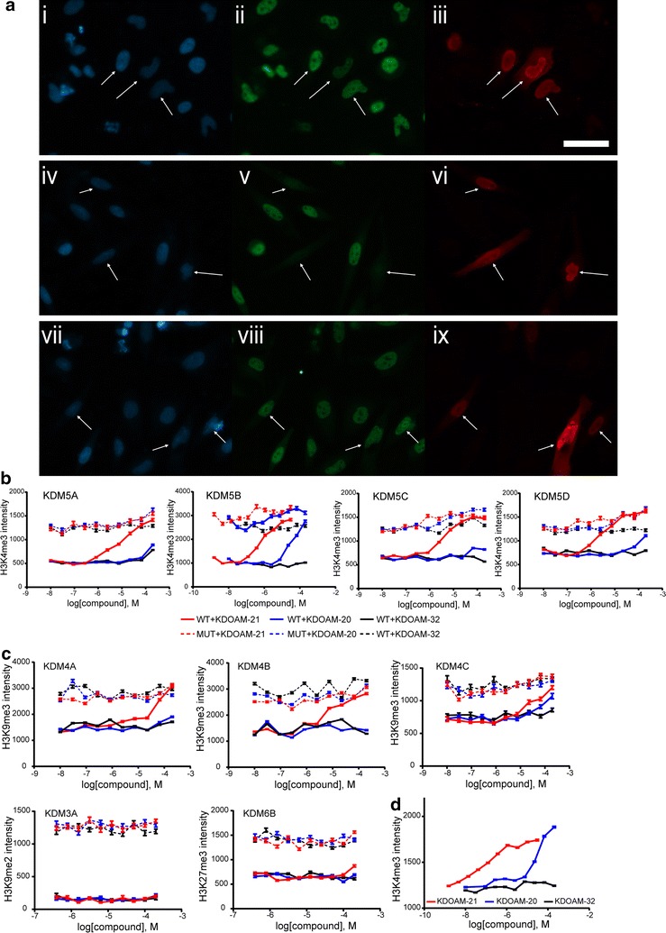
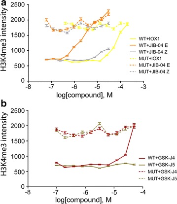
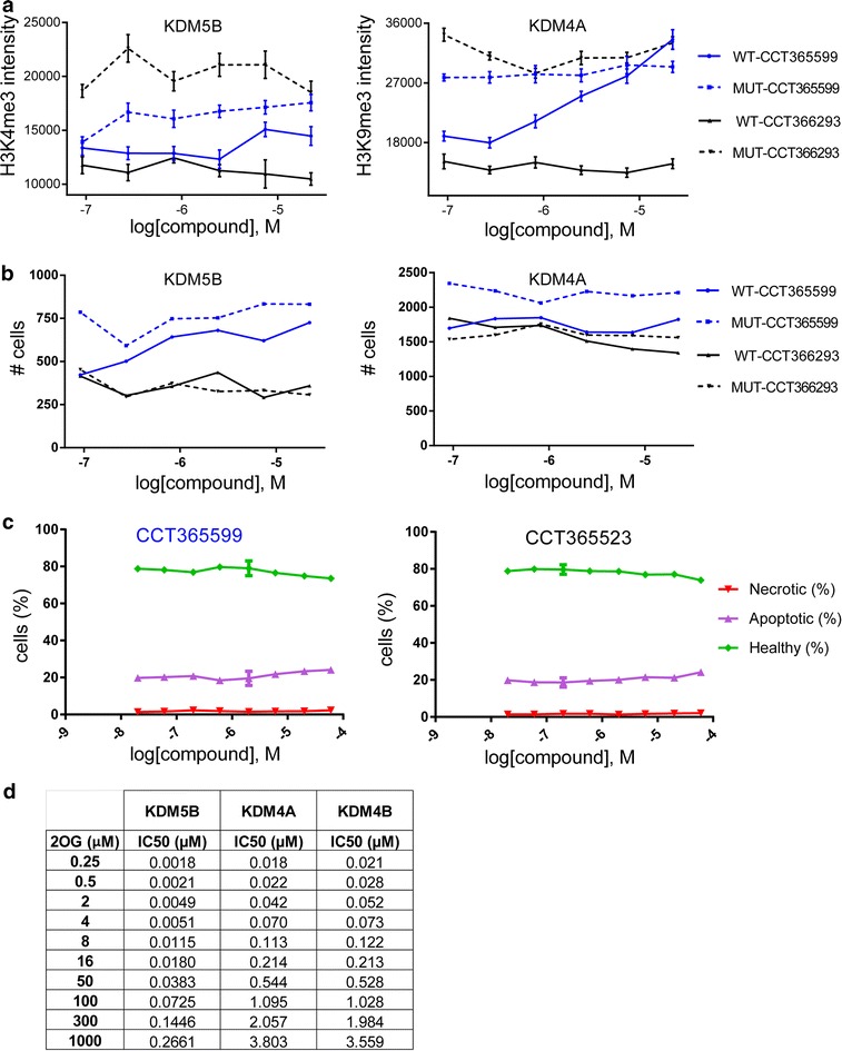
Results: A panel of assays for the Jumonji C subfamily of KDMs was developed to encompass all major branches of the JmjC phylogenetic tree. These assays compare compound activity against wild-type KDM proteins to a catalytically inactive version of the KDM, in which residues involved in the active-site iron coordination are mutated to inactivate the enzyme activity. These mutants are critical for assessing the specific effect of KDM inhibitors and for revealing indirect effects on histone methylation status. The reported assays make use of ectopically expressed demethylases, and we demonstrate their use to profile several recently identified classes of KDM inhibitors and their structurally matched inactive controls. The generated data correlate well with assay results assessing endogenous KDM inhibition and confirm the selectivity observed in biochemical assays with isolated enzymes. We find that both cellular permeability and competition with 2-oxoglutarate affect the translation of biochemical activity to cellular inhibition.

Conclusions: High-content-based immunofluorescence assays have been established for eight KDM members of the 2-oxoglutarate-dependent oxygenases covering all major branches of the JmjC-KDM phylogenetic tree. The usage of both full-length, wild-type and catalytically inactive mutant ectopically expressed protein, as well as structure-matched inactive control compounds, allowed for detection of nonspecific effects causing changes in histone methylation as a result of compound toxicity. The developed assays offer a histone lysine demethylase family-wide tool for assessing KDM inhibitors for cell activity and on-target efficacy. In addition, the presented data may inform further studies to assess the cell-based activity of histone lysine methylation inhibitors.

Keywords: 2-Oxoglutarate oxygenases; Apoptosis; Cell proliferation; Chromatin; Epigenetics; Histone lysine demethylase; Immunofluorescence; Toxicity.


Figures

Fig. 1
Fig. 1
Position of KDMs in phylogenetic tree of 2-OG-dependent oxygenases. a Phylogenetic tree of the catalytic domains of human 2-OG oxygenases adapted according to [5]. The different subfamilies of KDMs are indicated. Non-KDM oxygenases are in grey. Proteins for which IF assays are described herein are highlighted in red. b Domain organization of 2-OG-dependent oxygenases for which IF assays are presented. The identities of the different domains are: JmjC Jumonji C domain, JmjN Jumonji N domain, PHD plant homeodomain, TDR tudor domain, C5HC2 zinc finger C5HC2 type, LRR leucine-rich repeat, TCZ treble-clef zinc finger domain
Fig. 2
Fig. 2
Immunofluorescence assay assessing and comparing potencies of inhibitors in cells. a Widefield fluorescence imaging of HeLa cells after dosing with inhibitor, fixing and staining with i, iv, vii DAPI (blue), ii, v, viii histone antibody for H3K4me3 (green), and iii, vi, ix a FLAG-tag antibody that demarcates cells overexpressing KDM5B (red). Cells overexpressing the wild-type (WT) KDM were treated with the inhibitor KDOAM-21 (iiii) or with DMSO (ivvi); controls cells overexpressing an inactive mutant (MUT) were treated with DMSO (viiix). Arrowheads indicate KDM overexpressing cells. The scalebar represents 50 µm, b–d measurement of the average histone mark intensity in the transfected HeLa cells allows quantification of inhibitor potency against each target. KDOAM-21 (red), KDOAM-20 (blue) and the inactive control KDOAM-32 (black) were tested on multiple targets, b illustrates the characteristic dose–response curves of the potent KDOAM-21, weak KDOAM-20 and inactive KDOAM-32 compounds on KDM5 family members. The wild type (WT) and catalytically inactive mutant (MUT) of each target are shown as bold and dashed lines, respectively. The baseline for the mutant is of similar intensity as the nontransfected cells and approximately represents complete inhibition of the target demethylase activity, c selectivity of KDOAM-20 and KDOAM-21 against KDM5 is further demonstrated in cells by their weaker potency against other demethylases, i.e. members of the KDM3, KDM4 and KDM6 families. In d, KDOAM-21 and, to a lesser extent, KDOAM-20 are shown to have activity against endogenous KDMs as seen by changes in H3K4me3 in nontransfected cells. KDOAM-32 retains its inactivity
Fig. 3
Fig. 3
Effect of different KDM5 inhibitors on histone tail methylation status in HeLa cells. a Immunofluorescence assays of cells transfected with KDM5B and treated with the indicated inhibitors CPI-455 and KDOAM-32, respectively. Effect on H3K4me3 methylation is shown to be dose dependent in the upper panel. Number of cells in 20 fields, based on DAPI staining, is shown in the lower panel, b immunofluorescence assays of cells transfected with KDM5B and treated with inhibitors KDIPP15 or KDIPP51, as indicated. The dose-dependent effect on H3K4me3 methylation is shown in the upper panel. Number of cells in 20 fields, based on DAPI staining, is shown in the lower panel
Fig. 4
Fig. 4
Effect of various KDM inhibitors described in the literature on KDM5B-mediated H3K4me3 methylation in HeLa cells. a Effect of Pan-KDM inhibitors IOX1 [30] and JIB-04 E as well as inactive control compound JIB-04 Z [16] on KDM5B-mediated H3K4me3 demethylation, b effect of KDM6A/B inhibitor GSK-J4 and inactive control compound GSK-J5 [21, 31] on KDM5B-mediated H3K4me3 demethylation. Data represent the average and SEM of at least 100 cells
Fig. 5
Fig. 5
Apoptosis induced by different mechanisms is accompanied by methylation mark changes. a Images of HeLa cells treated with 100 nM paclitaxel for 24 h and stained with DAPI and a specific antibody for H3K4me3. The phase contrast image is shown to the left, blue DAPI nuclear stain in the middle and yellow H3K4me3 to the right. Arrows indicate apoptotic cells lacking the H3K4me3 mark, b number of HeLa cells treated with doxorubicin or paclitaxel in a dose-dependent manner based on counting of 12 fields, c immunofluorescence assay showing the effect of doxorubicin or paclitaxel treatment on H3K4me3 mark, d immunofluorescence assay showing the effect of doxorubicin or paclitaxel treatment on H3K27me3 mark, H3K9me2 mark and H3K36me2 mark, respectively
Fig. 6
Fig. 6
Cytotoxic activity of different KDM inhibitors in live cells. a High-content images of HeLa cells treated with 100 nM paclitaxel for 24 h and stained with Annexin V (green) and Yo-Pro (red). Apoptotic cells were defined as Annexin V positive with or without Yo-Pro 3 uptake; necrotic cells were defined as Yo-Pro 3 positive; and healthy cells were defined as Annexin V and Yo-Pro 3 negative, b percentage of healthy, apoptotic and necrotic HeLa cells treated with doxorubicin, paclitaxel or staurosporine in a dose-dependent manner, c percentage of healthy, apoptotic and necrotic HeLa cells treated with KDM inhibitors of the KDOAM series  or CPI-455 for 24 h in a dose-dependent manner, c immunofluorescence assay showing the effect of doxorubicin or paclitaxel treatment on H3K4me3 mark, d percentage of healthy, apoptotic and necrotic HeLa cells treated with compounds of the KDIPP series for 24 h in a dose-dependent manner, e percentage of healthy, apoptotic and necrotic HeLa cells treated with KDIPP15 (66 µM) or KDIPP51 (60 µM) for 24 h. Results are shown as mean ± SD from triplicates of two independent experiments
Fig. 7
Fig. 7
Cytotoxic activity of different KDM inhibitors in live cells and 2-OG dependency of KDM inhibition in vitro. Immunofluorescence assays of cells transfected with KDM5B or KDM4A and treated with the newly developed inhibitors CCT365599 and CCT366293, respectively. Effect on H3K4me3 methylation is shown in the left panel in a dose-dependent manner and on H3K9me3 in the right panel. Number of cells in 20 fields, based on DAPI staining, is shown in b, c. Percentage of healthy, apoptotic and necrotic HeLa cells treated with CCT365599 and CCT366293, respectively, in a dose-dependent manner, d IC50 values in dependency of different 2-OG concentrations determined in AlphaScreen assay for different KDMs

References

    1. Tessarz P, Kouzarides T. Histone core modifications regulating nucleosome structure and dynamics. Nat Rev Mol Cell Biol. 2014;15(11):703–708. doi: 10.1038/nrm3890. - DOI - PubMed
    1. Lawrence M, Daujat S, Schneider R. Lateral thinking: how histone modifications regulate gene expression. Trends Genet. 2016;32(1):42–56. doi: 10.1016/j.tig.2015.10.007. - DOI - PubMed
    1. Sen N. Epigenetic regulation of memory by acetylation and methylation of chromatin: implications in neurological disorders, aging, and addiction. Neuromol Med. 2015;17(2):97–110. doi: 10.1007/s12017-014-8306-x. - DOI - PubMed
    1. Muller S, Filippakopoulos P, Knapp S. Bromodomains as therapeutic targets. Expert Rev Mol Med. 2011;13:e29. doi: 10.1017/S1462399411001992. - DOI - PMC - PubMed
    1. Johansson C, Tumber A, Che K, Cain P, Nowak R, Gileadi C, Oppermann U. The roles of Jumonji-type oxygenases in human disease. Epigenomics. 2014;6(1):89–120. doi: 10.2217/epi.13.79. - DOI - PMC - PubMed

Publication types

MeSH terms