

Pulmonary Vein Thrombosis: A Recent Systematic Review
- PMID: 28265529
- PMCID: PMC5323025
- DOI: 10.7759/cureus.993
Pulmonary Vein Thrombosis: A Recent Systematic Review
Abstract
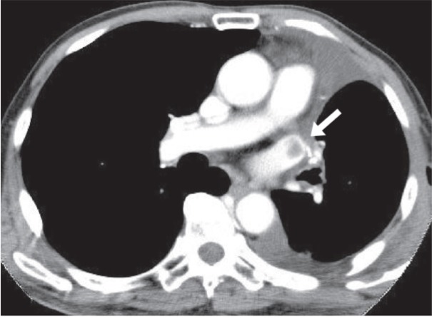
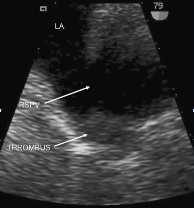
The pulmonary veins (PVs) are the most proximal source of arterial thromboembolism. Pulmonary vein thrombosis (PVT) is a rare but potentially lethal disease; its incidence is unclear, as most of the literature includes case reports. It most commonly occurs as a complica-tion of malignancy, post lung surgery, or atrial fibrillation and can be idiopathic in some cases. Most patients with PVT are commonly asymptomatic or have nonspecific symptoms such as cough, hemoptysis, and dyspnea from pulmonary edema or infarction. The thrombi are typically detected using a variety of imaging modalities including transesophageal echocardiogram (TEE), computed tomography (CT) scanning, magnetic resonance imaging (MRI), or pulmonary angiog-raphy. Treatment should be determined by the obstructing pathological finding and can include antibiotic therapy, anticoagulation, thrombectomy, and/or pulmonary resection. The delay in diagnosing this medical entity can lead to complications including pulmonary infarction, pulmonary edema, right ventricular failure, allograft failure, and peripheral embolism resulting in limb ischemia, stroke, and renal infarction (RI).
Keywords: pulmonary vein; thrombosis.
Conflict of interest statement
The authors have declared that no competing interests exist.
Figures

References
-
- Pulmonary vein thrombosis. Kim NH, Roldan CA, Shively BK. Chest. 1993;104:624–626. - PubMed
-
- Pulmonary vein thrombosis and peripheral embolization. Garcia M, Rodriguez L, Vandervoort P. http://www.ncbi.nlm.nih.gov/pubmed/8617103. Chest. 1996;109:846–847. - PubMed
-
- Pulmonary vein thrombosis associated with a large hiatal hernia. Saoraya J, Inboriboon PC. J Emerg Med. 2013;44:299–301. - PubMed
-
- Idiopathic pulmonary vein thrombosis. Mumoli N, Cei M. J Emerg Med. 2012;42:182–183. - PubMed
Publication types
LinkOut - more resources
Full Text Sources
Other Literature Sources
Research Materials