

Radiolabeled pertuzumab for imaging of human epidermal growth factor receptor 2 expression in ovarian cancer
- PMID: 28265738
- PMCID: PMC5471126
- DOI: 10.1007/s00259-017-3663-y
Radiolabeled pertuzumab for imaging of human epidermal growth factor receptor 2 expression in ovarian cancer
Abstract
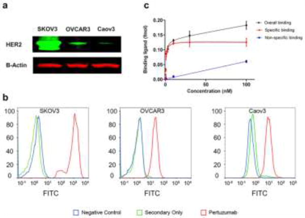
Purpose: Human epidermal growth factor receptor 2 (HER2) is over-expressed in over 30% of ovarian cancer cases, playing an essential role in tumorigenesis and metastasis. Non-invasive imaging of HER2 is of great interest for physicians as a mean to better detect and monitor the progression of ovarian cancer. In this study, HER2 was assessed as a biomarker for ovarian cancer imaging using 64Cu-labeled pertuzumab for immunoPET imaging.
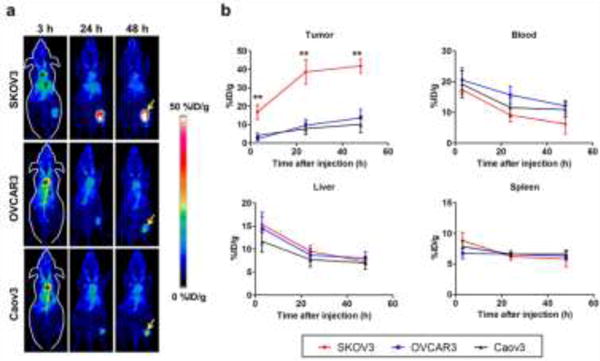
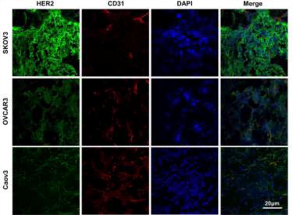
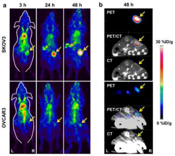
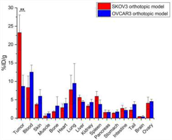
Methods: HER2 expression and binding were examined in three ovarian cancer cell lines (SKOV3, OVCAR3, Caov3) using in vitro techniques, including western blot and saturation binding assays. PET imaging and biodistribution studies in subcutaneous models of ovarian cancer were performed for non-invasive in vivo evaluation of HER2 expression. Additionally, orthotopic models were employed to further validate the imaging capability of 64Cu-NOTA-pertuzumab.
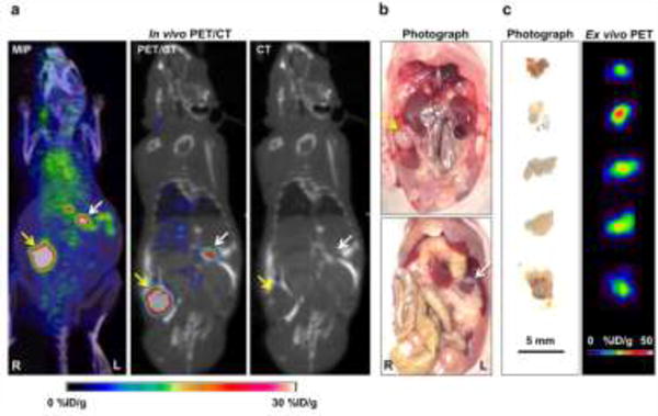
Results: HER2 expression was highest in SKOV3 cells, while OVCAR3 and Caov3 displayed lower HER2 expression. 64Cu-NOTA-pertuzumab showed high specificity for HER2 (Ka = 3.1 ± 0.6 nM) in SKOV3. In subcutaneous tumors, PET imaging revealed tumor uptake of 41.8 ± 3.8, 10.5 ± 3.9, and 12.1 ± 2.3%ID/g at 48 h post-injection for SKOV3, OVCAR3, and Caov3, respectively (n = 3). In orthotopic models, PET imaging with 64Cu-NOTA-pertuzumab allowed for rapid and clear delineation of both primary and small peritoneal metastases in HER2-expressing ovarian cancer.
Conclusions: 64Cu-NOTA-pertuzumab is an effective PET tracer for the non-invasive imaging of HER2 expression in vivo, rendering it a potential tracer for treatment monitoring and improved patient stratification.
Keywords: Human epidermal growth factor receptor 2 (HER2); Molecular imaging; Ovarian cancer; Pertuzumab; Positron emission tomography (PET).
Conflict of interest statement
Figures
Similar articles
-
Imaging of human epidermal growth factor receptor type 2 expression with 18F-labeled affibody molecule ZHER2:2395 in a mouse model for ovarian cancer.J Nucl Med. 2012 Jan;53(1):146-53. doi: 10.2967/jnumed.111.093047. Epub 2011 Dec 15. J Nucl Med. 2012. PMID: 22173842
-
Development and preclinical studies of 64Cu-NOTA-pertuzumab F(ab')2 for imaging changes in tumor HER2 expression associated with response to trastuzumab by PET/CT.MAbs. 2017 Jan;9(1):154-164. doi: 10.1080/19420862.2016.1255389. Epub 2016 Nov 4. MAbs. 2017. PMID: 27813707 Free PMC article.
-
64Cu-Labeled Trastuzumab Fab-PEG24-EGF Radioimmunoconjugates Bispecific for HER2 and EGFR: Pharmacokinetics, Biodistribution, and Tumor Imaging by PET in Comparison to Monospecific Agents.Mol Pharm. 2017 Feb 6;14(2):492-501. doi: 10.1021/acs.molpharmaceut.6b00963. Epub 2017 Jan 18. Mol Pharm. 2017. PMID: 28049295
-
Radiolabeled affibody-albumin bioconjugates for HER2-positive cancer targeting.Bioconjug Chem. 2011 Mar 16;22(3):413-21. doi: 10.1021/bc100432h. Epub 2011 Feb 7. Bioconjug Chem. 2011. PMID: 21299201 Free PMC article.
-
Exploring utilities of [ 64 Cu]Cu-DOTA-trastuzumab immunoPET in breast cancer: a systematic review and meta-analysis.Nucl Med Commun. 2025 Apr 1;46(4):277-284. doi: 10.1097/MNM.0000000000001949. Epub 2025 Jan 21. Nucl Med Commun. 2025. PMID: 39834168
Cited by
-
Dual-labeled pertuzumab for multimodality image-guided ovarian tumor resection.Am J Cancer Res. 2019 Jul 1;9(7):1454-1468. eCollection 2019. Am J Cancer Res. 2019. PMID: 31392081 Free PMC article.
-
Review: Radionuclide Molecular Imaging Targeting HER2 in Breast Cancer with a Focus on Molecular Probes into Clinical Trials and Small Peptides.Molecules. 2021 Oct 27;26(21):6482. doi: 10.3390/molecules26216482. Molecules. 2021. PMID: 34770887 Free PMC article. Review.
-
Imaging of Preclinical Endometrial Cancer Models for Monitoring Tumor Progression and Response to Targeted Therapy.Cancers (Basel). 2019 Nov 27;11(12):1885. doi: 10.3390/cancers11121885. Cancers (Basel). 2019. PMID: 31783595 Free PMC article. Review.
-
ImmunoPET imaging of CD38 expression in hepatocellular carcinoma using 64Cu-labeled daratumumab.Am J Transl Res. 2019 Sep 15;11(9):6007-6015. eCollection 2019. Am J Transl Res. 2019. PMID: 31632568 Free PMC article.
-
ImmunoPET: Antibody-Based PET Imaging in Solid Tumors.Front Med (Lausanne). 2022 Jun 28;9:916693. doi: 10.3389/fmed.2022.916693. eCollection 2022. Front Med (Lausanne). 2022. PMID: 35836956 Free PMC article. Review.
References
-
- Berchuck A, Kamel A, Whitaker R, Kerns B, Olt G, Kinney R, et al. Overexpression of Her-2/Neu Is Associated with Poor Survival in Advanced Epithelial Ovarian-Cancer. Cancer Res. 1990;50:4087–91. - PubMed
MeSH terms
Substances
Grants and funding
LinkOut - more resources
Full Text Sources
Other Literature Sources
Medical
Research Materials
Miscellaneous
