. 2017 Jun;230(6):842-858.
doi: 10.1111/joa.12601. Epub 2017 Mar 7.

A detailed musculoskeletal study of a fetus with anencephaly and spina bifida (craniorachischisis), and comparison with other cases of human congenital malformations

Affiliations

A detailed musculoskeletal study of a fetus with anencephaly and spina bifida (craniorachischisis), and comparison with other cases of human congenital malformations

Malak A Alghamdi et al. J Anat. 2017 Jun.

Abstract

Few descriptions of the musculoskeletal system of humans with anencephaly or spina bifida exist in the literature. Even less is published about individuals in which both phenomena occur together, i.e. about craniorachischisis. Here we provide a detailed report on the musculoskeletal structures of a fetus with craniorachischisis, as well as comparisons with the few descriptions for anencephaly and with musculoskeletal anomalies found in other congenital malformations. We focused in particular on the comparison with trisomies 13, 18, and 21 because neural tube defects have been associated with such chromosomal defects. Our results showed that many of the defects found in the fetus with craniorachischisis are similar not only to anomalies previously described in the available works on musculoskeletal phenotypes seen in fetuses with anencephaly and spina bifida, but also to a wide range of other different conditions/syndromes including trisomies 13, 18 and 21, and cyclopia. The fact that similar anomalies are seen commonly not only in a wide range of different syndromes, but also as variants of the normal human population and as the 'normal' phenotype of other animals, supports Pere Alberch's unfortunately named idea of a 'logic of monsters'. That is, it supports the idea that development is so constrained that both in 'normal' and abnormal development one sees certain outcomes being produced again and again because ontogenetic constraints only allow a few possible outcomes, thus also leading to cases where the anatomical defects of some organisms are similar to the 'normal' phenotype of other organisms. In fact, this applies not only to specific anomalies but also to general patterns, such as the fact that in pathological conditions affecting different regions of the body, one consistently sees more defects on the upper limbs than on the lower limbs. Such general patterns are, again, seen in the fetus examined for this study, which had 29 muscle anomalies on the right upper limb and 22 muscle anomalies on the left upper limb, vs. seven muscle anomalies on the right lower limb and two on the left lower limb. It is therefore hoped that this work, which is part of our effort to describe and compile information on human musculoskeletal defects found in a wide range of conditions, will contribute not only to a better understanding of craniorachischisis in particular and of human congenital malformations in general, but also to broader discussions on the fields of comparative anatomy, and developmental and evolutionary biology.

Keywords: anencephaly; birth defects; bones; comparative anatomy; craniorachischisis; fetus; human anatomy; muscles.


Figures

Figure 1
Figure 1
Illustration of the hinge points model of bending of the chick neural plate. Neuroepithelial cell wedging within the hinge points is indicated by red (median hinge point) and blue (dorsolateral hinge points). Arrows indicate mediolateral expansion of the epidermal ectoderm; single asterisk indicates furrowing associated with the median hinge point. Double asterisks indicate furrowing associated with the dorsolateral hinge points. ee, epidermal ectoderm; n, notochord (modified from Colas & Schoenwolf, 2001).
Figure 2
Figure 2
Diagram showing the two models of anterior neural tube closure: (A) the ‘zipper’ or continuous closure, (B) and the multi‐sites closure pattern. Closure I starts in the cervical region and extends bilaterally; closure II begins at the prosencephalic‐mesencephalic (forebrain‐midbrain) border and extends bilaterally; closure III is initiated adjacent to the stomodeum (precursor of the mouth and the anterior lobe of the pituitary gland) and extends caudally to meet closure II; and closure IV takes place above the rhombencephalon (hindbrain) and extends to meet closure II. (Modified from Golden & Chernoff, 1995).
Figure 3
Figure 3
Posterior view of the fetus with craniorachischisis, showing the NTD with open cranium and spinal cord (right figure), and CT scan of the skeletal system (left figure). Scale bar: 1 cm.
Figure 4
Figure 4
Anomalies of the craniorachischisis fetus in anterior view. Upper figure: CT scan of the head showing the facial bones and the associated deformities. Lower figure: in situ view of the specimen with obvious malformations indicated. Scale bar in the lower figure: 1 cm.
Figure 5
Figure 5
CT scans of the fetus with craniorachischisis, showing the bones of the cranial vault. In this and all subsequent figures, as well as in Fig. 4: Ant, anterior; Inf, inferior; Lat, lateral; Med, medial; Post, posterior; Sup, superior. Scale bar: 1 cm.
Figure 6
Figure 6
CT scans of anterior (upper figure) and lateral views (lower figures) of the fetus with craniorachischisis, showing the facial bones.
Figure 7
Figure 7
Anterior views of the neck of the fetus with craniorachischisis, showing absence of hyoid bone and presence of a hyoid arcade instead, which is somewhat similar to that often present in non‐human mammals (upper figure), and thyroid and cricoid cartilages that are apparently normal (lower figure). The hyoid arcade was connected to hypobranchial muscles, which are shown in more detail in Fig. 13. Lig, ligament; Lt, left; m, muscles; n, nerve. Scale bar: 1 cm.
Figure 8
Figure 8
CT of lateral view of the right upper limb of the fetus with craniorachischisis, showing bone malformations.
Figure 9
Figure 9
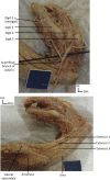
Anterior views of the right arm showing a dorsoepitrochlearis muscle – a muscle normally present in non‐human mammals – fused to the inner side of the pectoralis major (upper figure) and the short head of the biceps brachii, the coracobrachialis, and the pectoralis minor attached to the lesser tubercle of the humerus (lower figure). Scale bar in the upper figure: 1 cm.
Figure 10
Figure 10
Posterior view of the right forearm and hand of the fetus with craniorachischisis showing the superficial nerves and muscles and the digits. Scale bar: 1 cm.
Figure 11
Figure 11
Anterior view of the right hand of the fetus with craniorachischisis showing hypothenar muscles (upper figure) and dorsal (DAB) and palmar (PAD) interossei muscles (lower figure). Note that, contrary to the previous figures, the photographs, imaging, and labeling were done with software connected to microscope (see Material and Methods). AbdDM, abductor digiti minimi; ODM, opponens digiti minimi; FDM, flexor digiti minimi. Scale bar: 1 cm.
Figure 12
Figure 12
Lateral view of the face of the left side of the body fetus with craniorachischisis, showing some facial muscles. Lt, left side.
Figure 13
Figure 13
Anterior views of the neck of the fetus with craniorachischisis, showing the muscles of the neck, including the intermediate tendon between the anterior and posterior digastric muscles. Note that the right geniohyoid muscle is apparently missing. Lt, left; rt, right. Scale bar: 1 cm.
Figure 14
Figure 14
Inferior (upper figure) and anterior (lower figure) views of the fetus with craniorachischisis, showing the tongue and associated muscles, which include the pharyngeal palatoglossal muscle. Scale bar: 1 cm.

Similar articles

Cited by

References

    1. Alberch P (1989) The logic of monsters: evidence for internal constraint in development and evolution. Geobios 22, 21–57.
    1. Bardeen CR, Lewis WH (1901) Development of the limbs, body‐wall and back in man. Am J Anat 1, 1–35.
    1. Buckwalter JA, Flatt AE, Shurr DG, et al. (1981) The absent fifth metacarpal. J Hand Surg 6, 364–367. - PubMed
    1. Carlson BM (1981) Summary In: Morphogenesis and Pattern Formation (eds Connelly TG, Brinkley LL, Carlson BM.), pp. 289–293. New York: Raven Press.
    1. Christ B, Brand‐Saberi B (2004) Limb muscle development. Int J Dev Biol 46, 905–914. - PubMed

LinkOut - more resources