. 2017 Mar 7:7:43331.
doi: 10.1038/srep43331.

Efficient and rapid generation of large genomic variants in rats and mice using CRISMERE

Affiliations

Efficient and rapid generation of large genomic variants in rats and mice using CRISMERE

Marie-Christine Birling et al. Sci Rep. .

Abstract

Modelling Down syndrome (DS) in mouse has been crucial for the understanding of the disease and the evaluation of therapeutic targets. Nevertheless, the modelling so far has been limited to the mouse and, even in this model, generating duplication of genomic regions has been labour intensive and time consuming. We developed the CRISpr MEdiated REarrangement (CRISMERE) strategy, which takes advantage of the CRISPR/Cas9 system, to generate most of the desired rearrangements from a single experiment at much lower expenses and in less than 9 months. Deletions, duplications, and inversions of genomic regions as large as 24.4 Mb in rat and mouse founders were observed and germ line transmission was confirmed for fragment as large as 3.6 Mb. Interestingly we have been able to recover duplicated regions from founders in which we only detected deletions. CRISMERE is even more powerful than anticipated it allows the scientific community to manipulate the rodent and probably other genomes in a fast and efficient manner which was not possible before.


Conflict of interest statement

The authors declare no competing financial interests.

Figures

Figure 1
Figure 1. Genetic regions triplicated in DS mouse models.
The relative position of the mouse homologous regions on MMU16, −17, −10 and rat homologous region on RN011 and −20 to HSA21 are shown with the genes located at the borders of the genetic interval. In blue and grey, the position of the DS models (rat or mouse respectively) attempted in this paper.
Figure 2
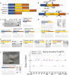
Figure 2. Targeted deletion, duplication and inversion for two rat genes.
(A) Variety of alleles detected in founders F0 and F1 when two pairs of sgRNAs for two sites flanking a region of interest are used. In blue the genomic region 5′ of the region of interest, in yellow the region of interest and in orange the genomic region 3′ of the region of interest. (B) Diagram for rat Cbs and Dyrk1a genes. Shown are the position of the primers used for genotyping, and position of the sgRNAs (C,D) concern Cbs. (C) Shown are deletion junction chromatograms of 3 different founders. Only one deleted allele was obtained in F0-274 (one band detected by PCR) whereas 2 deleted alleles were detected for founders F0-268 and -278 (2 bands observed by PCR and confirmed by a close analyze of the chromatograms). For founder F0-278, a bold arrow faint arrow shows the two distinct deletion alleles (see also Supplementary data S1). Both alleles passed germ line. (D) Shown are duplication and inversion junctions for Cbs amplified by PCR from ear clipping rat founder F0-264 with specific primers pairs. An example of sequence chromatograms of the inversion and duplication junctions is shown. The same inversion and duplication events were found independently on F1 rats (E,F) concern Dyrk1a (E). The duplication junctions amplified by PCR from ear clipping rat founders F0-384 and F0-386 with specific primers pairs are shown. Inversion junctions of F0-355 founder is shown as an example (see also Supplementary data S2) (F) Detection of rats with one copy of Dyrk1a. Appearance of a small founder F0 (yellow arrow) compared to its wt littermate. Monosomic rats for Dyrk1a were first detected by droplet digital PCR. Two ddPCRs (in the first and last exon) were performed in Dyrk1a gene and the loss of one complete copy was confirmed for F0-356, -368, -378 and F0-385. The junction for F0-378 is detected only with primers located farther from the sgRNAs and shows a more important deletion than expected (see Supplementary Fig. S1).
Figure 3
Figure 3. Targeted deletion and duplication for two rat genomic regions.
(A) Diagrams show both regions of interest (in orange, surrounded by 5′ (blue) and 3′ (brown) regions). Two pairs of sgRNAs flank both regions of interest. Shown are the position of the primers used for genotyping and position of the sgRNAs. (B) Targeted deletion of Umodl2-Prmt2 region (3.6 Mb) in founders. Shown the deletion junction chromatograms obtained after PCR amplification with specific primers pairs (F4 + R1) in rat founders F0-334 and F0-335. (C) Scheme of the 3.6 Mb genomic region with zoom at both extremities. Positions of the sgRNAs and PCR primers are illustrated. ddPCRs were performed on 9 genes (Abcg1, green; Pde9a, black; Cbs, blue; Sik1, brown; Trpm2, purple; Itgb2, pink; Slc19a1, dark blue; Lss, orange and S100b, red) distributed over the 3.6 Mb region. The region highlighted with a large light green arrow is found at the junction of the duplication. Oligonucleotides used (F and R) are shown (D) Droplet digital PCR results. Left panel of the graph shows some F0 results: a single copy of Cbs was detected in F0-782 whereas 1.8 copies of Cbs were detected in F0-802. On the right panal are F1 results: 1 or 3 copies of Cbs were clearly detected on the F0-802 first litter showing that both deletion and duplication went germ line. (E) Confirmation of the tandem duplication and deletion by junction sequencing. A 590 bp inversed fragment corresponding to the region 5′ of the first pair of sgRNA is found at the duplication junction. A small (70 bp) inversed sequence is found at the deletion junction (see Supplementary data S3D and E for details) (F) ddPCR results with 9 primer pairs on 2 F1 pups (291-DEL and 294-DUP) confirmed the deletion and duplication of the whole genomic region; a WT rat was taken as control (2 copies) (G) Targeted deletion and duplication of >24 Mb region in a stillborn rat F0. Shown are duplication and deletion junctions amplified by PCR on a stillborn rat F0 (Supplementary nts in gray). The gel pictures of both duplication and deletion junctions are presented.
Figure 4
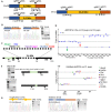
Figure 4. Mouse models obtained by CRISMERE.
Diagrams are as in Fig. 3. Shown are junctions amplified by PCR from mouse tail with specific primers. Examples of sequence chromatograms of the different junctions are shown. (A) Generation of monosomic Hmgn1 mouse line. The chromatogram of the deletion junction observed in F0-3 is shown (details in supplementary data S6). Germ line transmission was confirmed in 7 out of 15 offspring. (B,C) Deletion and duplication of mouse Tiam1 (226 kb). (B) Shown the chromatogram of the duplication junction observed in F0-14, the gel picture of the PCR products from F0-14 and a F1-30. Graph showing ddPCR results on both F0-14, 7 F1 offspring and a WT animal. F0-14 mosaicism is shown as 2.3 Tiam1 copies are detected with 2 distinct ddPCRs (5′ and 3′ of Tiam1, red and blue dots). Germ line transmission was confirmed by ddPCRs (3 copies detected in F1-30). (C) Shown the chromatogram of the deletion junction (primers F2 + R4) of F0-41 and gel picture with the PCR for F0-38, F0-40 and F0-41. Germ line transmission for the deletion was confirmed by ddPCR (F1-30 to F1-34; one copy detected) whereas the founder (F0-41) had a mosaic profile (1.7 copies of Tiam1). Note also a duplicated allele (3 copies of Tiam1) is also detected in F0-41 offspring (F1-28). A single copy of Tiam1 was detected in F0-38 by ddPCR (next graph) suggesting absence of mosaicism. The same ddPCR profile was observed for 2 of the offspring (F1-15 and -13). (D) Targeted deletion and inversion of the mouse genomic region ranging from Runx1 to Cbr1 (1 Mb). Shown the chromatogram of the deletion junction amplified by PCR from F0-32 tail (F1 + R3) with accompanying gel picture. Inversion of the whole region is confirmed by PCR amplification and Sanger sequencing of the 5′ junction on F0-7 (F1 + F4). Droplet digital PCRs on 3 genes distributed over the 1 Mb region (Runx1, blue; Gm28003, red and Cbr1, green) detected 3 copies in F0-138 as well as some of its offspring (F1-3, -6 and -7). A WT control (2 copies) and a deletion (1 copy) are included.
Figure 5
Figure 5. CRISpr MEdiated REarrangement mecanisms (CRISMERE) sgRNA pairs are illustrated by red arrows.
Chromosomes are represented by a thick gray line, the centromeres by a gray dot and the region of interest in yellow. (AC) Standard chromosomic recombination when Cas9 edits the genome in G1. After mitosis, two alleles distinct from the initial WT allele will be obtained (A) Intra-chromosomal recombination between two DSBs on a single chromosome (B) Trans-allelic recombination between two DSBs each on one of the two chromosome. (C) Trans-allelic recombination between (three or) four DSBs on the two chromosomes ending with head to head, tail to tail duplication (D) Schematic of the event that should take place in the eggs were Cas9 edits the genome in Cis configuration in G2 leading to monosomic and trisomic daughter cells after mitosis. Trans-allelic recombination between two DSBs on the two chromatids in G2 (similar to targeted asymmetric sister chromatid event of recombination TASCER).

Similar articles

Cited by

References

    1. Dierssen M., Herault Y. & Estivill X. Aneuploidy: from a physiological mechanism of variance to Down syndrome. Physiol Rev 89, 887–920 (2009). - PubMed
    1. Choong X. Y., Tosh J. L., Pulford L. J. & Fisher E. M. Dissecting Alzheimer disease in Down syndrome using mouse models. Front Behav Neurosci 9, 268 (2015). - PMC - PubMed
    1. Lana-Elola E. et al.. Genetic dissection of Down syndrome-associated congenital heart defects using a new mouse mapping panel. Elife 5 (2016). - PMC - PubMed
    1. Brault V. et al.. Opposite phenotypes of muscle strength and locomotor function in mouse models of partial trisomy and monosomy 21 for the proximal Hspa13-App region. PLoS Genet 11, e1005062 (2015). - PMC - PubMed
    1. Jiang X. et al.. Genetic dissection of the Down syndrome critical region. Hum Mol Genet (2015). - PMC - PubMed

Publication types