. 2017 Mar 7:7:43418.
doi: 10.1038/srep43418.

Mineral Surface Chemistry and Nanoparticle-aggregation Control Membrane Self-Assembly

Affiliations

Mineral Surface Chemistry and Nanoparticle-aggregation Control Membrane Self-Assembly

Nita Sahai et al. Sci Rep. .

Abstract

The self-assembly of lipid bilayer membranes to enclose functional biomolecules, thus defining a "protocell," was a seminal moment in the emergence of life on Earth and likely occurred at the micro-environment of the mineral-water interface. Mineral-lipid interactions are also relevant in biomedical, industrial and technological processes. Yet, no structure-activity relationships (SARs) have been identified to predict lipid self-assembly at mineral surfaces. Here we examined the influence of minerals on the self-assembly and survival of vesicles composed of single chain amphiphiles as model protocell membranes. The apparent critical vesicle concentration (CVC) increased in the presence of positively-charged nanoparticulate minerals at high loadings (mg/mL) suggesting unfavorable membrane self-assembly in such situations. Above the CVC, initial vesicle formation rates were faster in the presence of minerals. Rates were correlated with the mineral's isoelectric point (IEP) and reactive surface area. The IEP depends on the crystal structure, chemical composition and surface hydration. Thus, membrane self-assembly showed rational dependence on fundamental mineral properties. Once formed, membrane permeability (integrity) was unaffected by minerals. Suggesting that, protocells could have survived on rock surfaces. These SARs may help predict the formation and survival of protocell membranes on early Earth and other rocky planets, and amphiphile-mineral interactions in diverse other phenomena.


Conflict of interest statement

The authors declare no competing financial interests.

Figures

Figure 1
Figure 1. Mineral-vesicle interaction imaged by cryo-TEM.
Electron dense areas are the minerals (A) montmorillonite; (B) anatase; (C) goethite and (D) zincite. Suspensions were prepared by mixing 5 mg.mL−1 mineral in HEPES buffer (pH 7.1) with 75 mM DA micelles (100 mM for zincite). The pH was adjusted to 7.8 with NaOH and the suspension was interacted overnight on a rotor/mixer. Vesicle sizes range from 50 to 1000 nm.
Figure 2
Figure 2. Initial rates of vesicle formation normalized to the mineral surface area (r0) as a function of IEP of the minerals.
(A) DA vesicle formation from micelles at final pH 7.1 ± 0.1; (B) DA/DOH vesicle formation from rehydrating lipid thin films at pH 8.1 ± 0.2. Error bars represent the standard deviation of the slope calculated using the LINEST function of excel 2013 (see Methods and Extended Data Figures 3 and 4 for details). (C,D) Extent of initial rate enhancement per m2 of a mineral as compared to the no-mineral control. The dashed blue and red lines, respectively, indicate the trends for the nano and micro-meter sized particle.
Figure 3
Figure 3. Schematic of the proposed model for mineral enhancing rate of vesicle formation.
In the first step, bilayers formed by rehydration of lipid thin films or micelles rapidly adsorb on mineral surfaces forming islands that partially coat the mineral. A greater amount of adsorption of negatively-charged lipids occurs on positively-charged mineral than on negatively-charged minerals. In the second step, the lipid islands partially-coating the mineral act as a template for further attachment of lipid from solution and self-assemble to form vesicles. Thus, the lipid islands catalyze vesicle formation. The more the adsorbed lipid, the greater is the observed rate-enhancement effect. The adsorbed islands may stack up several nanometers away from the surface.
Figure 4
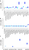
Figure 4. Summary of mineral effects on CVC of DA/DOH (2:1).
CVC as determined by (A) DLS and (B,C) fluorescence, at pH 8.1 and particle loadings of 0.1 and 1.0 mg.mL−1. Little or no effect was observed except in the case of siderite, γ-alumina and goethite at high loading; (C) effect of settling of positively-charged minerals compared to negatively-charged silica which provides a positive control. See Methods and Extended Data Figures 7 and 8 for details.

Similar articles

Cited by

References

    1. Costerton J. W. et al.. Bacterial biofilms in nature and disease. Annu. Rev. Microbiol. 41, 435–464 (1987). - PubMed
    1. Rao S. Acid stress and aquatic microbial interactions (CRC Press, 1989).
    1. Nealson K. H. Sediment bacteria: who’s there, what are they doing, and what’s new? Annu. Rev. Earth Planet. Sci. 25, 403–434 (1997). - PubMed
    1. Deamer D. W. & Oro J. Role of lipids in prebiotic structures. Biosystems 12, 167–175 (1980). - PubMed
    1. Oró J., Miller S. L. & Lazcano A. The origin and early evolution of life on Earth. Annual Annu. Rev. Earth Planet. Sci. 18, 317 (1990). - PMC - PubMed

Publication types

LinkOut - more resources