. 2017 Mar 7:8:14612.
doi: 10.1038/ncomms14612.

Misrouting of v-ATPase subunit V0a1 dysregulates lysosomal acidification in a neurodegenerative lysosomal storage disease model

Affiliations

Misrouting of v-ATPase subunit V0a1 dysregulates lysosomal acidification in a neurodegenerative lysosomal storage disease model

Maria B Bagh et al. Nat Commun. .

Abstract

Defective lysosomal acidification contributes to virtually all lysosomal storage disorders (LSDs) and to common neurodegenerative diseases like Alzheimer's and Parkinson's. Despite its fundamental importance, the mechanism(s) underlying this defect remains unclear. The v-ATPase, a multisubunit protein complex composed of cytosolic V1-sector and lysosomal membrane-anchored V0-sector, regulates lysosomal acidification. Mutations in the CLN1 gene, encoding PPT1, cause a devastating neurodegenerative LSD, INCL. Here we report that in Cln1-/- mice, which mimic INCL, reduced v-ATPase activity correlates with elevated lysosomal pH. Moreover, v-ATPase subunit a1 of the V0 sector (V0a1) requires palmitoylation for interacting with adaptor protein-2 (AP-2) and AP-3, respectively, for trafficking to the lysosomal membrane. Notably, treatment of Cln1-/- mice with a thioesterase (Ppt1)-mimetic, NtBuHA, ameliorated this defect. Our findings reveal an unanticipated role of Cln1 in regulating lysosomal targeting of V0a1 and suggest that varying factors adversely affecting v-ATPase function dysregulate lysosomal acidification in other LSDs and common neurodegenerative diseases.


Conflict of interest statement

The authors declare no competing financial interests.

Figures
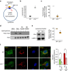
Figure 1
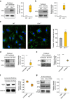
Figure 1. Palmitoylation of V0a1 of v-ATPase and regulation of lysosomal acidification.
(a) Schematic diagram showing the localization of the cytosolic V1 sector (yellow) and V0 sector (blue) of v-ATPase on lysosomal membrane. The V1 sector hydrolyses ATP to ADP generating energy for the lysosomal membrane-anchored V0 sector to translocate protons from the cytosol to the lysosomal lumen for acidification. (b) Lysosomal pH in WT and Cln1−/− neurons. Lysosomal pH was measured using Oregon green dextran and TMR dextran as described in the Methods. Note that compared with WT neurons the lysosomal pH in Cln1−/− cells is significantly elevated (WT: ∼4.88±0.05 versus Cln1−/−: ∼5.96±0.02; n=6, **P<0.01). (c) Enzymatic activity of v-ATPase in purified lysosomal fractions from brain tissues of WT and Cln1−/− mice (n=4), *P<0.05. (d) Western blot analysis of total lysates of HEK-293 cells transfected with the empty vector (mock control), V0a1, mutant (Cys24Ser) V0a1 or mutant (Cys25Ser) V0a1 constructs. ABE assay was used to confirm that Cys-25 but not Cys-24 in V0a1 undergoes palmitoylation in V0a1. (e) Levels of V0a1 in lysosomal fractions isolated from the brain tissues of WT and Cln1−/− mice (n=4, **P<0.01. (f) Confocal imaging of primary neurons from WT and Cln1−/− mice showing colocalization of V0a1 with Lamp2. Colocalization between V0a1 and Lamp2 was assessed using the Manders' colocalization coefficients M1 (green) and M2 (red), which, respectively, represent the overlap with green pixels as the denominator and vice versa (n=23 for WT and n=27 for Cln1−/−, ***P<0.001), scale bars, 5 μm.
Figure 2
Figure 2. Palmitoylation of V0a1 promotes its interaction with AP-2.
(a) Antibody to AP-2 pulls down V0a1 in total brain lysates from WT and Cln1−/− mice (n=4, *P<0.05). (b) Antibody to V0a1 pulls down AP-2 in total brain lysates from WT and Cln1−/− mice (n=4, **P<0.01). (c) Confocal imaging of cultured neurons isolated from WT and Cln1−/−mouse brains were performed to determine interaction of V0a1 with AP-2 by PLA reaction (n=129 for WT and n=62 for Cln1−/−, ***P<0.001). (d,e) Pull-down experiments using either AP-2- or V0a1 antibody were performed to detect V0a1 and AP-2, respectively, in total lysates from untreated and bromopalmitate-treated (Br-palm) WT brain slices (n=5, **P<0.01). (f) Western blot analysis and quantitation of V0a1 in isolated lysosomal fractions from untreated and bromopalmitate (Br-palm)-treated WT brain slices (n=4, *P<0.05). (g) HEK-293 cells were transfected with WT GFP-V0a1 and mutant (Cys25Ser) GFP-V0a1-construct and pull-down experiments were performed using AP-2 antibody to detect GFP-V0a1 (n=4), *P<0.05, scale bars, 20 μm.
Figure 3
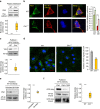
Figure 3. In Cln1−/− mice V0a1 is misrouted to plasma membrane preventing its interaction with AP-3.
(a) Western blot analysis and densitometric quantitation of V0a1 in isolated plasma membrane fraction from WT and Cln1−/− mouse brain (n=4, *P<0.05). (b) Localization of V0a1 in the plasma membrane in WT and Cln1−/− neurons using Na+, K+-ATPase as membrane marker. Colocalization between V0a1 and Na+, K+-ATPase was assessed using the Manders' colocalization coefficients M1 and M2 (n=18 for WT and n=22 for Cln1−/−, ***P<0.001; scale bars, 5 μm. (c) Pull-down assay with AP-3 antibody detects V0a1 in total brain lysates from WT and Cln1−/− mouse brain (n=4, *P<0.05). (d) Confocal imaging of PLA reaction showing V0a1 and AP-3δ interaction in neurons isolated from WT and Cln1−/−mouse brain (n=188 for WT and n=158 for Cln1−/−, ***P<0.001); scale bars, 20 μm. (e) Pull-down assay with AP-3 antibody using total lysates from untreated (lane 1) and bromopalmitate-treated (lane 2) WT brain slices to detect V0a1 and its densitometric quantitation (n=4, *P<0.05). (f) HEK-293 cells were transfected with WT GFP-V0a1 and GFP-V0a1-Cys25Ser mutant construct, and pull-down experiments were conducted with AP-3 antibody to detect GFP-V0a1,*P<0.05(n=4).
Figure 4
Figure 4. Endosomal and lysosomal localization of Ppt1.
(a) Colocalization of Ppt1 with lysosome marker (LAMP 1) in WT and Cln1−/− brain cells (n=34). (b) Colocalization of Ppt1 with early endosome marker (EEA1) in WT and Cln1−/− cells (n=6). (c) Colocalization of Ppt1 with late endosome marker (Rab 9) in WT and Cln1−/− cells (n=5). Colocalization of Ppt1 with the endosomal markers in WT cells was assessed using the Manders' colocalization coefficients M1 and M2. Scale bars, 5 μm.
Figure 5
Figure 5. Localization of V0a1 in the Golgi and in early endosome.
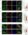
(a) Confocal imaging colocalizing the Golgi marker (GM-130) with V0a1 in cortical neurons from WT and Cln1−/− mouse brain (n=26 for WT and 20 for Cln1−/−). (b) Colocalization of V0a1 with early sorting endosome (Rab-5) using confocal imaging of WT and Cln1−/− neurons (n=31 for WT and 27 for Cln1−/−, **P<0.01, ***P<0.001). (c) Colocalization of V0a1 with early endosome marker (EEA1) in WT and Cln1−/− neuronal cells (n=12 for WT and 10 for Cln1−/−, ***P<0.001). Scale bars, 5 μm.
Figure 6
Figure 6. Endosomal localization of V0a1 and AP-2.
(a) Colocalization of V0a1 with Rab11 (Recycling endosome marker) using confocal imaging of WT and Cln1−/− neurons (n=27 for WT and 24 for Cln1−/−, ***P<0.001). (b) Colocalization of V0a1 with late endosome marker (Rab 9) in isolated neurons from WT and Cln1−/−mouse brain (n=41 for WT and 31 for Cln1−/−, ****P<0.0001). (c) Colocalization of AP-2 with Rab11 in WT and Cln1−/− neurons (n=14 for WT and n=19 for Cln1−/−). *P<0.05, **P<0.01. Scale bars, 5 μm.
Figure 7
Figure 7. S-acylated V0a1 is a potential substrate of Ppt1.
(a) Ppt1 activity in various subcellular fractions (n=4). (b) Confocal imaging of cultured neurons from WT and Cln1−/−mice showing colocalization of Ppt1 immunoreactivity with that of V0a1 in WT neurons only; scale bar, 5 μm. (c) Proximity ligation assay with mouse-V0a1 and rabbit-Ppt1 antibody to show possible interaction between V0a1 and Ppt1. The red dots are the positive signals showing V0a1–Ppt1 interaction in WT cells. Cells from Cln1−/−mice lacking Ppt1 and WT cells with no primary antibody served as the controls (n=252 for WT and n=102 for Cln1−/−). Scale bars, 20 μm. (d) Pull-down experiment with Ppt1 antibody using lysates of HEK-293 cells transfected with FLAG-tagged WT-V0a1, C25A-, C25S- and C58A-mutant constructs.
Figure 8
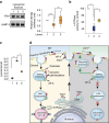
Figure 8. Restoration of near-normal lysosomal pH by NtBuHA in Cln1−/− mice and schematic explaining how V0a1 may be misrouted to plasma membrane in Cln1−/− mice.
(a) Western blot analysis and densitometric quantitation to show that NtBuHA treatment of Cln1−/−mice restores near-normal level of V0a1 in brain, WT (lane 1) versus untreated Cln1−/− (lane 2), ***P<0.001 and untreated Cln1−/− (lane 2) versus NtBuHA-treated Cln1−/− (lane3), **P<0.01, n=8. (b) v-ATPase activity in isolated lysosomal fractions from WT (lane1), untreated (lane 2) and NtBuHA-treated (lane 3) Cln1−/−mouse brain, WT (lane1) versus untreated Cln1−/− (lane 2), *P<0.05 and untreated Cln1−/− (lane 2) versus NtBuHA-treated Cln1−/− (lane3), *P<0.05, n=4. (c) Primary neuronal cells isolated from Cln1−/−mouse brain were treated with a thioesterase-mimetic, NtBuHA, for 7 days and lysosomal pH was measured with Oregon green-dextran and TMR dextran, WT (lane1) versus untreated Cln1−/− (lane 2), *P<0.05 and untreated Cln1−/− (lane 2) versus NtBuHA-treated Cln1−/− (lane3), *P<0.05, n=4. (d) Schematic representation of endosomal sorting and trafficking of V0a1 in WT (left panel) and Cln1−/− (right panel) mice. Note that in Cln1−/− cells the V0a1 fails to dissociate from AP-2, preventing it from interacting with AP-3, which is required for its transport from the sorting endosome to the late endosomal/lysosomal membrane; consequently, the V0a1–AP-2 complex is misrouted via recycling endosome to the plasma membrane. This defect impairs lysosomal v-ATPase activity, thereby dysregulating lysosomal acidification in neurons of Cln1−/− mice.

Similar articles

Cited by

References

    1. de Duve C. & Wattiaux R. Functions of lysosomes. Annu. Rev. Physiol. 28, 435–492 (1966). - PubMed
    1. Mellman I., Fuchs R. & Helenius A. Acidification of the endocytic and exocytic pathways. Annu. Rev. Biochem. 55, 663–700 (1986). - PubMed
    1. Futerman A. H. & van Meer G. The cell biology of lysosomal storage disorders. Nat. Rev. Mol. Cell Biol. 5, 554–565 (2004). - PubMed
    1. Platt F. M., Boland B. & van der Spoel A. C. The cell biology of disease: lysosomal storage disorders: the cellular impact of lysosomal dysfunction. J. Cell Biol. 199, 723–734 (2012). - PMC - PubMed
    1. Holopainen J. M., Saarikoski J., Kinnunen P. K. & Järvelä I. Elevated lysosomal pH in neuronal ceroid lipofuscinoses. Eur. J. Biochem. 268, 5851–5856 (2001). - PubMed

Publication types

Substances

LinkOut - more resources