. 2017 Mar 7:7:43502.
doi: 10.1038/srep43502.

Experimental Evaluation of Kidney Regeneration by Organ Scaffold Recellularization

Affiliations

Experimental Evaluation of Kidney Regeneration by Organ Scaffold Recellularization

Andrea Remuzzi et al. Sci Rep. .

Abstract

The rising number of patients needing renal replacement therapy, alongside the significant clinical and economic limitations of current therapies, creates an imperative need for new strategies to treat kidney diseases. Kidney bioengineering through the production of acellular scaffolds and recellularization with stem cells is one potential strategy. While protocols for obtaining organ scaffolds have been developed successfully, scaffold recellularization is more challenging. We evaluated the potential of in vivo and in vitro kidney scaffold recellularization procedures. Our results show that acellular scaffolds implanted in rats cannot be repopulated with host cells, and in vitro recellularization is necessary. However, we obtained very limited and inconsistent cell seeding when using different infusion protocols, regardless of injection site. We also obtained experimental and theoretical data indicating that uniform cell delivery into the kidney scaffolds cannot be obtained using these infusion protocols, due to the permeability of the extracellular matrix of the scaffold. Our results highlight the major physical barriers that limit in vitro recellularization of acellular kidney scaffolds and the obstacles that must be investigated to effectively advance this strategy for regenerative medicine.


Conflict of interest statement

The authors declare no competing financial interests.

Figures

Figure 1
Figure 1. Explanted renal scaffolds.
(a) Photograph of the transplanted decellularized kidney before (left) and after (middle) unclamping of left renal artery and vein showing homogeneous perfusion of the graft without signs of bleeding. (a, right) Photograph of longitudinal sections of transplanted scaffold after three weeks within Lewis recipient rats. (be) Hematoxylin and eosin staining of the explanted scaffolds demonstrates the presence of intense inflammatory cell infiltration, especially in the renal pericapsular areas and the remaining structures are filled with clotted material (b and c, 3 and 7 days, respectively). At 21 days the acellular scaffolds show less cell infiltration and some ECM degradation (d and e). (b,c,d) Scale bar, 50 μm. (e) Scale bar, 1000 μm.
Figure 2
Figure 2. Repopulation of kidney scaffolds with mES cells via RA and RV for 24 hours.
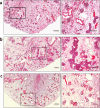
Hematoxylin and eosin staining of representative kidney scaffold seeded with mES cells via the RA (ac) shows that cells are located into glomerular (a and b, left) and occasionally in peritubular capillaries (a and b, right). (a) Scale bar, 10 μm. (b) Scale bar, 50 μm. (c) Mosaic view of a transversal cross section of kidney recellularized by RA demonstrating a homogeneous repopulation of glomeruli and vascular structures in the cortical region. Scale bar, 250 μm. (df) Hematoxylin and eosin staining of kidney scaffold seeded with mES cells via RV shows that glomeruli are devoid of cells (d and e, left). mES cells are preferentially located in peritubular capillaries in the medulla (d and e, right). (d) Scale bar, 10 μm. (e) Scale bar, 50 μm. (f) Mosaic view of a transversal cross-section of kidney recellularized via RV with cells only focally an sparsely located within the scaffold. Scale bar, 250 μm.
Figure 3
Figure 3. Recellularization of kidney scaffolds with mES via RA and U for 24 hours and quantification of seeded cells in kidney scaffolds.
(ac) Hematoxylin and eosin staining of kidney scaffold seeded with mES cells via RA and U shows that cells are located in glomerular and vascular structures in the cortical region (a and b, left), while cells infused through the collecting system partially reached tubular compartments (a and b, right). (a) Scale bar, 10 μm. (b) Scale bar, 50 μm. (c) Mosaic view of a transversal cross section of recellularized kidney demonstrating that most cells were confined to the glomerular capillary level and very few cells reached the tubular compartment. Scale bar, 500 μm. (d) Volume density of seeded cells in cortical (upper) and medullar (lower) regions after infusion of mES cells with different experimental protocols (n = 5 for RA; n = 3 for RV, RA + U, RA + RV + U and HP) and different time points compared to native untreated kidneys (n = 3).
Figure 4
Figure 4. Recellularization of kidney scaffolds with mES via RA, RV and U for 24 hours and 72 hours.
(ac) Hematoxylin and eosin staining of kidney scaffold seeded with mES cells via the RA, RV, and U shows cells located in glomerular, vascular and tubular structures, at 24 hours. (c) Mosaic view of a transversal cross section of recellularized kidney demonstrating that most cells were confined to the cortical region and very few cells reached the medullar region. (df) Hematoxylin and eosin staining of kidney scaffold seeded with mES cells via RA, RV and U and continuously perfused with recirculating culture medium for 72 hours. Cells are located in glomerular, vascular structures with a pattern similar to 24 hours. (f) Mosaic view of a transversal cross section of recellularized kidney. (a and d) Scale bar, 10 μm. (b and d) Scale bar, 50 μm. (c and f) Scale bar, 250 μm.
Figure 5
Figure 5. Recellularization of kidney scaffolds with mES via RA at high pressure (HP).
Mosaic view of transversal cross sections of hematoxylin and eosin stained recellularized kidneys 24 hours, 72 hours and 7 days after seeding. (a) The cells have major distribution within the tubules at 24 hours post-seeding despite being injected through the arterial vasculature. (b) The repopulation of scaffolds was increased 72 hours post-seeding, (c) while there was a decrease in cell numbers after 7 days of perfusion. (a,b,c, left) Scale bar, 250 μm. (a,b,c, right) Scale bar, 125 μm.
Figure 6
Figure 6. Fluid flow and velocity distribution in acellular kidney scaffolds.
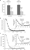
(a) Measurements of perfusion medium exiting from the renal vein as compared to fluid exiting from the ureter and kidney capsule during arterial infusion. (b) Calculated fluid velocity of cell perfusion medium along the entire scaffold, from renal artery to the renal vein for infusion of cell medium at 1.0 ml/min. (c) Calculated fluid velocity in the scaffold circulation for infusion of cell medium at 20 ml/min.

References

    1. Thomas B. et al.. Maintenance Dialysis throughout the World in Years 1990 and 2010. J Am Soc Nephrol 26, 2621–2633 (2015). - PMC - PubMed
    1. Liyanage T. et al.. Worldwide access to treatment for end-stage kidney disease: a systematic review. Lancet 385, 1975–1982 (2015). - PubMed
    1. Couser W. G., Remuzzi G., Mendis S. & Tonelli M. The contribution of chronic kidney disease to the global burden of major noncommunicable diseases. Kidney Int 80, 1258–1270 (2011). - PubMed
    1. Winkelmayer W. C., Weinstein M. C., Mittleman M. A., Glynn R. J. & Pliskin J. S. Health economic evaluations: the special case of end-stage renal disease treatment. Med Decis Making 22, 417–430 (2002). - PubMed
    1. Hart A. et al.. Kidney. Am J Transplant 16 Suppl 2, 11–46 (2016). - PMC - PubMed

Publication types