. 2017 Mar 7:7:43733.
doi: 10.1038/srep43733.

Heterozygous mutation of eEF1A1b resulted in spermatogenesis arrest and infertility in male tilapia, Oreochromis niloticus

Affiliations

Heterozygous mutation of eEF1A1b resulted in spermatogenesis arrest and infertility in male tilapia, Oreochromis niloticus

Jinlin Chen et al. Sci Rep. .

Abstract

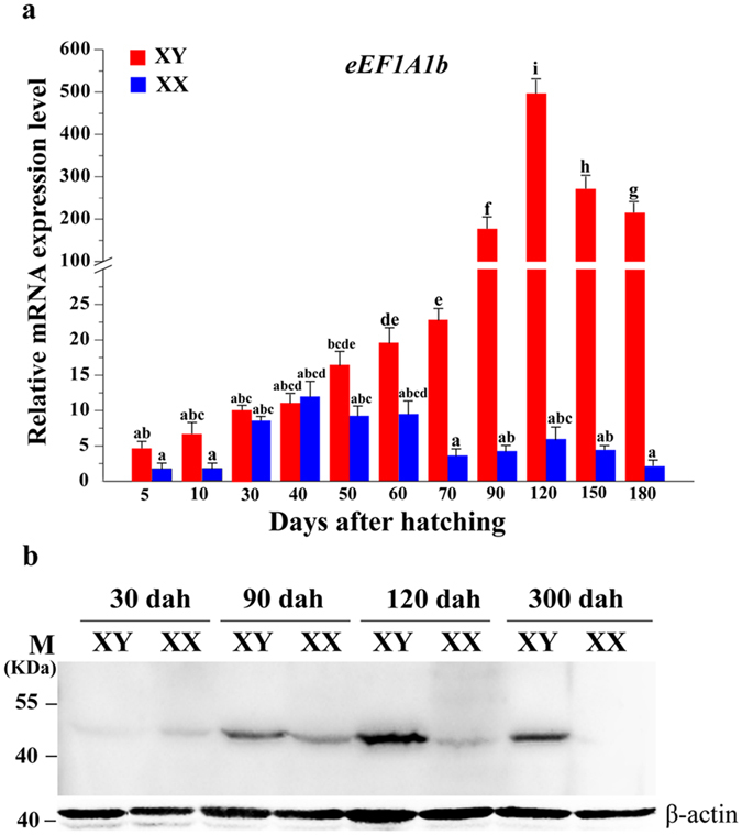
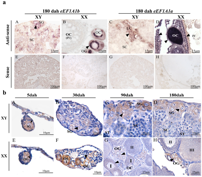
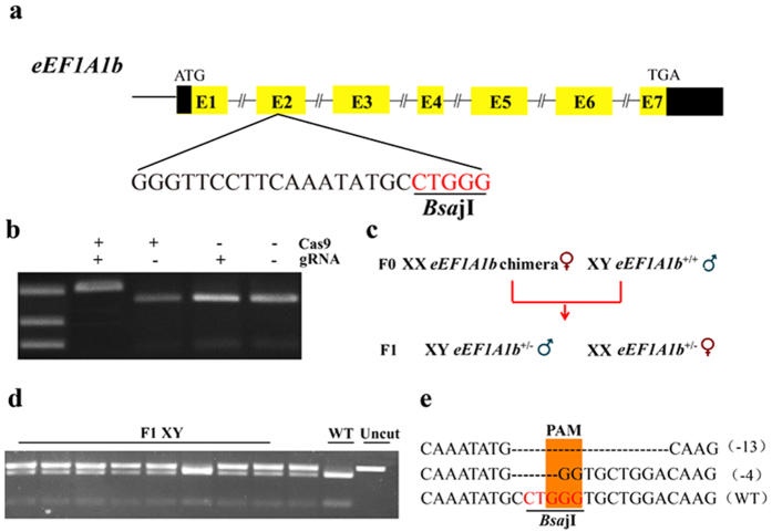
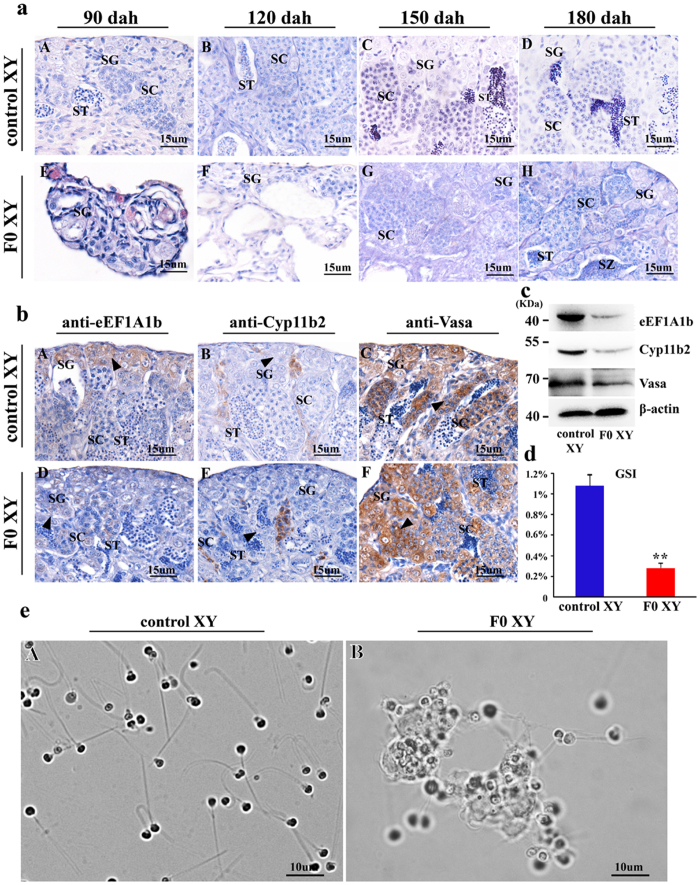
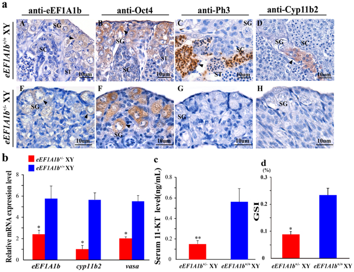
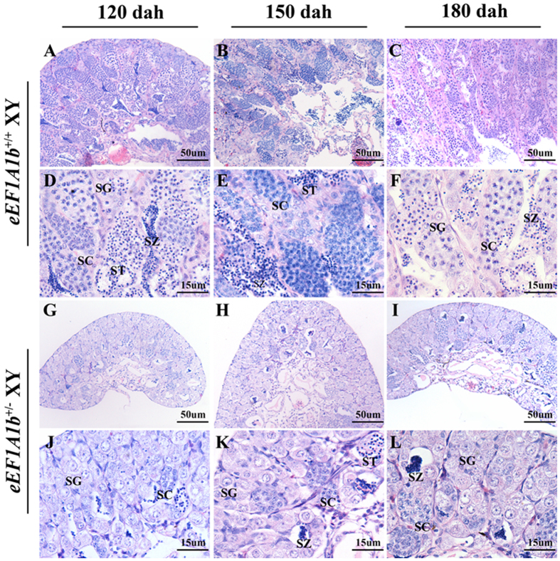
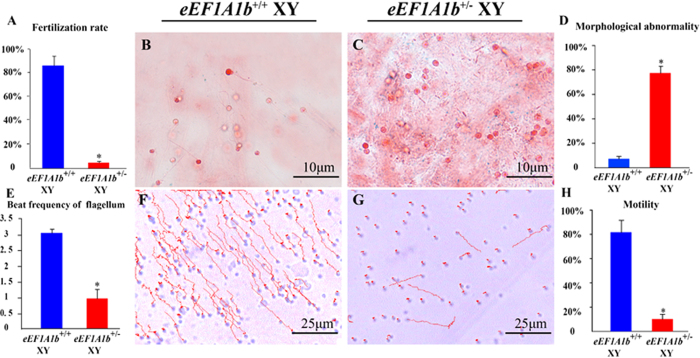
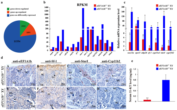
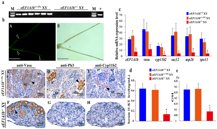
Eukaryotic elongation factor 1 alpha (eEF1A) is an essential component of the translational apparatus. In the present study, eEF1A1b was isolated from the Nile tilapia. Real-time PCR and Western blot revealed that eEF1A1b was expressed highly in the testis from 90 dah (days after hatching) onwards. In situ hybridization and immunohistochemistry analyses showed that eEF1A1b was highly expressed in the spermatogonia of the testis. CRISPR/Cas9 mediated mutation of eEF1A1b resulted in spermatogenesis arrest and infertility in the F0 XY fish. Consistently, heterozygous mutation of eEF1A1b (eEF1A1b+/-) resulted in an absence of spermatocytes at 90 dah, very few spermatocytes, spermatids and spermatozoa at 180 dah, and decreased Cyp11b2 and serum 11-ketotestosterone level at both stages. Further examination of the fertilization capacity of the sperm indicated that the eEF1A1b+/- XY fish were infertile due to abnormal spermiogenesis. Transcriptomic analyses of the eEF1A1b+/- testis from 180 dah XY fish revealed that key elements involved in spermatogenesis, steroidogenesis and sperm motility were significantly down-regulated compared with the control XY. Transgenic overexpression of eEF1A1b rescued the spermatogenesis arrest phenotype of the eEF1A1b+/- testis. Taken together, our data suggested that eEF1A1b is crucial for spermatogenesis and male fertility in the Nile tilapia.


Conflict of interest statement

The authors declare no competing financial interests.

Figures

Figure 1
Figure 1. Expression patterns of eEF1A1b in developing gonads.
(a) Ontogenetic expression of eEF1A1b in tilapia gonads analyzed by real-time PCR. Data are expressed as mean ± SD of three different gonadal pools at each developmental stage. Different letters indicate statistical differences (P < 0.05) as determined by one-way ANOVA followed by post hoc test. (b) Expression of eEF1A1b in gonads of XX and XY fish at 30, 90, 120 and 300 dah by Western blot. Lanes 1, 3, 5 and 7, proteins extracted from testes; lanes 2, 4, 6 and 8, proteins extracted from ovaries. eEF1A1b was detected in both testis and ovary with significant higher expression in testis. 0.2 micrograms of protein were added per lane.
Figure 2
Figure 2. Cellular localization of eEF1A1b in tilapia testis and ovary at different developmental stages by ISH and IHC.
(a) Cell type expressing eEF1A1a and eEF1A1b in tilapia gonads by ISH. eEF1A1b was detected in spermatogonia of the testis, while in the oogonia and phase I oocytes of the ovary at (A and B). eEF1A1a was detected in somatic cell of the testis, while in the oocytes and somatic cells of the ovary (C and D). (b) Cell type expressing eEF1A1b in tilapia gonads by IHC. Consistent with in situ hybridization results, eEF1A1b was detected in the spermatogonia of the testis from 5 to 180 dah (A–D), while in the oogonia of ovary at 5 dah, later in the oogonia and phase I oocytes of the ovary from 30 to 180 dah (E-H). SG, spermatogonia; SC, spermatocytes; ST, spermatids; OG, oogonia; OC, oocytes; I-IV, phase I to phase IV oocytes; Arrowheads indicate the positive signal.
Figure 3
Figure 3. Design of CRISPR/Cas9 targeting eEF1A1b and establishment of the eEF1A1b mutation line.
(a,b) Disruption of tilapia eEF1A1b by CRISPR/Cas9. Gene structure of eEF1A1b showing the target site and the BsajI restriction site. 300 ng/μl of Cas9 mRNA and 150 ng/μl of gRNA were co-injected into one-cell stage embryos. At 72 hours after injection, 20 embryos were randomly selected and pooled to extract their genomic DNA for PCR amplification. The indels were confirmed by two assays, restriction enzyme digestion and Sanger sequencing. The Cas9 mRNA and gRNA were added as indicated. Clear undigested band was detected in embryos injected with both Cas9 mRNA and gRNA compared with the control. (c) Schematic diagram showing the breeding plan of F0 to F1 fish. (d) eEF1A1b+/− F1 generation detected by restriction enzyme digestion. (e) Sanger sequencing results from the uncleaved bands were listed. Deletions are marked by dashes, and the PAM is marked in light orange. Numbers to the right of the sequences indicate the loss or gain of bases for each allele, with the number of bases inserted (+) or deleted (−) indicated in parentheses. WT, wild type.
Figure 4
Figure 4. Effects of eEF1A1b deficiency on spermatogenesis and fertility in F0.
(a) Histological observations of testis from F0 and control XY fish at 90, 120, 150 and 180 dah. eEF1A1b deficiency resulted in spermatogenesis arrest. All kinds of germ cells including spermatogonia, spermatocytes, spermatids and spermatozoa were present at the control testis at 90, 120, 150 and 180 dah (A-D). In contrast, in the F0 testis only spermatogonia were observed at 90 and 120 dah, spermatocytes appeared at 150 dah, and spermatids and spermatozoa appeared at 180 dah (E-H). (b) Expression of eEF1A1b, Cyp11b2 and Vasa in the F0 and control XY fish by IHC. By IHC, eEF1A1b was observed in the control testis (A) while almost undetectable in F0 testis (D). Reduced Cyp11b2 expression was observed in Leydig cells in the F0 testis (B) compared with the control testis (E). Vasa positive signals were detected in both the control and F0 testis (C and F). Arrowheads indicate the positive signal. SG, spermatogonia; SC, spermatocytes; ST, spermatids; SZ, spermatozoa. (c) Expression of eEF1A1b, Cyp11b2 and Vasa in the F0 and control XY fish by Western blot. (d) Gonadal somatic index (GSI) of the F0 and control XY fish. (e) Morphology and motility of sperm from 180 dah F0 and control XY fish. The sperm from the control fish were with normal flagella, and displayed vigorous flagella activity and progressive movement (A); while the sperm from F0 fish were characterized by short or absent flagella. The motility of short-flagella sperms was weak, and the flagella-less sperms were stuck together, showing no motility (B). F0, eEF1A1b deficiency.
Figure 5
Figure 5. Effects of the heterozygous mutation of eEF1A1b on spermatogenesis and fertility.
(a) IHC analyses of eEF1A1b, Oct 4 (spermatogonia maker), Ph3 (spermatocyte maker) and Cyp11b2 expression in the testis of eEF1A1b+/− and eEF1A1b+/+ XY fish at 90 dah. eEF1A1b and Oct 4, Ph3, Cyp11b2 were observed in the spermatogonia, spermatocytes and Leydig cells, respectively, of the eEF1A1b+/+ testis. In contrast, Oct 4 and reduced expression of eEF1A1b were observed in the spermatogonia, while no Ph3 and Cyp11b2 expression was detected in the eEF1A1b+/− testis. Arrowheads indicate the positive signal. SG, spermatogonia; SC, spermatocytes; ST, spermatids. (b) Expression of eEF1A1b, cyp11b2 and vasa in the eEF1A1b+/− and eEF1A1b+/+ XY fish at 90 dah by real-time PCR. (c) Serum 11-KT levels of the eEF1A1b+/− and eEF1A1b+/+ XY fish. (d) GSI of eEF1A1b+/− and eEF1A1b+/+ XY fish at 90 dah. Results were expressed as mean ± SD. Different letters indicate statistical differences at P < 0.05 as determined by one-way ANOVA followed by post hoc test.
Figure 6
Figure 6. Histological observations of testis from eEF1A1b+/ XY fish at 120, 150 and 180 dah.
Abundant spermatocytes, spermatids and spermatozoa were observed in the testis of eEF1A1b+/+ XY fish at 120, 150 and 180 dah (A-F), while just very few spermatocytes were observed at 120 dah, and a markedly reduced number of spermatocytes, spermatids and spermatozoa were observed at 150 and 180 dah in the testis of eEF1A1b+/− XY fish (G-L). D-F and J-L, Higher magnification of the A-C and G-I, respectively. SG, spermatogonia; SC, spermatocytes; ST, spermatids; SZ, spermatozoa.
Figure 7
Figure 7. Sperm analyses of eEF1A1b+/ XY fish at 180 dah.
The fertilization rate of eEF1A1b+/− XY fish was significantly lower than that of eEF1A1b+/+ XY fish (A). Sperm of eEF1A1b+/+ XY fish were with normal flagella, while most sperm of eEF1A1b+/− XY fish were flagella-less, and the ratio of sperm with morphological abnormalities to the total sperm was higher in eEF1A1b+/− XY fish than in eEF1A1b+/+ XY fish (B-D). The beat frequency of sperm flagella was higher in eEF1A1b+/+ XY fish than in eEF1A1b+/− XY fish (E). And the ratio of forward motility sperm was higher in eEF1A1b+/+ XY fish than in eEF1A1b+/− XY fish (F-H). The red line indicates the trajectories of sperm. Results were expressed as mean ± SD. Different letters indicate statistical differences at P < 0.05 as determined by one-way ANOVA followed by post hoc test.
Figure 8
Figure 8. Transcriptomic analyses of gene expression profiles in eEF1A1b+/ testis at 180 dah.
(a) Testis expressed genes were divided into three parts: 1522 down-regulated genes, 1891 up-regulated genes, and 11554 non-differentially expressed genes. (b) Validation of genes with disrupted expression files from transcriptome data by real-time PCR. All examined genes displayed similar expression profiles to those from the transcriptome data. (c) Validation of genes with disrupted expression files from transcriptome data by IHC. Arrowheads indicate the positive signal. SG, spermatogonia; SC, spermatocytes; ST, spermatids; SZ, spermatozoa; LC, Leydig cells. (d) Serum 11-KT levels of the eEF1A1b+/− and eEF1A1b+/+ XY fish.
Figure 9
Figure 9. Transgene mediated rescue of spermatogenesis in eEF1A1b+/ XY fish.
(a) Confirmation of eEF1A1b transgene insertion by genomic PCR. Lane 1 and 14, DNA marker; Lane 2-7, XY fish carrying eEF1A1b-transgene; Lane 8-13, XY control fish; Lane 15, empty plasmid as template. (b) A, the GFP signals in the testis of eEF1A1b+/−/TG XY fish in a dark field; Arrowheads indicate the positive signal. B, bright field; C-H, IHC analyses of Vasa, PH3 and Cyp11b2 expression in the testis of eEF1A1b+/−/TG and eEF1A1b+/− XY fish; SG, spermatogonia; SC, spermatocytes; ST, spermatids. (c) Expression of eEF1A1b, cyp11b2, vasa, suz12, usp26 and spo11 in the eEF1A1b+/−/TG, eEF1A1b+/− and eEF1A1b+/+ XY fish by real-time PCR. (d) Serum 11-KT levels of the eEF1A1b+/−/TG, eEF1A1b+/− and eEF1A1b+/+ XY fish. (e) GSI of eEF1A1b+/−/TG, eEF1A1b+/− and eEF1A1b+/+ XY fish. Results were expressed as mean ± SD. Different letters indicate statistical differences at P < 0.05 as determined by one-way ANOVA followed by post hoc test.

Similar articles

Cited by

References

    1. Mateyak M. K. & Kinzy T. G. eEF1A: thinking outside the ribosome. Journal of Biological Chemistry 285, 21209–21213 (2010). - PMC - PubMed
    1. Koji N., Masataka K., Shigekazu N. & Yoshito K. Structure of the two genes coding for polypeptide chain elongation factor lα (EF-lα) froni Saccharomyces cerevisiae. Gene 45, 265–273 (1986). - PubMed
    1. Hovemann B., Richter S., Walldorf U. & Cziepluch C. Two genes encode related cytoplasmic elongation factors 1α (EF-1α) in Drosophila melanogaster with continuous and stage specific expression. Nucleic Acids Research 16, 3175–3194 (1988). - PMC - PubMed
    1. Danforth B. N. & Ji S. Elongation factor-1 alpha occurs as two copies in bees: implications for phylogenetic analysis of EF-1 alpha sequences in insects. Molecular biology and evolution 15, 225–235 (1998). - PubMed
    1. Gao D., Li Z., Murphy T. & Sauerbier W. Structure and transcription of the gene for translation elongation factor 1 subunit alpha of zebrafish (Danio rerio). Biochimica et Biophysica Acta (BBA)-Gene Structure and Expression 1350, 1–5 (1997). - PubMed

Publication types

MeSH terms

Substances