. 2017 Mar 7:7:43846.
doi: 10.1038/srep43846.

Screening with an NMNAT2-MSD platform identifies small molecules that modulate NMNAT2 levels in cortical neurons

Affiliations

Screening with an NMNAT2-MSD platform identifies small molecules that modulate NMNAT2 levels in cortical neurons

Yousuf O Ali et al. Sci Rep. .

Erratum in

Abstract

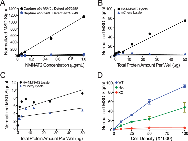
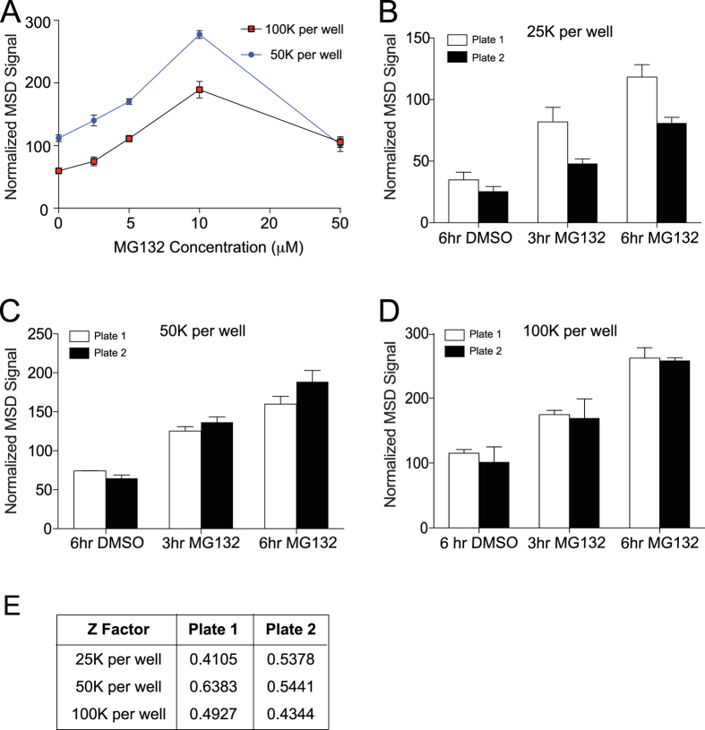
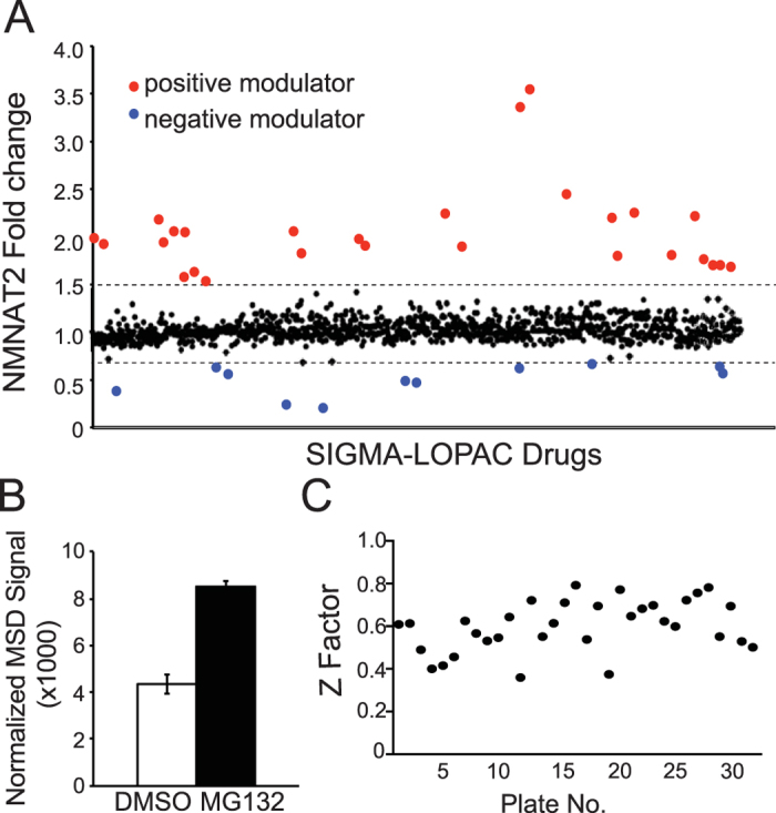
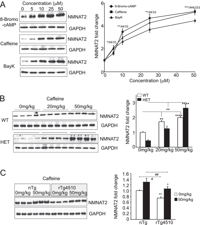
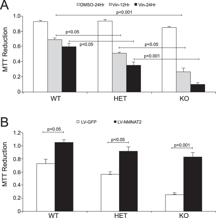
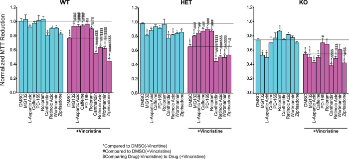
Nicotinamide mononucleotide adenylyl transferase 2 (NMNAT2) is a key neuronal maintenance factor and provides potent neuroprotection in numerous preclinical models of neurological disorders. NMNAT2 is significantly reduced in Alzheimer's, Huntington's, Parkinson's diseases. Here we developed a Meso Scale Discovery (MSD)-based screening platform to quantify endogenous NMNAT2 in cortical neurons. The high sensitivity and large dynamic range of this NMNAT2-MSD platform allowed us to screen the Sigma LOPAC library consisting of 1280 compounds. This library had a 2.89% hit rate, with 24 NMNAT2 positive and 13 negative modulators identified. Western analysis was conducted to validate and determine the dose-dependency of identified modulators. Caffeine, one identified NMNAT2 positive-modulator, when systemically administered restored NMNAT2 expression in rTg4510 tauopathy mice to normal levels. We confirmed in a cell culture model that four selected positive-modulators exerted NMNAT2-specific neuroprotection against vincristine-induced cell death while four selected NMNAT2 negative modulators reduced neuronal viability in an NMNAT2-dependent manner. Many of the identified NMNAT2 positive modulators are predicted to increase cAMP concentration, suggesting that neuronal NMNAT2 levels are tightly regulated by cAMP signaling. Taken together, our findings indicate that the NMNAT2-MSD platform provides a sensitive phenotypic screen to detect NMNAT2 in neurons.


Conflict of interest statement

The authors declare no competing financial interests.

Figures

Figure 1
Figure 1. Development of an NMNAT2-MSD platform specifically detecting NMNAT2 with high sensitivity.
(A) Ab110040 antibody as capture and sulfotagged Ab56980 antibody as the detection antibody pair, but not in reverse order provides, a strong NMNAT2-MSD signal and a linear detection with increasing concentrations of recombinant NMNAT2 protein. (B) Dose-dependent MSD signals for cell lysates of Cos-7 cells transfected with HA-NMNAT2 or mCherry. (C) Low NMNAT2-MSD signals for Cos-7 cells transfected with HA-NMNAT2 or mCherry when the order of capture and detection antibody was reversed. (D) NMNAT2-MSD signals detected from DIV 14 NMNAT2 WT, HET and KO neurons plated at the indicated densities.
Figure 2
Figure 2. The NMNAT2-MSD platform detects dynamic changes in NMNAT2 protein levels in primary cortical neuron cultures in specific manner.
(A) NMNAT2-MSD signals from cortical neurons treated with the indicated concentrations of MG132. (BD) NMNAT2-MSD signals for neurons treated with 3 or 6 hrs of MG132. Two separate plates were examined. Neurons were plated at 25,000 (B), 50,000 (C), or 100,000 (D) cells per well of 96 well plates. (E) Summary of z factors for MG132-enhancement of NMNAT2 levels as measured by MSD.
Figure 3
Figure 3. Identification of NMNAT2 modulators from the Sigma-LOPAC library screen using the NMNAT2-MSD platform.
(A) NMNAT2 fold change from drug treatment of wild type cortical neurons plated in 96 well plates at density of 50 K per well and treated with 10 μM of the library compound for 6 hours. Fold change is represented as change over DMSO control. (B) Summary for NMNAT2-MSD signals detected from neurons treated with DMSO (negative control) and MG132 (positive control) from all the plates included in the data analysis. (C) Z factor distribution for all the assay plates included for data analysis.
Figure 4
Figure 4. Caffeine increases NMNAT2 expression in vitro and in rTg4510 tauopathy mice.
(A) Western blots show NMNAT2 and GAPDH levels in DIV14 cortical neurons after 6 hrs of treatment with 8-Bromo-cAMP, caffeine or Bay-K. Summary for the dose-dependent impact of 8-Bromo-cAMP, caffeine or Bay-K on NMNAT2 levels. *Caffeine/#BayK/$8-bromo-cAMP, */#/$p < 0.05, **/##/$$p < 0.001, ***/###/$$$p < 0.0001 when compared to DMSO control for respective drugs. (B) NMNAT2 levels in the cortex of NMNAT2 WT and HET mice after saline or caffeine treatment (two independent experiments with n = 8 animals per treatment group). (C) NMNAT2 levels in the cortex of rTg4510 mice and controls after saline or caffeine treatment (repeated in two trials, with n = 6 total animals per treatment group). Bar graphs were plotted with mean ± sem; statistical significance was assessed by one-way ANOVA, *p < 0.05, **p < 0.001 (Complete original blots provided in Supplemental Figure S7).
Figure 5
Figure 5. NMNAT2 protects against vincristine induced cell death.
MTT assay shows reduced viability of NMNAT2 HET and KO neurons after vincristine exposure. (A) DIV14 NMNAT2 WT, HET, KO neurons were treated with DMSO or 10 uM vincristine for 12 or 24 hours. Cell viability was assessed with a MTT reduction assay. Experiment was repeated 3 times in triplicates. (B) NMNAT2 WT, HET, KO neurons were transduced with either LV-GFP or LV-NMNAT2 at DIV2. At DIV14, these neurons were treated with 10 uM vincristine for 24 hours. Experiment was repeated 3 times in triplicates. Statistical significance assessed using One-way ANOVA, followed by posthoc Tukey analysis.
Figure 6
Figure 6. NMNAT2-specific neuroprotection assay.
Cell viability was evaluated by MTT reduction. DIV14 NMNAT2 WT, HET, or KO neurons were pretreated with DMSO or 10 uM of the indicated compounds for 6 hours and then exposed to either DMSO (blue) or drug + vincristine (magenta) for additional 6 hours. Statistical significance within each group was assessed by two-tailed T-test, and intergroup differences were assessed by one-way ANOVA. *Compared to DMSO only; #compared to DMSO + vincristine; $compared to drug + DMSO. */#/$p < 0.05, **/##/$$p < 0.001 ***/###/$$$p < 0.0001.

Similar articles

Cited by

References

    1. Coleman M. P. & Freeman M. R. Wallerian degeneration, wld(s), and nmnat. Annu Rev Neurosci 33, 245–267, doi: 10.1146/annurev-neuro-060909-153248 (2010). - DOI - PMC - PubMed
    1. Ali Y. O., Li-Kroeger D., Bellen H. J., Zhai R. G. & Lu H. C. NMNATs, evolutionarily conserved neuronal maintenance factors. Trends Neurosci 36, 632–640, doi: 10.1016/j.tins.2013.07.002 (2013). - DOI - PMC - PubMed
    1. D’Angelo I. et al. Structure of nicotinamide mononucleotide adenylyltransferase: a key enzyme in NAD(+) biosynthesis. Structure 8, 993–1004 (2000). - PubMed
    1. Bork P., Holm L., Koonin E. V. & Sander C. The cytidylyltransferase superfamily: identification of the nucleotide-binding site and fold prediction. Proteins 22, 259–266, doi: 10.1002/prot.340220306 (1995). - DOI - PubMed
    1. Lau C., Niere M. & Ziegler M. The NMN/NaMN adenylyltransferase (NMNAT) protein family. Front Biosci (Landmark Ed) 14, 410–431 (2009). - PubMed

Publication types

MeSH terms

Substances

LinkOut - more resources