. 2017 Mar 7:7:43199.
doi: 10.1038/srep43199.

Commensal gut bacteria modulate phosphorylation-dependent PPARγ transcriptional activity in human intestinal epithelial cells

Affiliations

Commensal gut bacteria modulate phosphorylation-dependent PPARγ transcriptional activity in human intestinal epithelial cells

Malgorzata Nepelska et al. Sci Rep. .

Abstract

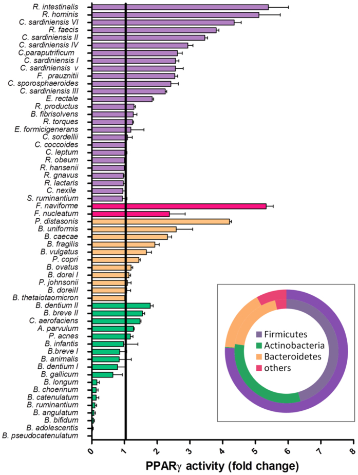
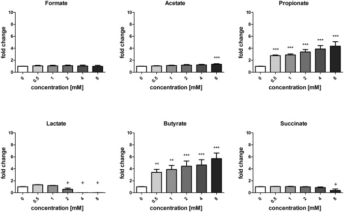
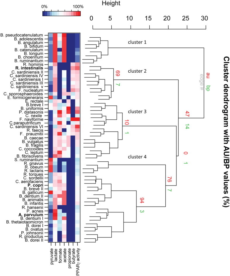
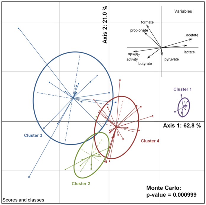
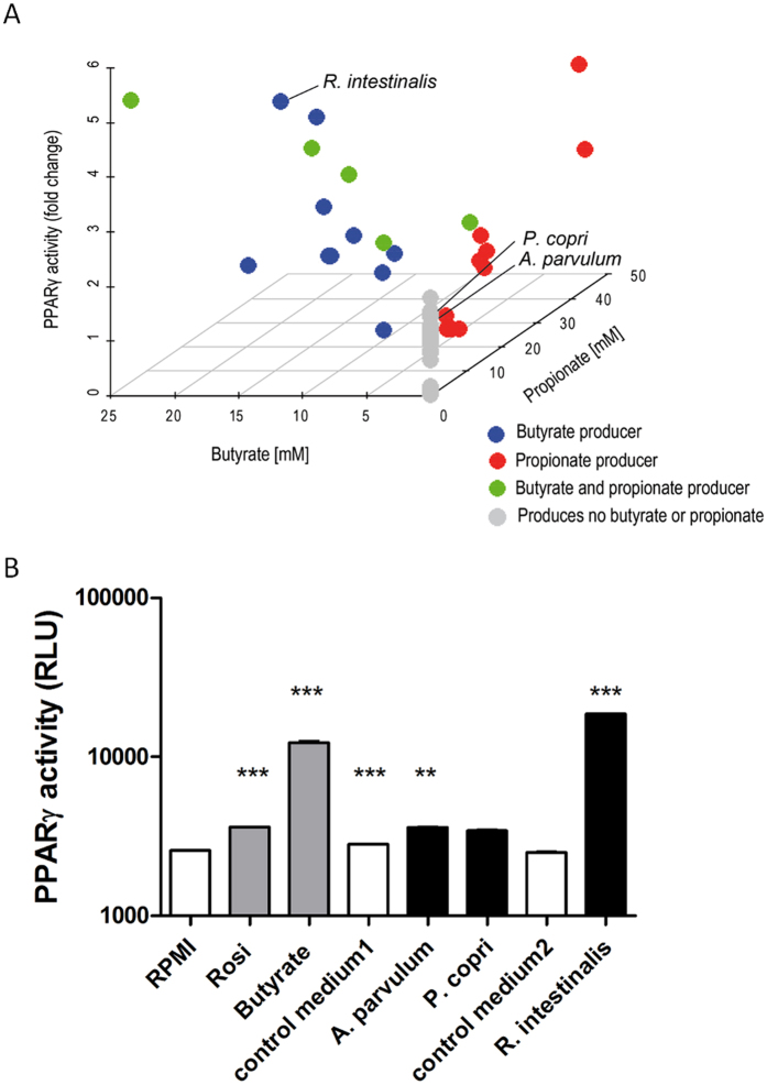
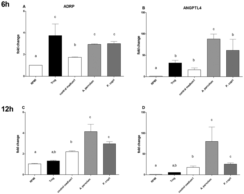
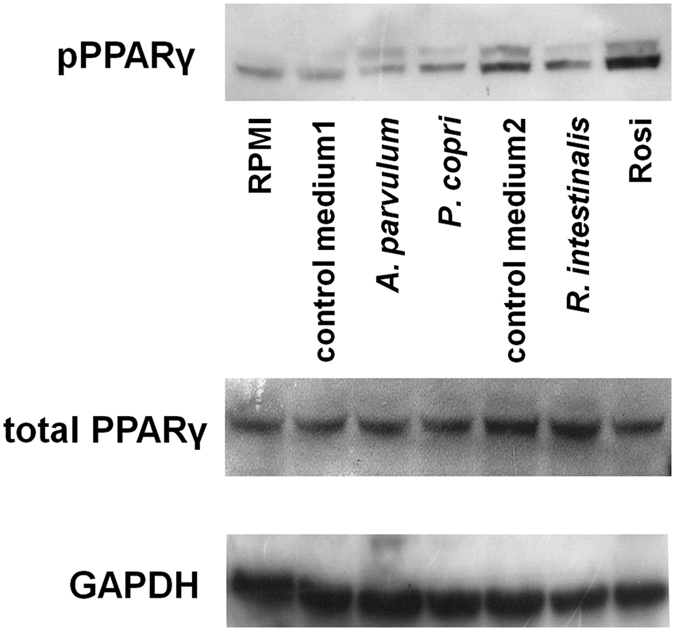
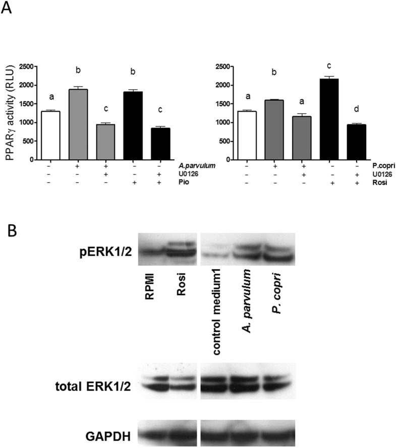
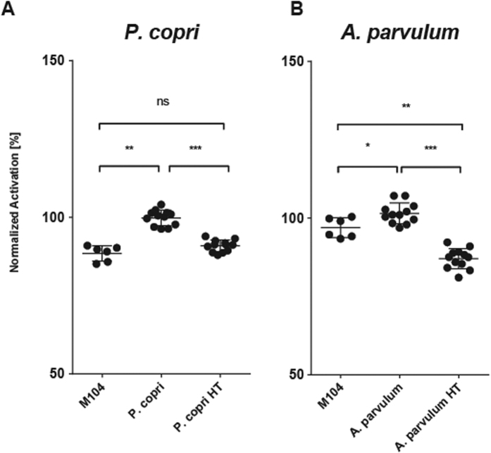
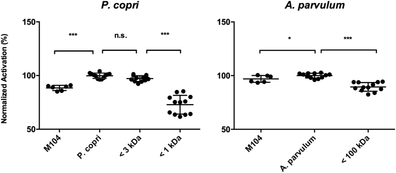
In healthy subjects, the intestinal microbiota interacts with the host's epithelium, regulating gene expression to the benefit of both, host and microbiota. The underlying mechanisms remain poorly understood, however. Although many gut bacteria are not yet cultured, constantly growing culture collections have been established. We selected 57 representative commensal bacterial strains to study bacteria-host interactions, focusing on PPARγ, a key nuclear receptor in colonocytes linking metabolism and inflammation to the microbiota. Conditioned media (CM) were harvested from anaerobic cultures and assessed for their ability to modulate PPARγ using a reporter cell line. Activation of PPARγ transcriptional activity was linked to the presence of butyrate and propionate, two of the main metabolites of intestinal bacteria. Interestingly, some stimulatory CMs were devoid of these metabolites. A Prevotella and an Atopobium strain were chosen for further study, and shown to up-regulate two PPARγ-target genes, ANGPTL4 and ADRP. The molecular mechanisms of these activations involved the phosphorylation of PPARγ through ERK1/2. The responsible metabolites were shown to be heat sensitive but markedly diverged in size, emphasizing the diversity of bioactive compounds found in the intestine. Here we describe different mechanisms by which single intestinal bacteria can directly impact their host's health through transcriptional regulation.


Conflict of interest statement

The authors declare no competing financial interests.

Figures

Figure 1
Figure 1. Effect of conditioned media (CM) on transcriptional activity of PPARγ in HT-29-PPARγ reporter cells.
Activation expressed as the fold increase towards its control (growth-medium) are represented as bar plot. Bacteria are sorted by response in decreasing order and grouped by phylum (violet = Firmicutes, pink = Fusobacteria, yellow = Bacteroidetes, green = Actinobacteria). Distribution of the bacterial phyla is represented in the doughnut chart on the bottom right. The inner circle represents the screened collection, the outer circle the phyla representation in the human colon based on the MetaHIT data.
Figure 2
Figure 2. Dose-response of organic acids and SCFA on PPARγ activation.
Fold change represents the readout of the reporter cell line (relative light units: RLU) normalized to the non-treated control. HT-29-PPARγ cells were exposed to formate, succinate, lactate, acetate, propionate, butyrate in concentrations rising from 0.5 to 8 mM for 24 h. Data are represented as mean ± standard error of the mean (SEM) of triplicate measurement of three independent experiments. ***P < 0.001, **P < 0.005, compared with the control (Student’s t-test). +Significant decrease due to impaired cell viability.
Figure 3
Figure 3. Bacterial species and their OAs pattern cluster with PPARγ response.
Clustering using pvclust with AU (Approximately Unbiased) p-value and BP (Bootstrap Probability). The heat-map represents the PPARγ response and OA concentrations in percent of the highest value for the respective variable.
Figure 4
Figure 4. Inter-class PCA using classes defined by previous cluster analysis.
The PCA separates the 4 clusters significantly (Monte-Carlo test: p = 0.000999). Variables are plotted on the same components (upper right of the graph) showing their importance in the separation of the clusters.
Figure 5
Figure 5
SCFA independent activators of PPARγ (A) PPARγ activation plotted against butyrate and propionate concentrations. CMs containing butyrate but no propionate are represented in blue, CMs containing propionate but no butyrate are represented in red, CMs containing both propionate and butyrate are represented in green and CMs deprived of these two SCFA are represented in grey. (B) Activation of PPARγ pathway by chosen bacteria on colonic reporter cell line HT-29-PPARγ. Rosiglitazone (Rosi, 5 μM) is used as control for activation. Control medium 1 is the medium used to culture of A. parvulum and P. copri (M104). Control medium 2 is the medium used to culture R. intestinalis (M58). Data are represented as mean ± standard error of the mean (SEM) of triplicate measurement of a representative of three independent experiments. ***P < 0.001, **P < 0.005, compared with the control media (Student’s t-test).
Figure 6
Figure 6. Transcriptional regulation of PPARγ target genes upon stimulation with chosen CMs.
Up-regulation of mRNA for ADRP (A,C) and ANGPTL4 (B,D) by chosen CMs. The expression determined by Quantitative real-time PCR on total RNA extracted from cells exposed to CMs for 6 h (A,B) and 12 h (C,D) higher and lower panel respectively. Expression is represented as fold change compared to the absence of any stimulation (RPMI cell culture medium only). Data are represented as mean ± standard error of the mean (SEM) of triplicates of one representative experiment of three independent repetitions. Data were analyzed applying an ANOVA test followed by a post-hoc Tuckey test. Bars superscripted with different letters have a difference of at least p < 0.05.
Figure 7
Figure 7. Phosphorylation of PPARγ by A. parvulum and P. copri supernatants.
A. parvulum, P. copri and rosiglitazone (10 μM) induce PPARγ phosphorylation as compared to its growth medium M104. The medium M58 used for the culture of R. intestinalis shows strong activation capacity itself and therefore no increase of PPARγ phosphorylation can be observed. The nuclear fraction proteins were blotted (Western Blot) for phosphorylated PPARγ. Total PPARγ was used as control for phosphorylated PPARγ. GAPDH was used as loading control.
Figure 8
Figure 8. Implication of ERK in the activation of PPARγ.
(A) The A. parvulum and P. copri -induced signal were inhibited using the specific inhibitor for MEK/ERK (U0126) on HT-29-PPARγ cells. Different letters indicate statistically different results (p < 0.05). (B) The implication of MEK/ERK in PPARγ activation by A. parvulum and P. copri was confirmed on the protein level showing phosphorylation of ERK1/2. Total ERK1/2 was used as control for phosphorylated PPARγ. GAPDH was used as loading control.
Figure 9
Figure 9. Loss of PPARγ activation upon heat treatment of conditioned media.
P. copri (A) and A. parvulum (B) conditioned media were incubated for 10 min at 100 °C to test the heat-stability of the PPARγ activating compound. In comparison with the culture medium M104 both P. copri (A) and A. parvulum (B) show significant activation of CM. The activation of PPARγ is lost after the heat treatment (HT). Experiments were performed in triplicates using two independent bacterial cultures and normalized to the activating CMs indicated as 100%. Data were analyzed applying an ANOVA test followed by a post-hoc Tuckey test. Significance levels are indicated as follows: ***P < 0.001, **P < 0.005, *P < 0.05.
Figure 10
Figure 10. Identification of the size of PPARγ activating compound in the CMs.
A. parvulum and P. copri CMs were fractions into fractions >100 kDa, >50 kDa, >30 kDa, >10 kDa, >3 kDa and >1 kDa. The lowest fraction showing activation on PPARγ reporter cell lines and the highest fraction showing loss of activity are represented for P. copri (A) and A. parvulum (B). The activating compound produced by P. copri is smaller than 3 kDa and bigger then 1 kDa. A. parvulum loses activity significantly after filtering using a 100 kDa filter. Experiments were performed in triplicates using two independent bacterial cultures and normalized to the activating CMs. Data were analyzed applying an ANOVA test followed by a post-hoc Tuckey test. Significance levels are indicated as follows: ***P < 0.001, **P < 0.005, *P < 0.05.

Similar articles

Cited by

References

    1. Sekirov I., Russell S. L., Antunes L. C. M. & Finlay B. B. Gut Microbiota in Health and Disease. Physiol. Rev. 90, 859–904 (2010). - PubMed
    1. Cerf-Bensussan N. & Gaboriau-Routhiau V. The immune system and the gut microbiota: friends or foes? Nat. Rev. Immunol. 10, 735–44 (2010). - PubMed
    1. Cummings J. H. Microbial Digestion of Complex Carbohydrates in Man. Proc. Nutr. Soc. 43, 35–44 (1984). - PubMed
    1. Albert M. J., Mathan V. I. & Baker S. J. Vitamin B12 synthesis by human small intestinal bacteria. Nature 283, 781–782 (1980). - PubMed
    1. Bäckhed F. et al. The gut microbiota as an environmental factor that regulates fat storage. Proc. Natl. Acad. Sci. USA 101, 15718–23 (2004). - PMC - PubMed

Publication types

MeSH terms